. 2013 Aug;38(4):2611-20.
doi: 10.1111/ejn.12259. Epub 2013 May 28.

Fear extinction deficits following acute stress associate with increased spine density and dendritic retraction in basolateral amygdala neurons

Affiliations

Fear extinction deficits following acute stress associate with increased spine density and dendritic retraction in basolateral amygdala neurons

Mouna Maroun et al. Eur J Neurosci. 2013 Aug.

Abstract

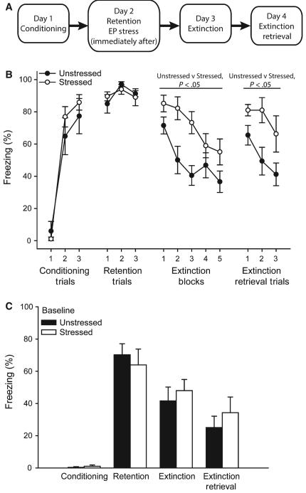
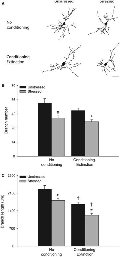
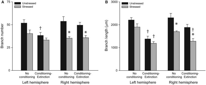
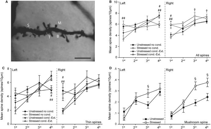
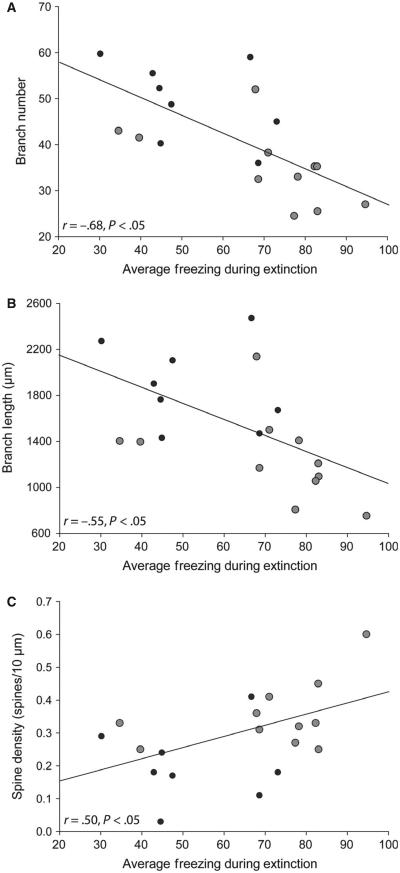
Stress-sensitive psychopathologies such as post-traumatic stress disorder are characterized by deficits in fear extinction and dysfunction of corticolimbic circuits mediating extinction. Chronic stress facilitates fear conditioning, impairs extinction, and produces dendritic proliferation in the basolateral amygdala (BLA), a critical site of plasticity for extinction. Acute stress impairs extinction, alters plasticity in the medial prefrontal cortex-to-BLA circuit, and causes dendritic retraction in the medial prefrontal cortex. Here, we examined extinction learning and basolateral amygdala pyramidal neuron morphology in adult male rats following a single elevated platform stress. Acute stress impaired extinction acquisition and memory, and produced dendritic retraction and increased mushroom spine density in basolateral amygdala neurons in the right hemisphere. Unexpectedly, irrespective of stress, rats that underwent fear and extinction testing showed basolateral amygdala dendritic retraction and altered spine density relative to non-conditioned rats, particularly in the left hemisphere. Thus, extinction deficits produced by acute stress are associated with increased spine density and dendritic retraction in basolateral amygdala pyramidal neurons. Furthermore, the finding that conditioning and extinction as such was sufficient to alter basolateral amygdala morphology and spine density illustrates the sensitivity of basolateral amygdala morphology to behavioral manipulation. These findings may have implications for elucidating the role of the amygdala in the pathophysiology of stress-related disorders.

Keywords: amygdala; dendritic spine; emotional learning; rat.


Figures

F<sc>IG</sc>. 1
FIG. 1
Acute stress impairs extinction. (A) Schematic of experimental design. (B) Percentage freezing (mean ± standard error of the mean) in response to a conditioned stimulus during conditioning, fear retention, extinction and extinction retrieval in rats that were either unstressed (n = 8) or stressed (n = 10) or after fear retention testing. (C) Percentage freezing during the 120-s baseline period during conditioning, fear retention, extinction and extinction retrieval in rats that were either stressed or unstressed after fear retention testing.
F<sc>IG</sc>. 2
FIG. 2
Acute stress produces dendritic retraction in basolateral amygdala (BLA) pyramidal neurons. (A) Reconstructions of representative BLA pyramidal neurons from unstressed vs. stressed rats that were either non-conditioned or underwent fear conditioning and extinction. Neurons are at or near the mean for each group. Scale bar: 50 μm. (B) Total branch number [mean ± standard error of the mean (SEM)] for BLA pyramidal neurons in unstressed (black bars) vs. stressed (gray bars) rats that were either non-conditioned or underwent fear conditioning and extinction. *P < 0.05 relative to unstressed within the behavioral testing condition. †p < 0.05 relative to non-conditioned within the stress condition. (C) Total branch length (mean ± SEM) for BLA pyramidal neurons in unstressed (black bars) vs. stressed (gray bars) rats that were either non-conditioned or underwent fear conditioning and extinction. *P < 0.05 relative to unstressed within the testing condition. †P < 0.05 relative to untested within the stress condition. Unstressed, no conditioning, n = 5. Stressed, no conditioning, n = 4. Unstressed, conditioning and extinction, n = 8. Stressed, conditioning and extinction, n = 10.
F<sc>IG</sc>. 3
FIG. 3
Acute stress produces dendritic retraction in the right basolateral amygdala (BLA), whereas fear conditioning and extinction produces dendritic retraction in the left BLA. (A) Total branch number [mean ± standard error of the mean (SEM)] for right and left BLA pyramidal neurons in unstressed (black bars) vs. stressed (gray bars) rats that were either non-conditioned or underwent fear conditioning and extinction. *P < 0.05 relative to unstressed within the learning condition. †P < 0.05 relative to untested within the stress condition. (B) Total branch length (mean ± SEM) for right and left BLA pyramidal neurons in unstressed (black bars) vs. stressed (gray bars) rats that were either non-conditioned or underwent fear conditioning and extinction. *P < 0.05 relative to unstressed within the learning condition. †P < 0.05 relative to untested within the stress condition. Unstressed, no conditioning, n = 5. Stressed, no conditioning, n = 4. Unstressed, conditioning and extinction, n = 8. Stressed, conditioning and extinction, n = 10.
F<sc>IG</sc>. 4
FIG. 4
Acute stress and fear conditioning and extinction alter spine density on basolateral amygdala (BLA) pyramidal neurons. (A) Digital micrograph demonstrating different spine types on a pyramidal neuron in the BLA. Several photomicrographs were taken at different Z-levels, and merged to increase the number of spines in focus. B, branched; M, mushroom; S, stubby; T, thin. Scale bar: 5 μm. (B) Spine density [mean ± standard error of the mean (SEM)] on first-order to fourth-order branches of BLA pyramidal neurons in the right and left hemispheres in unstressed vs. stressed rats that were either non-conditioned or underwent fear conditioning and extinction. †P < 0.05 for unstressed vs. stressed non-conditioned rats. #P < 0.05 for unstressed non-conditioned rats vs. unstressed rats that underwent conditioning and extinction. ##P < 0.05 for stressed non-conditioned rats vs. stressed rats that underwent conditioning and extinction. (C) Thin spine density (mean ± SEM) on first-order to fourth-order branches of BLA pyramidal neurons in the right and left hemispheres in unstressed vs. stressed rats that were either non-conditioned or underwent fear conditioning and extinction. #P < 0.05 for unstressed non-conditioned rats vs. unstressed rats that underwent conditioning and extinction. ##P < 0.05 for stressed non-conditioned rats vs. stressed rats that underwent conditioning and extinction. (D) Mushroom spine density (mean ± SEM) on first-order to fourth-order branches of BLA pyramidal neurons in the right and left hemispheres in unstressed vs. stressed rats collapsed across learning conditions. §P < 0.05 for unstressed vs. stressed rats. Unstressed, no conditioning, n = 5. Stressed, no conditioning, n = 4. Unstressed, conditioning and extinction, n = 8. Stressed, conditioning and extinction, n = 10.
F<sc>IG</sc>. 5
FIG. 5
Stress-induced dendritic remodeling and alterations in mushroom spine density correlate with extinction deficits. (A) Linear regression for average basolateral amygdala (BLA) branch number for unstressed (black dots; n = 8) and stressed (gray dots; n = 10) rats vs. percentage freezing during extinction (averaged across trials). (B) Linear regression for average BLA dendritic length for unstressed (black dots) and stressed (gray dots) rats vs. percentage freezing during extinction (averaged across trials). (C) Linear regression for average mushroom spine density on third-order and fourth-order branches for unstressed (black dots) and stressed (gray dots) rats vs. percentage freezing during extinction (averaged across trials).

References

    1. Akirav I, Maroun M. The role of the medial prefrontal cortex-amygdala circuit in stress effects on the extinction of fear. Neural Plast. 2007;2007:1–11. - PMC - PubMed
    1. Akirav I, Segev A, Motanis H, Maroun M. D-cycloserine into the BLA reverses the impairing effects of exposure to stress on the extinction of contextual fear, but not conditioned taste aversion. Learn. Memory. 2009;16:682–686. - PubMed
    1. Baran SE, Armstrong CE, Niren DC, Hanna JJ, Conrad CD. Chronic stress and sex differences on the recall of fear conditioning and extinction. Neurobiol. Learn. Mem. 2009;91:323–332. - PMC - PubMed
    1. Berridge CW, Mitton E, Clark W, Roth RH. Engagement in a non-escape (displacement) behavior elicits a selective and lateralized suppression of frontal cortical dopaminergic utilization in stress. Synapse. 1999;32:187–197. - PubMed
    1. Blanchard DC, Blanchard RJ. Innate and conditioned reactions to threat in rats with amygdaloid lesions. J. Comp. Physiol. Psych. 1972;81:281–290. - PubMed

Publication types