. 2014 Feb;22(2):208-15.
doi: 10.1038/ejhg.2013.108. Epub 2013 May 29.

Novel form of X-linked nonsyndromic hearing loss with cochlear malformation caused by a mutation in the type IV collagen gene COL4A6

Affiliations

Novel form of X-linked nonsyndromic hearing loss with cochlear malformation caused by a mutation in the type IV collagen gene COL4A6

Simone Rost et al. Eur J Hum Genet. 2014 Feb.

Abstract

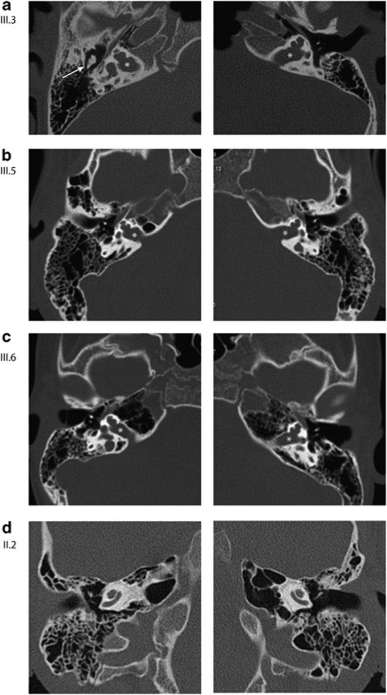
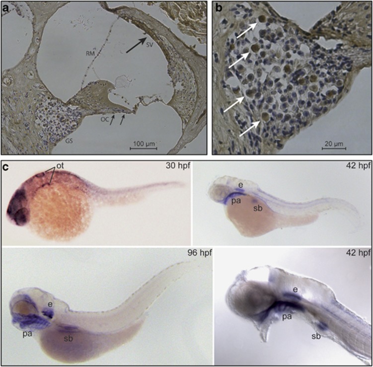
Hereditary hearing loss is the most common human sensorineural disorder. Genetic causes are highly heterogeneous, with mutations detected in >40 genes associated with nonsyndromic hearing loss, to date. Whereas autosomal recessive and autosomal dominant inheritance is prevalent, X-linked forms of nonsyndromic hearing impairment are extremely rare. Here, we present a Hungarian three-generation family with X-linked nonsyndromic congenital hearing loss and the underlying genetic defect. Next-generation sequencing and subsequent segregation analysis detected a missense mutation (c.1771G>A, p.Gly591Ser) in the type IV collagen gene COL4A6 in all affected family members. Bioinformatic analysis and expression studies support this substitution as being causative. COL4A6 encodes the alpha-6 chain of type IV collagen of basal membranes, which forms a heterotrimer with two alpha-5 chains encoded by COL4A5. Whereas mutations in COL4A5 and contiguous X-chromosomal deletions involving COL4A5 and COL4A6 are associated with X-linked Alport syndrome, a nephropathy associated with deafness and cataract, mutations in COL4A6 alone have not been related to any hereditary disease so far. Moreover, our index patient and other affected family members show normal renal and ocular function, which is not consistent with Alport syndrome, but with a nonsyndromic type of hearing loss. In situ hybridization and immunostaining demonstrated expression of the COL4A6 homologs in the otic vesicle of the zebrafish and in the murine inner ear, supporting its role in normal ear development and function. In conclusion, our results suggest COL4A6 as being the fourth gene associated with X-linked nonsyndromic hearing loss.


Figures

Figure 1
Figure 1
(a) Pedigree of the family. Black squares indicate affected male subjects with severe hereditary hearing loss, the gray square (II.1) indicates a male with acquired deafness, circles with black dots show mutation carriers who are healthy to date (unshaded) or who are affected mildly to moderately (shaded). The detected nucleotides at position c.1771 of the COL4A6 gene are given for every family member: G or GG means hemi- or homozygous for the wild-type sequence, GA means c.1771G>A heterozygous and A denotes the mutation in hemizygous form. (b) Representative pure tone audiometry of eight family members. The female subjects II.2 and III.4 show regular thresholds at their current age. The females II.6 (at 41 years of age) and III.10 (at 7 years of age) show normal thresholds of their right ears, and suffer from low-grade conductive hearing loss of maximum 25 dB at 1 kHz (II.6) and a maximum of 20 dB between 0.5 and 1 kHz (III.10) on their left side. Female II.4 (at present) expresses bilateral moderate mixed hearing loss of maximum 35 dB (right) and 40 dB (left) at 1 kHz, and an additional conductive component of maximum 25 dB (right) at 2 kHz and 30 dB (left) at 250 Hz and 1 kHz. Thresholds of patients III.3 (at 8 years), III.5 (at 24 years) and III.6 (at 12 years) at their latest follow-up, showing only residual hearing in single frequencies (red—right side, blue—left side, circles and crosses—air conduction, arrow heads—bone conduction, vertical arrows—frequency not heard). (c) Behavior audiometry under best aided conditions (bilateral hearing aids) of the index patient III.3 at the age of 3 years prior to cochlea implantation of the left ear (dashed lines), and at the age of 8 years before cochlear implantation of the right ear. On the left: speech audiometry ‘Mainzer Kindertest' (solid diamonds) and ‘Freiburger monosyllables' (solid boxes), on the right: functional gain using hearing aids measured with warble thresholds (red—right ear, blue—left ear).
Figure 2
Figure 2
High-resolution imaging of the temporal bone of the index patient (III.3) at the age of 3 years, of his affected cousins (III.5 and III.6) and his healthy mother (II.2). Both sides are shown in each case. (ac) Axial HRCT scans showing the isolated malformed cochlea, with incomplete partition of the cochlea and incomplete separation from the internal auditory canal (asterisks). The vestibular labyrinth is normal. The mastoid cavity and the middle ear ossicles appear regular (white arrow indicating malleo-incudial complex). (d) Representative images from a HRCT scan of the temporal bone in the mother of the index patient demonstrating a regular cochlea.
Figure 3
Figure 3
(a) Multiple sequence alignment of 80 amino acids encompassing glycine-591 of the human COL4A6 protein and orthologous proteins from other species using the software tool Multalign (http://multalin.toulouse.inra.fr/multalin/). Glycine-591 is perfectly conserved (black vertical bar; note that amino acid 591 is shifted by 19 positions to 610 in the alignment due to insertions in COL4A6 of the other species; red letters: high consensus, blue: low consensus, black: neutral). (b) Structural comparison of wild-type COL4A6 (upper part) with the Gly591Ser variant (lower part). Left subfigures illustrate estimated melting temperatures in degrees Celsius along the COL4A6 amino acid sequence. The respective mutation site is highlighted with an arrow. The Gly591Ser mutation seriously affects the melting temperature of the natural model. In the three-dimensional models (right subfigures), the COL4A6 chain is displayed in blue, whereas the white ribbons are the corresponding COL4A5 chains in the heterotrimerical structure. Atoms and bonds are colored in red, in case they clash or have unfavorable contacts with atoms of the other helices (van der Waals overlap >0.6 Å). The pairings of clashing atoms are indicated by yellow lines. In the wild-type COL4A6, at position 591, Gly has no side chain at all and yields a clash-free conformation of the three chains. By contrast, the mutation to Ser produces an atypical large side-chain structure at the same position. In the model of the triple-helical collagen structure, this side chain has several hard clashes that reduce the stability of the interchain conformation and likely triggers disarrangement of the quaternary structure.
Figure 4
Figure 4
COL4A6 expression in the mouse inner ear and in the zebrafish embryo. (a) Thin sections of a demineralized murine inner ear were incubated with rabbit polyclonal Col4A6 antibody M137. COL4A6 is strongly expressed at membranous and osseous structures at the stria vascularis of the spiral ligament (black arrow), which were more intensely stained as compared with the basilar membrane underlying the organ of Corti (small black arrows). GS: ganglion spirale, OC: organ of Corti, RM: Reissner's membrane, SV: stria vascularis. (b) At higher magnification, a very distinct and pronounced reactivity was seen in a subgroup of ganglia cells of the ganglion spirale (white arrows). (c) Whole-mount in situ hybridization on zebrafish embryos of different developmental stages (e: ear, hpf: hours post fertilization, ot: otic vesicle, pa: pharyngeal arch, sb: swim bladder).

References

    1. Bayazit YA, Yilmaz M. An overview of hereditary hearing loss. ORL J Otorhinolaryngol Relat Spec. 2006;68:57–63. - PubMed
    1. Yan D, Liu XZ. Genetics and pathological mechanisms of Usher syndrome. J Hum Genet. 2010;55:327–335. - PMC - PubMed
    1. Kokotas H, Petersen MB, Willems PJ. Mitochondrial deafness. Clin Genet. 2007;71:379–391. - PubMed
    1. Kelsell DP, Dunlop J, Stevens HP, et al. Connexin 26 mutations in hereditary non-syndromic sensorineural deafness. Nature. 1997;387:80–83. - PubMed
    1. Tekin M, Arnos KS, Pandya A. Advances in hereditary deafness. Lancet. 2001;358:1082–1090. - PubMed

MeSH terms

Supplementary concepts

LinkOut - more resources