. 2013 Aug;61(8):555-70.
doi: 10.1369/0022155413493912. Epub 2013 May 28.

Systematic characterization of myocardial inflammation, repair, and remodeling in a mouse model of reperfused myocardial infarction

Affiliations

Systematic characterization of myocardial inflammation, repair, and remodeling in a mouse model of reperfused myocardial infarction

Panagiota Christia et al. J Histochem Cytochem. 2013 Aug.

Abstract

Mouse models of myocardial infarction are essential tools for the study of cardiac injury, repair, and remodeling. Our current investigation establishes a systematic approach for quantitative evaluation of the inflammatory and reparative response, cardiac function, and geometry in a mouse model of reperfused myocardial infarction. Reperfused mouse infarcts exhibited marked induction of inflammatory cytokines that peaked after 6 hr of reperfusion. In the infarcted heart, scar contraction and chamber dilation continued for at least 28 days after reperfusion; infarct maturation was associated with marked thinning of the scar, accompanied by volume loss and rapid clearance of cellular elements. Echocardiographic measurements of end-diastolic dimensions correlated well with morphometric assessment of dilative remodeling in perfusion-fixed hearts. Hemodynamic monitoring was used to quantitatively assess systolic and diastolic function; the severity of diastolic dysfunction following myocardial infarction correlated with cardiomyocyte hypertrophy and infarct collagen content. Expression of molecular mediators of inflammation and cellular infiltration needs to be investigated during the first 72 hr, whereas assessment of dilative remodeling requires measurement of geometric parameters for at least four weeks after the acute event. Rapid initiation and resolution of the inflammatory response, accelerated scar maturation, and extensive infarct volume loss are important characteristics of infarct healing in mice.

Keywords: cardiac remodeling; cardiomyocyte; cytokine; fibroblast; inflammation; myocardial infarction.


Conflict of interest statement

Declaration of Conflicting Interests: The author(s) declared no potential conflicts of interest with respect to the research, authorship, and/or publication of this article.

Figures

Figure 1.
Figure 1.
Morphometric analysis of remodeling-associated parameters in infarcted mouse hearts. After 1 hr coronary occlusion and 7 days (same was done for 3 and 28 days) of reperfusion, the infarcted heart was perfusion-fixed and sectioned from base to apex (A-M) at 250-µm intervals. Ten 5-µm sections were cut at each interval and the first section was stained for hematoxylin/eosin (A-M); thus, each partition corresponded to a 300-µm interval. Left ventricular volumes were quantitated by adding the volumes corresponding to each 300-µm partition. To assess the left ventricular end-diastolic volume (LVEDV), the left ventricular chamber area (LVCA) was traced and measured at each interval (N). The formula LVEDV = 1.33(LVCA1 + LVCA2 + . . . + LVCAn) × 300 µm was used. The infarct volume was assessed by tracing the infarct area (IA; arrows) for each interval (O). The size of the infarct was assessed as the ratio: infarct volume (IV)/left ventricular volume (LVV). Note the midmyocardial location of the infarct that extends from the level of the papillary muscles (D) to the true apex (M). Bar = 1 mm.
Figure 2.
Figure 2.
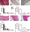
Reperfused murine myocardial infarcts exhibit marked and progressive contraction. A-C: Hematoxylin eosin staining. After 3 days of reperfusion (A) most dead cardiomyocytes in the infarcted mouse heart are replaced by granulation tissue (arrows). After 7 (B) to 28 (C) days of reperfusion, the healing scar becomes much thinner and less cellular. After 28 days of reperfusion, only a thin strip of collagen-based scar remains in the area of the infarct (C, arrows). D: Quantitative analysis of the size of the infarct shows marked contraction of the scar after 7 and 28 days of reperfusion (**p<0.01 vs 3-day infarcts). E: Quantitation of the infarct volume shows marked tissue loss after 7–28 days of reperfusion (**p<0.01 vs 3-day infarcts). F: In contrast, the volume of the noninfarcted septum progressively increases after 7–28 days of reperfusion reflecting hypertrophic remodeling of the surviving myocardial segments (*p<0.05, **p<0.01 vs sham) (sham n=8, 3d n=8, 7d n=13, 28d n=12). G-J: Representative images illustrate increase in septal thickness after 3 (H), 7 (I), and 28 (J) days of reperfusion in comparison with sham myocardium (G). Bars A-C = 0.1 mm; G-J = 100 µm.
Figure 3.
Figure 3.
Reperfused infarction in the mouse is associated with marked but transient infiltration of the infarct with neutrophils and macrophages. A-C: Neutrophil immunohistochemistry in the infarcted mouse heart after 3 days (A), 7 days (B), and 28 days of reperfusion (C). D: Neutrophil density in the infarcted area (I) is markedly higher than in remote noninfarcted areas (R) (^^p<0.01 vs corresponding R) after 3 days of reperfusion, but decreases significantly after 7–28 days as the postinfarction inflammatory reaction resolves (**p<0.01 vs 3-day infarcts). Neutrophil infiltration in the peri-infarct area (PI) is low and the density is comparable with the remote remodeling myocardium (R). E. Quantitative analysis of the total number of neutrophils in the infarcted heart. Intense infiltration of the infarct with neutrophils after 3 days of reperfusion is followed by marked reduction after 7–28 days (**p<0.01 vs 3-day infarct). F-H: Immunohistochemical identification of macrophages using staining for Mac-2 after 3 days (I), 7 days (J) and 28 days of reperfusion (K). I; Macrophage density in the infarcted and remodeling heart. Macrophage density in the infarcted area (I) after 3–7 days of reperfusion is significantly higher than in remote noninfarcted areas (R) (^^p<0.01 vs corresponding R). Macrophage density in the infarct is significantly reduced as the scar matures after 7–28 days of reperfusion (**p<0.01 vs 3-day infarct). Macrophage density in the peri-infarct area (PI) is higher than that in the remodeling myocardium (R) after 3 days of reperfusion (^^p<0.01 vs corresponding R), but is significantly reduced after 7–28 days of reperfusion (**p<0.01 vs corresponding 3-day peri-infarct area density). J: Quantitative analysis of total infarct macrophage numbers showed that the marked accumulation of macrophages in the infarct after 3d of reperfusion is followed by a progressive reduction in macrophage numbers after 7–28 days (**p<0.01 vs 3-day infarct) (sham n=8, 3d n=8, 7d n=13, 28d n=12). Bars A-C; F-H = 0.1 mm.
Figure 4.
Figure 4.
The fibrotic response in reperfused murine myocardial infarction. A-C: Staining for α-smooth muscle actin identifies infarct myofibroblasts as spindle-shaped cells located outside the media of vessels (arrows) after 3 days (A), 7 days (B) and 28 days (C) of reperfusion. D-E: High magnification images show α-SMA immunoreactivity in infarct myofibroblasts after 3 days of reperfusion (arrows). Smooth muscle cells also showed intense α-SMA staining (arrowheads). F-H: Sirius red staining identifies the collagen network in the healing infarct after 3 days (F), 7 days (G) and 28 days (H) of reperfusion. I: Quantitative analysis of myofibroblast density in the infarct (I), peri-infarct area (PI) and remote remodeling myocardium (R). Myofibroblast density in the infarct is significantly higher in the infarcted area after 3–28 days of reperfusion as compared with corresponding non-infarcted remote remodeling areas (^^p<0.01 vs corresponding R). Infarct myofibroblast density peaks after 3 days of reperfusion and is significantly decreased as the scar matures (*p<0.05, **p<0.01 vs 3-day infarcts). Myofibroblast density in the peri-infarct area after 3 and 7 days of reperfusion is higher than that in remote areas (^^p<0.01, ^p<0.05 vs corresponding remote myocardium). J: Quantitation of the total number of infarct myofibroblasts in the infarct shows a progressive reduction in infarct myofibroblasts as the scar matures (**p<0.01 vs 3-day infarcts). K: Quantitative analysis of the collagen-stained area in the infarcted myocardium. Collagen content is markedly higher in the infarcted area (I) as compared with the remote (R) remodeling myocardium (^^p<0.01 vs corresponding R). As the scar matures after 28 days of reperfusion, collagen content in the infarcted area increases (*p<0.05 vs 7-day infarct). After 7–28 days of reperfusion, the peri-infarct (PI) area has significantly higher collagen content than the remote remodeling myocardium (^^p<0.01 vs corresponding R) (sham n=8, 3d n=8, 7d n=13, 28d n=12). Bars A-C, F-H = 100 µm; D-E = 40 µm.
Figure 5.
Figure 5.
Time course of cytokine and extracellular matrix protein upregulation in reperfused mouse infarcts. A-C. Myocardial mRNA expression of the proinflammatory cytokines TNF-α (A), IL-1β (B) and IL-6 (C) is markedly upregulated after 6 hr of reperfusion (**p<0.01 vs sham). Peak IL-1β and IL-6 levels are increased more than 20-fold in the infarcted heart; in contrast, TNF-α induction is less impressive. Proinflammatory cytokine mRNA expression is reduced after 24–72 hr of reperfusion (^^p<0.01 vs 6 hr reperfusion interval), suggesting rapid repression of the inflammatory response. D-F: TGF-β isoform upregulation in the infarcted myocardium. TGF-β1 (D) and TGF-β2 (E) show rapid but persistent induction after 6–72 hr of reperfusion. In contrast, TGF-β3 (F) mRNA levels progressively increase, showing a late peak after 72 hr of reperfusion (^^p<0.01 vs 6 hr reperfusion interval). G: IL-10 mRNA expression also shows a rapid and prolonged upregulation. H: M-CSF levels increase after 6 hr of reperfusion, but are significantly reduced after 72 hr. I-K: Upregulation of structural (collagen) and matricellular (TSP-1 and OPN) extracellular matrix proteins in the infarcted myocardium. Collagen α1 chain (I) and TSP-1 levels (J) significantly increase after 72 hr of reperfusion. OPN (K) shows a rapid upregulation after 6 hr of reperfusion followed by a progressive increase in expression during the transition to the proliferative phase of healing (sham n=14, 6 hr n=16, 24 hr n=14, 72 hr n=12). L-O: Immunohistochemical staining for IL-1β in sham (L) and infarcted mouse hearts. IL-1β expression is negligible in sham myocardium (L), but is markedly increased in the infarct after 3 days of reperfusion (M) and is localized in cells with morphological characteristics of inflammatory leukocytes and vascular cells. Some cardiomyocytes in the infarct also exhibit IL-1β immunoreactivity. After 7 (N) and 28 days (O) of reperfusion, IL-1β staining in the infarct is significantly reduced. Bars L-O = 10 µm.
Figure 6.
Figure 6.
Time course of adverse remodeling of the infarcted mouse heart. A: Morphometric analysis using perfusion-fixation demonstrated that the ventricle dilates significantly after 7 days of reperfusion and exhibits further enlargement after 28 days. B: Quantitative analysis showed that left ventricular end-diastolic volume (LVEDV) significantly increases after 7–28 days of reperfusion (*p<0.05, **p<0.01 vs sham). C: Morphometrically derived LVEDV correlated well with LVEDD measurements obtained through echocardiography in the same animals (r = 0.74, p<0.01) (sham n=8, 3d n=8, 7d n=13, 28d n=12). D Serial echocardiographic assessment of left ventricular remodeling following infarction. Representative images are shown at baseline and after 7 and 28 days of reperfusion. Quantitative analysis of the findings is shown in Table 1 (n=10). Bars A = 0.5 mm; D = 1 mm.
Figure 7.
Figure 7.
Cardiomyocyte hypertrophy in the remodeling noninfarcted myocardium. Cardiomyocyte cross-sectional area was assessed in the noninfarcted subendocardial and subepicardial areas using WGA lectin staining. Representative stained sections from the anterior infarcted wall (A-D) and the posterior wall (E-H) are shown is a sham animal (A, E) and in infarcted mice after 3 days (B, F), 7 days (C, G), and 28 days of reperfusion (D, H). I-J: Quantitative analysis showed that cardiomyocyte cross-sectional area (CSA) in the noninfarcted subepicardium of the anterior wall significantly increased after 3–28 days of reperfusion (**p<0.01 vs sham) J: In the noninfarcted posterior wall, CSA increased after 7–28 days of reperfusion (sham n=8, 3d n=8, 7d n=13, 28d n=12). Bars A-H = 20 µm.
Figure 8.
Figure 8.
Relationship among diastolic dysfunction, fibrosis, and hypertrophy in the infarcted heart. A: There was a good correlation (r = 0.34, p<0.05, n=36) between dp/dtmax, assessed through invasive hemodynamic monitoring, and echocardiographically measured fractional shortening (FS). B. –dp/dtmax an indicator of diastolic function had a strong negative correlation with cardiomyocyte cross-sectional area (CSA) (r = −0.62, p<0.01, n=34), indicating a close relation between hypertrophic remodeling and impaired diastolic function. C. A significant correlation between dp/dtmax and collagen content in the infarcted heart was also noted (r = −0.44, p<0.05, n=23). dp/dtmax, peak first derivative of ventricular pressure; –dp/dtmax, peak negative value of the first derivative of ventricular pressure.

References

    1. Bogen DK, Rabinowitz SA, Needleman A, McMahon TA, Abelmann WH. 1980. An analysis of the mechanical disadvantage of myocardial infarction in the canine left ventricle. Circ Res. 47:728–741 - PubMed
    1. Bujak M, Dobaczewski M, Chatila K, Mendoza LH, Li N, Reddy A, Frangogiannis NG. 2008. Interleukin-1 receptor type I signaling critically regulates infarct healing and cardiac remodeling. Am J Pathol. 173:57–67 - PMC - PubMed
    1. Bujak M, Dobaczewski M, Gonzalez-Quesada C, Xia Y, Leucker T, Zymek P, Veeranna V, Tager AM, Luster AD, Frangogiannis NG. 2009. Induction of the CXC chemokine interferon-gamma-inducible protein 10 regulates the reparative response following myocardial infarction. Circ Res. 105:973–983 - PMC - PubMed
    1. Bujak M, Ren G, Kweon HJ, Dobaczewski M, Reddy A, Taffet G, Wang XF, Frangogiannis NG. 2007. Essential role of Smad3 in infarct healing and in the pathogenesis of cardiac remodeling. Circulation. 116:2127–2138 - PubMed
    1. Chen W, Frangogiannis NG. 2013. Fibroblasts in post-infarction inflammation and cardiac repair. Biochim Biophys Acta. 1833:945–953 - PMC - PubMed

Publication types