

A new approach to aesthetic maxillofacial surgery: surgical treatment of unilateral exophthalmos due to maxillary sinus mucocele
- PMID: 23714910
- PMCID: PMC3671482
- DOI: 10.1097/SCS.0b013e318287d154
A new approach to aesthetic maxillofacial surgery: surgical treatment of unilateral exophthalmos due to maxillary sinus mucocele
Abstract
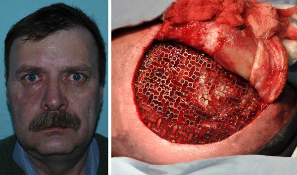
Maxillary sinus mucocele, known as a rare condition, can cause major therapeutic difficulties, especially when it invades the orbit leading to exophthalmia. Treatment is very difficult because the eye globe has to be repositioned, and the facial symmetry needs to be reconstructed as a result of malar bone invasion. This article reports the case of a 54-year-old patient with unilateral exophthalmia caused by the evolution of a maxillary mucocele that extended toward the orbit after destroying the malar bone and the orbital floor. The treatment consisted of a 1-step restoration of both the orbit floor and the malar bone using a temporomandibular flap composed of 2 bone fragments. Lipostructure and a titanium mesh to reconstruct the calvarial defect were necessary to restore facial aesthetics after placing back the eye globe in its initial site. After surgery, the patient followed a complex rehabilitation program including massage kinesiotherapy and psychological consultation and support. These had an essential contribution to the successful final outcome in terms of psychological impact, functionality, and aesthetics.
Figures


Similar articles
-
[Unilateral pseudotumoral exophthalmos in ethmoid-sphenoid-maxillary mucocele].Oftalmologia. 1994 Apr-Jun;38(2):114-7. Oftalmologia. 1994. PMID: 8186203 Romanian.
-
Maxillary sinus mucocele with orbital complications.Ann Ital Chir. 2018 Apr 9;7:S2239253X18028190. Ann Ital Chir. 2018. PMID: 29661986
-
Orbital complications of infected mucocele in the paranasal sinuses.Auris Nasus Larynx. 2020 Dec;47(6):990-995. doi: 10.1016/j.anl.2020.05.012. Epub 2020 Jun 11. Auris Nasus Larynx. 2020. PMID: 32536502
-
Osteoplasty flap technique for repair of latent (30-year) post-traumatic frontal sinus mucocele: case report and review of the literature.J Oral Maxillofac Surg. 2012 Sep;70(9):2092-6. doi: 10.1016/j.joms.2011.10.015. Epub 2012 Apr 26. J Oral Maxillofac Surg. 2012. PMID: 22542331 Review.
-
Massive proptosis of the globe.J Oral Maxillofac Surg. 2000 Jul;58(7):794-9. doi: 10.1053/joms.2000.7268. J Oral Maxillofac Surg. 2000. PMID: 10883696 Review. No abstract available.
Cited by
-
Reconstruction of Large Orbital Floor Defect Caused by Maxillary Sinus Mucocele.Arch Craniofac Surg. 2017 Sep;18(3):197-201. doi: 10.7181/acfs.2017.18.3.197. Epub 2017 Sep 26. Arch Craniofac Surg. 2017. PMID: 29090202 Free PMC article.
References
-
- Shinder R, Al-Zubidi N, Esmaeli B. Survey of orbital tumors at a comprehensive cancer center in the United States. Head Neck. 2011;33:610–614. - PubMed
-
- Palmer-Hall AM, Anderson SF. Paraocular sinus mucoceles. J Am Optom Assoc. 1997;68:725–733. - PubMed
-
- Cairns BE. Pathophysiology of TMD pain – basic mechanisms and their implications for pharmacotherapy. Journal of Oral Rehabilitation. 2010;37:391–410. - PubMed
-
- Kaltreider SA, Dortzbach RK. Destructive cysts of the maxillary sinus affecting the orbit. Arch Ophthalmol. 1988;106:1398–1402. - PubMed
-
- Som PM, Shugar JM. Antral mucoceles: a new look. J Comput Assist Tomogr. 1980;4:484–488. - PubMed
Publication types
MeSH terms
Substances
Grants and funding
LinkOut - more resources
Full Text Sources
Other Literature Sources
Medical
