

Memory reorganization following anterior temporal lobe resection: a longitudinal functional MRI study
- PMID: 23715092
- PMCID: PMC3673465
- DOI: 10.1093/brain/awt105
Memory reorganization following anterior temporal lobe resection: a longitudinal functional MRI study
Abstract
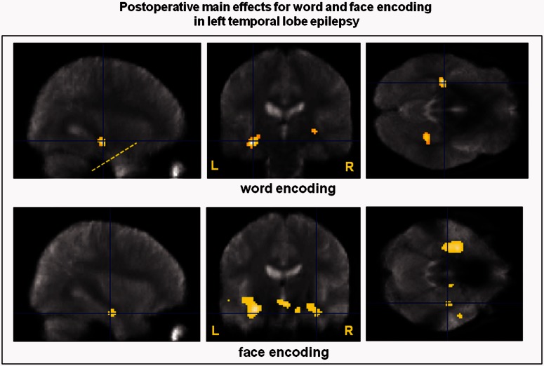
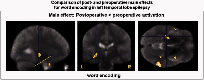
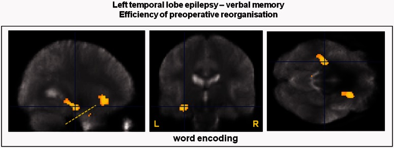
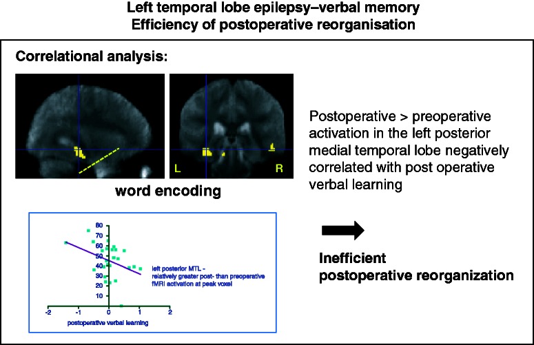
Anterior temporal lobe resection controls seizures in 50-60% of patients with intractable temporal lobe epilepsy but may impair memory function, typically verbal memory following left, and visual memory following right anterior temporal lobe resection. Functional reorganization can occur within the ipsilateral and contralateral hemispheres. We investigated the reorganization of memory function in patients with temporal lobe epilepsy before and after left or right anterior temporal lobe resection and the efficiency of postoperative memory networks. We studied 46 patients with unilateral medial temporal lobe epilepsy (25/26 left hippocampal sclerosis, 16/20 right hippocampal sclerosis) before and after anterior temporal lobe resection on a 3 T General Electric magnetic resonance imaging scanner. All subjects had neuropsychological testing and performed a functional magnetic resonance imaging memory encoding paradigm for words, pictures and faces, testing verbal and visual memory in a single scanning session, preoperatively and again 4 months after surgery. Event-related analysis revealed that patients with left temporal lobe epilepsy had greater activation in the left posterior medial temporal lobe when successfully encoding words postoperatively than preoperatively. Greater pre- than postoperative activation in the ipsilateral posterior medial temporal lobe for encoding words correlated with better verbal memory outcome after left anterior temporal lobe resection. In contrast, greater postoperative than preoperative activation in the ipsilateral posterior medial temporal lobe correlated with worse postoperative verbal memory performance. These postoperative effects were not observed for visual memory function after right anterior temporal lobe resection. Our findings provide evidence for effective preoperative reorganization of verbal memory function to the ipsilateral posterior medial temporal lobe due to the underlying disease, suggesting that it is the capacity of the posterior remnant of the ipsilateral hippocampus rather than the functional reserve of the contralateral hippocampus that is important for maintaining verbal memory function after anterior temporal lobe resection. Early postoperative reorganization to ipsilateral posterior or contralateral medial temporal lobe structures does not underpin better performance. Additionally our results suggest that visual memory function in right temporal lobe epilepsy is affected differently by right anterior temporal lobe resection than verbal memory in left temporal lobe epilepsy.
Keywords: anterior temporal lobe resection; functional MRI; temporal lobe epilepsy; verbal memory; visual memory.
Figures
References
-
- Alpherts WC, Vermeulen J, van Rijen PC, da Silva FH, van Veelen CW. Verbal memory decline after temporal epilepsy surgery? A 6-year multiple assessments follow-up study. Neurology. 2006;67:626–31. - PubMed
-
- Alpherts WC, Vermeulen J, van Rijen PC, da Silva FH, van Veelen CW. Standard versus tailored left temporal lobe resections: differences in cognitive outcome? Neuropsychologia. 2008;46:455–60. - PubMed
-
- Baxendale S, Thompson P. Defining meaningful postoperative change in epilepsy surgery patients: measuring the unmeasurable? Epilepsy Behav. 2005;6:207–11. - PubMed
-
- Baxendale S, Thompson P, Harkness W, Duncan J. Predicting memory decline following epilepsy surgery: a multivariate approach. Epilepsia. 2006;47:1887–94. - PubMed
Publication types
MeSH terms
Grants and funding
LinkOut - more resources
Full Text Sources
Other Literature Sources
Medical
