

Increased gyrification, but comparable surface area in adolescents with autism spectrum disorders
- PMID: 23715094
- PMCID: PMC3673467
- DOI: 10.1093/brain/awt106
Increased gyrification, but comparable surface area in adolescents with autism spectrum disorders
Abstract
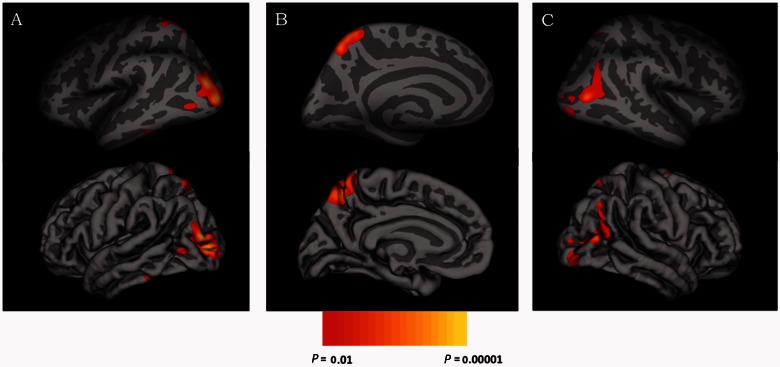
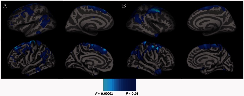
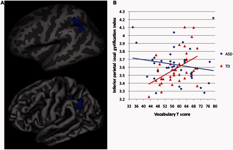
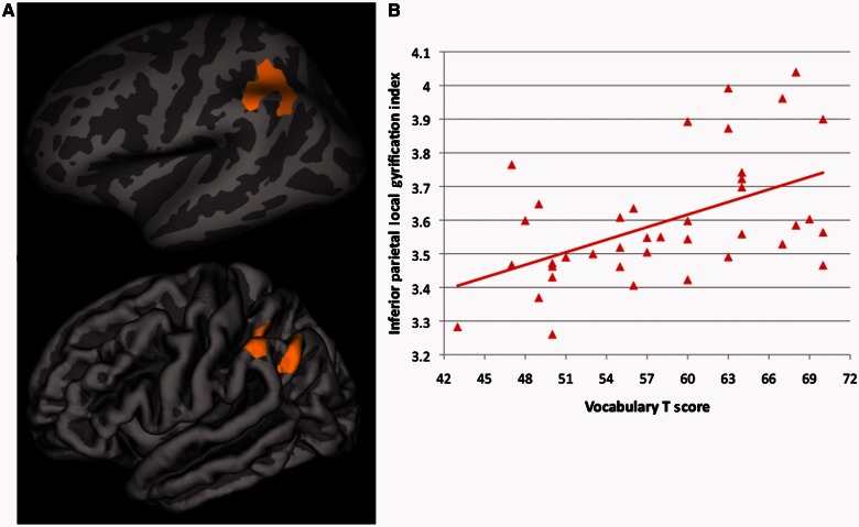
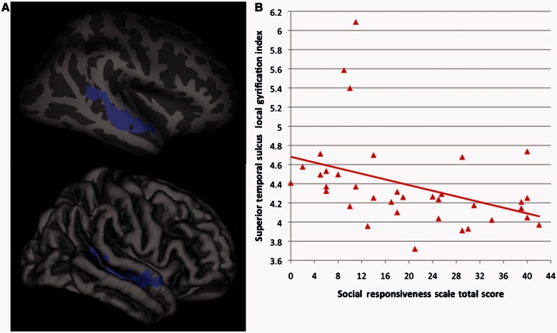
Autism spectrum disorders are associated with atypically excessive early brain growth. Recent studies suggest that later cortical development, specifically cortical thickness, during adolescence and young adulthood is also aberrant. Nevertheless, previous studies of other surface-based metrics (e.g. surface area and gyrification) at high-resolution in autism spectrum disorders are limited. Forty-one males with autism spectrum disorders and 39 typically developing males matched on age (mean ≈ 17; range = 12-24 years) and IQ (mean ≈ 113; range = 85-143) provided high-resolution 3 T anatomical magnetic resonance imaging scans. The FreeSurfer image analysis suite quantified vertex-level surface area and gyrification. There were gyrification increases in the autism spectrum disorders group (relative to typically developing subjects) localized to bilateral posterior cortices (cluster corrected P < 0.01). Furthermore, the association between vocabulary knowledge and gyrification in left inferior parietal cortex (typically developing group: positive correlation; autism spectrum disorders group: no association) differed between groups. Finally, there were no group differences in surface area, and there was no interaction between age and group for either surface area or gyrification (both groups showed decreasing gyrification with increasing age). The present study complements and extends previous work by providing the first evidence of increased gyrification (though no differences in surface area) at high resolution among adolescents and young adults with autism spectrum disorders and by showing a dissociation in the relationship between vocabulary and gyrification in autism spectrum disorders versus typically developing subjects. In contrast with previous findings of age-related cortical thinning in this same autism spectrum disorders sample, here we find that increases in gyrification are maintained across adolescence and young adulthood, implicating developmentally dissociable cortical atypicalities in autism spectrum disorders.
Keywords: MRI; autism; cortical folding; gyrification; surface area.
Figures
References
-
- Bailey A, Luthert P, Dean A, Harding B, Janota I, Montgomery M, et al. A clinicopathological study of autism. Brain. 1998;121:889–905. - PubMed
-
- Bonnici HM, William T, Moorhead J, Stanfield AC, Harris JM, Owens DG, et al. Pre-frontal lobe gyrification index in schizophrenia, mental retardation and comorbid groups: an automated study. Neuroimage. 2007;35:648–54. - PubMed
-
- Booth R, Wallace GL, Happé F. Connectivity and the corpus callosum in autism spectrum conditions: insights from comparison of autism and callosal agenesis. Prog Brain Res. 2011;189:303–17. - PubMed
Publication types
MeSH terms
Grants and funding
LinkOut - more resources
Full Text Sources
Other Literature Sources
