Review
. 2013 Jun;140(12):2576-85.
doi: 10.1242/dev.092270.

Stem cells in retinal regeneration: past, present and future

Affiliations
Review

Stem cells in retinal regeneration: past, present and future

Conor M Ramsden et al. Development. 2013 Jun.

Abstract

Stem cell therapy for retinal disease is under way, and several clinical trials are currently recruiting. These trials use human embryonic, foetal and umbilical cord tissue-derived stem cells and bone marrow-derived stem cells to treat visual disorders such as age-related macular degeneration, Stargardt's disease and retinitis pigmentosa. Over a decade of analysing the developmental cues involved in retinal generation and stem cell biology, coupled with extensive surgical research, have yielded differing cellular approaches to tackle these retinopathies. Here, we review these various stem cell-based approaches for treating retinal diseases and discuss future directions and challenges for the field.

Keywords: A ge-related macular degeneration; Clinical trials; Retinitis pigmentosa; Stargardt's disease; Stem cells.


Figures
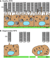
Fig. 1.
Fig. 1.
Healthy and degenerated retinal pigment epithelium. (A) Healthy retinal pigment epithelium (RPE) also showing the outer segments (OS, grey) of the photoreceptors. (B) Degenerated RPE is discontinuous, shows the loss of adherence (yellow bars) to Bruch's membrane (BM, green) and the loss of tight junctions (red bars) between RPE cells. Photoreceptor loss also occurs due, in part, to the inability of the degenerated RPE to phagocytose the photoreceptor outer segments, as illustrated by the lack of phagosomes (Ph) within the RPE cells.
Fig. 2.
Fig. 2.
The effects of retinal pigment epithelium damage on visual field. (A) The visual fields in normal subjects. (B) The visual field in the case of macular diseases, such as age-related macular degeneration and Stargardt's disease. (C) The tunnel visual field experienced in individuals with retinitis pigmentosa.
Fig. 3.
Fig. 3.
Phenotypic characterisation of human embryonic stem cell-derived retinal pigment epithelium. (A) Electron microscopy of human embryonic stem cell (hESC)-derived retinal pigment epithelium (RPE) showing features typical of native polarised RPE, such as basal nucleus (Nuc) location, presence of melanocytes (white arrowhead) and apical microvilli (black arrow). (B) Immunocytochemistry showing the pigmented monolayer of hESC-derived RPE cells with hexagonal shape, as demarcated by the tight junction protein ZO1 in green and the intercellular gap junction protein connexion 43 in red.
Fig. 4.
Fig. 4.
Photoreceptor rescue using human embryonic stem cell-derived retinal pigment epithelium patches. (A) The proposed mechanism of photoreceptor rescue using a retinal pigment epithelium (RPE) patch graft of human embryonic stem cell (hESC)-derived RPE cultured on a plastic polymer substrate (red bracket) that is transplanted between the native, degenerated RPE (blue bracket) and photoreceptor outer segments. (B) The pigmented RPE monolayer on the plastic polymer, ready for implantation. (C) Histological section of a patch graft (red bracket) in a normal porcine eye, lying over the healthy native pig RPE (yellow bracket). BM, Bruch's membrane; OS, outer segments; Ph, phagosome; PP, plastic polymer; RPE, retinal pigment epithelium.

References

    1. Age-Related Eye Disease Study Research Group (2001). A randomized, placebo-controlled, clinical trial of high-dose supplementation with vitamins C and E, beta carotene, and zinc for age-related macular degeneration and vision loss: AREDS report no. 8. Arch. Ophthalmol. 119, 1417-1436 - PMC - PubMed
    1. Allikmets R., Singh N., Sun H., Shroyer N. F., Hutchinson A., Chidambaram A., Gerrard B., Baird L., Stauffer D., Peiffer A., et al. (1997). A photoreceptor cell-specific ATP-binding transporter gene (ABCR) is mutated in recessive Stargardt macular dystrophy. Nat. Genet. 15, 236-246 - PubMed
    1. Araki R., Uda M., Hoki Y., Sunayama M., Nakamura M., Ando S., Sugiura M., Ideno H., Shimada A., Nifuji A., et al. (2013). Negligible immunogenicity of terminally differentiated cells derived from induced pluripotent or embryonic stem cells. Nature 494, 100-104 - PubMed
    1. Arnhold S., Klein H., Semkova I., Addicks K., Schraermeyer U. (2004). Neurally selected embryonic stem cells induce tumor formation after long-term survival following engraftment into the subretinal space. Invest. Ophthalmol. Vis. Sci. 45, 4251-4255 - PubMed
    1. Arnhold S., Absenger Y., Klein H., Addicks K., Schraermeyer U. (2007). Transplantation of bone marrow-derived mesenchymal stem cells rescue photoreceptor cells in the dystrophic retina of the rhodopsin knockout mouse. Graefe's Arch. Clin. Exp. Ophthalmol. 245, 414-422 - PubMed

Publication types

MeSH terms