. 2013 Sep;27(9):3775-85.
doi: 10.1096/fj.12-225649. Epub 2013 May 28.

Steroids as γ-secretase modulators

Affiliations

Steroids as γ-secretase modulators

Joo In Jung et al. FASEB J. 2013 Sep.

Abstract

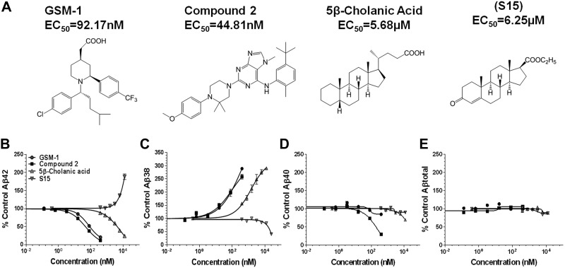
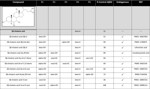
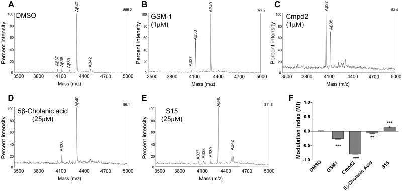
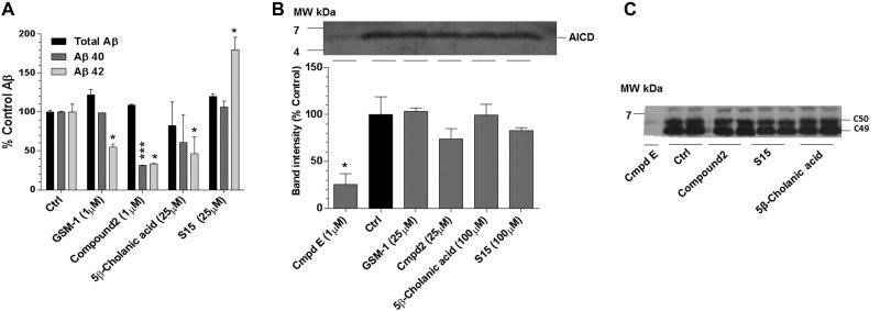
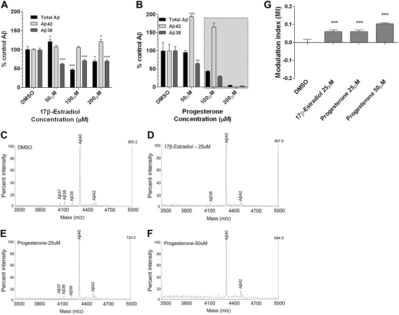
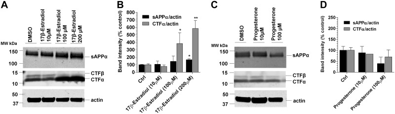
Aggregation and accumulation of Aβ42 play an initiating role in Alzheimer's disease (AD); thus, selective lowering of Aβ42 by γ-secretase modulators (GSMs) remains a promising approach to AD therapy. Based on evidence suggesting that steroids may influence Aβ production, we screened 170 steroids at 10 μM for effects on Aβ42 secreted from human APP-overexpressing Chinese hamster ovary cells. Many acidic steroids lowered Aβ42, whereas many nonacidic steroids actually raised Aβ42. Studies on the more potent compounds showed that Aβ42-lowering steroids were bonafide GSMs and Aβ42-raising steroids were inverse GSMs. The most potent steroid GSM identified was 5β-cholanic acid (EC50=5.7 μM; its endogenous analog lithocholic acid was virtually equipotent), and the most potent inverse GSM identified was 4-androsten-3-one-17β-carboxylic acid ethyl ester (EC50=6.25 μM). In addition, we found that both estrogen and progesterone are weak inverse GSMs with further complex effects on APP processing. These data suggest that certain endogenous steroids may have the potential to act as GSMs and add to the evidence that cholesterol, cholesterol metabolites, and other steroids may play a role in modulating Aβ production and thus risk for AD. They also indicate that acidic steroids might serve as potential therapeutic leads for drug optimization/development.

Keywords: Alzheimer's disease; Aβ; amyloid; cholesterol metabolites.


Figures

Figure 1.
Figure 1.
γ-Secretase modulatory activity of select compounds in CHO-2B7 cells. A) Chemical structures of the various GSM compounds tested, along with their calculated EC50 for Aβ42 modulation determined in triplicate. GSM-1 (piperidine acetic acid–type), EC50 = 92.17 nM; Cmpd2 (piperazinyl pyrimidine type), EC50 = 44.81 nM; S529, EC50 = 5.68 μM; and 4-androsten-3-one-17β-carboxylic acid ethyl ester (S15), 50% increase at 6.25 μM. B–E) Concentration-response curves for Aβ42 (B), Aβ38 (C), Aβ40 (D) and total Aβ (E). Aβ levels from DMSO-treated cells (n=6) served as the control.
Figure 2.
Figure 2.
Markush structure of acidic steroid/sterol and associated structure-activity relationships. Effects of the various S529 derivatives, some of which are known endogenous steroidal metabolites (indicated by a checkmark) on Aβ42 production are summarized as compared to the DMSO vehicle-treated cells.
Figure 3.
Figure 3.
Pattern of Aβ isoforms after MALDI-TOF analysis from conditioned medium from GSM-treated cells. A–E) Effect of various GSM treatments on Aβ isoforms with representative Aβ spectra from 2–3 experiments with 3 replicates/experiment: DMSO (A), GSM-1 (B), Cmpd2 (C), S529 (D), and S15 (E). Identity of the Aβ peptide is noted above the appropriate peak and stacked bar graph, demonstrating the ratio of each peak height to the sum of all the peaks. F) Modulation index (MI) reflecting overall shifting in each Aβ profile induced by the treatments was calculated. As an example, the MI is determined by comparison of the DMSO-treated group to the drug-treated group such that the peak height for the longer Aβ species, Aβ42, is 0.08, and the sum of the peaks for Aβ37, Aβ38, and Aβ39 is 0.17, with the difference being 0.09. With the use of the same calculation, the difference between the longer and shorter Aβ peptides for GSM-1 is 0.35. This value is normalized with the value from DMSO treatment, resulting in MI = −0.26. Negative value indicates GSM activity; positive value indicates iGSM activity. **P < 0.01, ***P < 0.001.
Figure 4.
Figure 4.
In vitro γ-secretase activity assay shows a direct association of each GSM and iGSM in APP processing. A) Graph shows that each treatment was capable of modulating γ-secretase cleavage activity in a cell-free assay using carbonate-extracted membranes from H4-APP695wt-overexpressing cells. Control (Ctrl) refers to the net activity of membranes incubated for 2 h with DMSO (T2) after the subtraction of the basal γ-secretase activity (T0). To measure basal γ-secretase activity, samples were treated with compound E, an irreversible pan-γ-secretase inhibitor, at the starting point T0 of membrane incubation (61). Each sample is then normalized to the control. Each group is normalized to percentage of control (n=2); graph is representative of 4 assays. *P<0.05, ***P<0.001; unpaired, 2-tailed Student's t test. B) AICD immunoblot, along with its quantitation is shown after each drug treatment. AICD is markedly reduced or not detected after compound E treatment. GSM-1, Cmpd2, S529, and S15 treatments generated AICD with no significant quantitative changes. *P < 0.05; 1-way ANOVA with Dunnett's multiple comparison test. C) With the use of bicine urea gels, AICD fragments can be separated into C49 and C50 after each drug treatment (62). No significant changes are detected in either the level or position of ε-cleavage or after GSM-1, Cmpd 2, S15, or S529 treatment consistent with modulatory activity (33). MW, molecular weight.
Figure 5.
Figure 5.
Estrogen and progesterone show inverse GSM activity. A, B) Dose-response curves of 17β-estradiol (A) and progesterone (B) in CHO-2B7 cells show varying effects on Aβ42, Aβ38, and total Aβ levels. iGSM response of 17β-estradiol is weak but notable at 200 μM (note that the total Aβ increase at 50 μM is not typical for these studies). Effects of progesterone >50 μM (grayed area) are rejected due to cellular toxicity. C–F) IP/MS analyses of 17β-estradiol (D) and progesterone (E, F) in CHO-2B7 APP-overexpressing cells. DMSO (C) is used as the control to compare Aβ spectra of estrogen and progesterone-treated group (n=3). Species of Aβ peptides are marked above the each peak. G) MI for 17β-estradiol and progesterone. ***P < 0.001; 1-way ANOVA with Dunnett's multiple comparison test.
Figure 6.
Figure 6.
α-Secretase assay was performed for 17β-estradiol and progesterone. Immunoblots show minimal change in sAPPα and increase in CTFα in 17β-estradiol-treated cells (A) while sAPPα and CTFα levels did not change in progesterone-treated cells (C). Each blot was quantified (B, D). DMSO served as the control; results are representative of 2–3 repeats of 3 individual experiments. *P < 0.05, **P < 0.01; 1-way ANOVA with Dunnett's multiple comparison test.

References

    1. Golde T. E., Eckman C. B., Younkin S. G. (2000) Biochemical detection of Aβ isoforms: implications for pathogenesis, diagnosis, and treatment of Alzheimer's disease. Biochim. Biophys. Acta 1502, 172–187 - PubMed
    1. Teller J. K., Russo C., DeBusk L. M., Angelini G., Zaccheo D., Dagna-Bricarelli F., Scartezzini P., Bertolini S., Mann D. M., Tabaton M., Gambetti P. (1996) Presence of soluble amyloid beta-peptide precedes amyloid plaque formation in Down's syndrome. Nat. Med. 2, 93–95 - PubMed
    1. Russo C., Saido T. C., DeBusk L. M., Tabaton M., Gambetti P., Teller J. K. (1997) Heterogeneity of water-soluble amyloid β-peptide in Alzheimer's disease and Down's syndrome brains. FEBS Lett. 409, 411–416 - PubMed
    1. Moore B., Chakrabarty P., Levites Y., Kukar T., Baine A.-M., Moroni T., Ladd T., Das P., Dickson D., Golde T. (2012) Overlapping profiles of Abeta peptides in the Alzheimer's disease and pathological aging brains. Alzheimers Res. Ther. 4, 18. - PMC - PubMed
    1. Wang R., Sweeney D., Gandy S., Sisodia S. (1996) The profile of soluble amyloid beta protein in cultured cell media. Detection and quantification of amyloid beta protein and variants by immunoprecipitation-mass spectrometry. J. Biol. Chem. 271, 31894–31902 - PubMed

Publication types

LinkOut - more resources