

Homozygous mutations in a predicted endonuclease are a novel cause of congenital dyserythropoietic anemia type I
- PMID: 23716552
- PMCID: PMC3762094
- DOI: 10.3324/haematol.2013.089490
Homozygous mutations in a predicted endonuclease are a novel cause of congenital dyserythropoietic anemia type I
Abstract
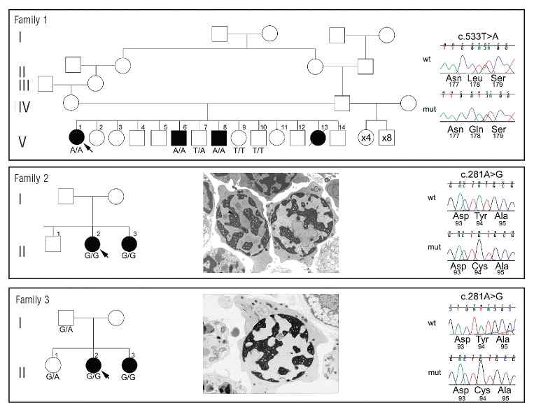
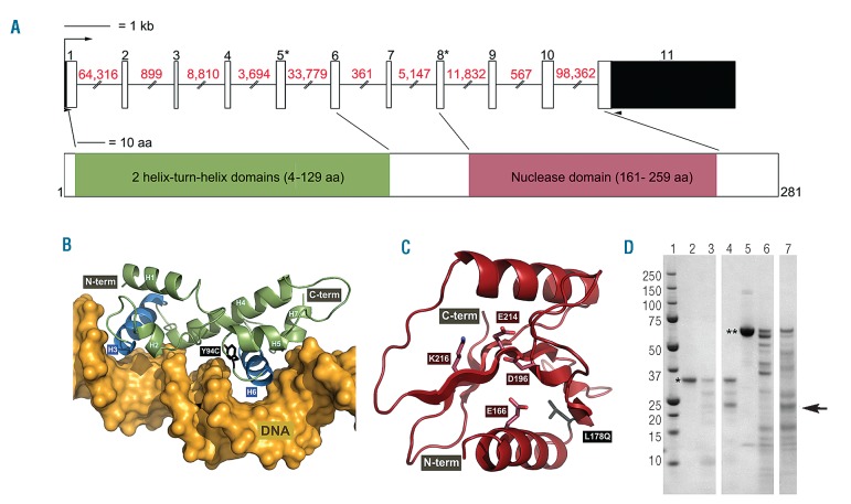
The congenital dyserythropoietic anemias are a heterogeneous group of rare disorders primarily affecting erythropoiesis with characteristic morphological abnormalities and a block in erythroid maturation. Mutations in the CDAN1 gene, which encodes Codanin-1, underlie the majority of congenital dyserythropoietic anemia type I cases. However, no likely pathogenic CDAN1 mutation has been detected in approximately 20% of cases, suggesting the presence of at least one other locus. We used whole genome sequencing and segregation analysis to identify a homozygous T to A transversion (c.533T>A), predicted to lead to a p.L178Q missense substitution in C15ORF41, a gene of unknown function, in a consanguineous pedigree of Middle-Eastern origin. Sequencing C15ORF41 in other CDAN1 mutation-negative congenital dyserythropoietic anemia type I pedigrees identified a homozygous transition (c.281A>G), predicted to lead to a p.Y94C substitution, in two further pedigrees of SouthEast Asian origin. The haplotype surrounding the c.281A>G change suggests a founder effect for this mutation in Pakistan. Detailed sequence similarity searches indicate that C15ORF41 encodes a novel restriction endonuclease that is a member of the Holliday junction resolvase family of proteins.
Figures
References
-
- Heimpel H, Wendt F. Congenital dyserythropoietic anemia with karyorrhexis and multinuclearity of erythroblasts. Helvetica Medica Acta. 1968;34(2):103–15 - PubMed
-
- Renella R, Wood WG. The congenital dyserythropoietic anemias. Hematol Oncol Clin North Am. 2009;23(2):283–306 - PubMed
-
- Heimpel H, Matuschek A, Ahmed M, Bader-Meunier B, Colita A, Delaunay J, et al. Frequency of congenital dyserythropoietic anemias in Europe. Eur J Haematol. 2010;85(1):20–5 - PubMed
Publication types
MeSH terms
Substances
Grants and funding
- MC_UU_12009/1/MRC_/Medical Research Council/United Kingdom
- RC2 HL102926/HL/NHLBI NIH HHS/United States
- 095552/WT_/Wellcome Trust/United Kingdom
- MC_U137761446/MRC_/Medical Research Council/United Kingdom
- 090532/WT_/Wellcome Trust/United Kingdom
- 092809/WT_/Wellcome Trust/United Kingdom
- RC2 HL103010/HL/NHLBI NIH HHS/United States
- RC2 HL102923/HL/NHLBI NIH HHS/United States
- UC2 HL102926/HL/NHLBI NIH HHS/United States
- MC_UU_12021/1/MRC_/Medical Research Council/United Kingdom
- UC2 HL103010/HL/NHLBI NIH HHS/United States
- MC_UU_12010/6/MRC_/Medical Research Council/United Kingdom
- RC2 HL102924/HL/NHLBI NIH HHS/United States
- UC2 HL102923/HL/NHLBI NIH HHS/United States
- UC2 HL102924/HL/NHLBI NIH HHS/United States
- RC2 HL102925/HL/NHLBI NIH HHS/United States
- UC2 HL102925/HL/NHLBI NIH HHS/United States
LinkOut - more resources
Full Text Sources
Other Literature Sources
Molecular Biology Databases
