. 2013 Sep;98(9):1353-8.
doi: 10.3324/haematol.2012.080424. Epub 2013 May 28.

Mouse gene targeting reveals an essential role of mTOR in hematopoietic stem cell engraftment and hematopoiesis

Affiliations

Mouse gene targeting reveals an essential role of mTOR in hematopoietic stem cell engraftment and hematopoiesis

Fukun Guo et al. Haematologica. 2013 Sep.

Abstract

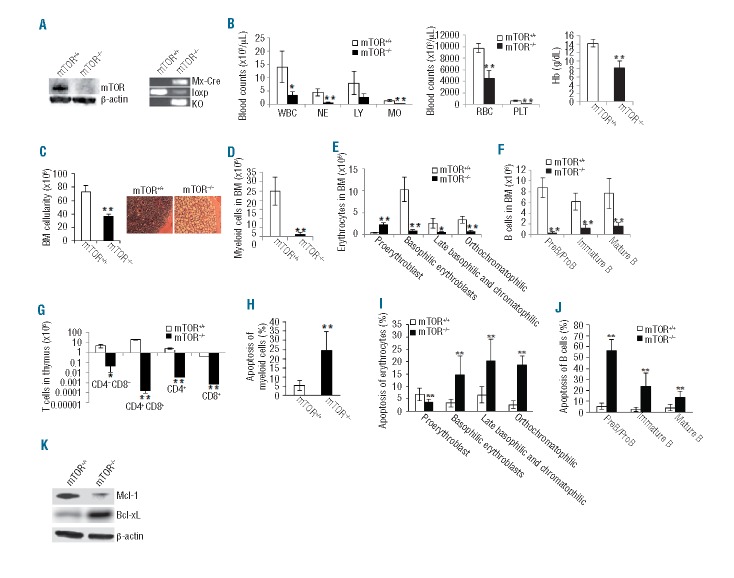
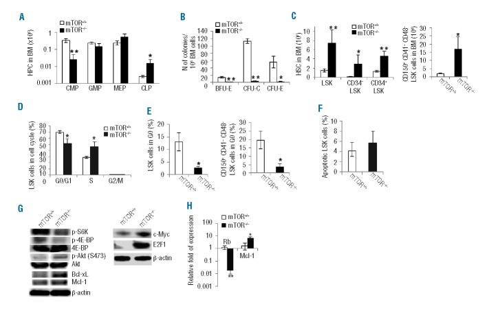
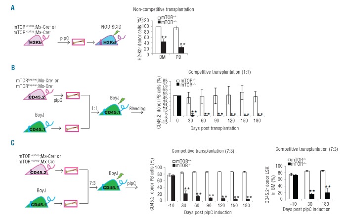
mTOR integrates signals from nutrients and growth factors to control protein synthesis, cell growth, and survival. Although mTOR has been established as a therapeutic target in hematologic malignancies, its physiological role in regulating hematopoiesis remains unclear. Here we show that conditional gene targeting of mTOR causes bone marrow failure and defects in multi-lineage hematopoiesis including myelopoiesis, erythropoiesis, thrombopoiesis, and lymphopoiesis. mTOR deficiency results in loss of quiescence of hematopoietic stem cells, leading to a transient increase but long-term exhaustion and defective engraftment of hematopoietic stem cells in lethally irradiated recipient mice. Furthermore, ablation of mTOR causes increased apoptosis in lineage-committed blood cells but not hematopoietic stem cells, indicating a differentiation stage-specific function. These results demonstrate that mTOR is essential for hematopoietic stem cell engraftment and multi-lineage hematopoiesis.


Figures

Figure 1.
Figure 1.
mTOR deficiency causes impaired hematopoiesis. (A) Western blotting of mTOR protein (left) and PCR genotyping mTOR loxP and knockout (KO) alleles (right) in bone marrow (BM) cells. The Mx1-Cre allele was also genotyped. (B) Blood count parameters. WBC: white blood cell; NE: neutrophil; LY: lymphocyte; MO: monocyte; RBC: red blood cell; PLT: platelet; Hb: hemoglobin. (C) Total number of BM cells (left) and hematoxylin and eosin staining of bone sections (right). (D) The number of myeloid cells (Gr1+Mac1+) in BM. (E) The numbers of erythroid lineages in BM were quantified by FACS. Erythroid cells at different developmental stages include proerythroblasts (Ter119med CD71hi), basophilic erythroblasts (Ter119hiCD71hi), late basophilic and chromatophilic erythroblasts (Ter119hiCD71med), and orthochromatophilic erythroblasts (Ter119hiCD71lo). (F) The numbers of B-cell lineage in BM were analyzed by FACS. B cells at different developmental stages include preB/proB (B220loIgM), immature B (B220loIgM+), and mature B(B220hiIgM+). (G) The numbers of T-cell lineage in thymus were analyzed by FACS. (H–J) The percentages of apoptotic myeloid cells (H), erythrocytes (I) and B cells (J) were analyzed by Annexin V staining. (K) Western blotting of Mcl-1 and Bcl-xL in lineage positive (Lin+) BM cells. Mouse number for each group n=5. *P<0.05; **P<0.01. Error bars represent mean ± s.d.
Figure 2.
Figure 2.
Deletion of mTOR affects HSC and HPC homeostasis. (A) The numbers of common myeloid progenitors (CMPs) (LinSca1c-Kit+CD34+CD16/CD32mid), granulocyte-macrophage progenitors (GMPs) (LinSca1c-Kit+CD34+ CD16/CD32hi), megakaryocyte-erythroid progenitors (MEPs) (LinSca1c-Kit+CD34CD16/CD32lo), and common lymphoid progenitors (CLP) (LinIL7R+Sca1medc-Kitmed-hi) in bone marrow (BM) were analyzed by FACS. (B) The colony-forming activities of CFU-E, CFU-C and BFU-E were examined using the total BM cells. (C) The numbers ofLSK (Lin scal+c-kit+), CD34LSK, CD34+LSK, and CD150+CD41CD48LSK cells in BM were analyzed by FACS. (D) Cell cycle profile of LSK cells. LSK cells were labeled with BrdU in vivo, followed by BrdU and 7-AAD staining and FACS analysis (G0/G1:BrdU7-AADlo; S:BrdU+; G2/M:BrdU7-AADhi). (E) The percentages of LSK progenitor cells and CD150+CD41-CD48-LSK stem cells in G0 phase (pyronin Y-Hoechst 33342lo) of cell cycle were determined. (F) The percentage of apoptotic LSK cells was analyzed by Annexin V staining. (G) Western blot analysis of the signaling activities in Lin- cells. (H) Quantitative RT-PCR analysis of LSK cells for the expression of indicated genes. n=3–5 in each test group. *P<0.05; **P<0.01. Error bars represent mean ± s.d.
Figure 3.
Figure 3.
mTOR deficiency causes an impaired repopulating potential of HSCs. (A) Left: schematics of donor transplantation into NOD-SCID mice. Right: bone marrow (BM) cells from pIpC-treated donor mice were transplanted into sub-lethally irradiated NOD/SCID mice. The donor-derived cells in BM and peripheral blood (PB) of recipient mice were analyzed identified by H2-Kb+ staining. (B) Left hand side: schematics of competitive donor transplantation into syngeneic BoyJ mice. Right: BM cells from pIpC-treated donor mice (C57Bl/6) were mixed at 1:1 ratio with BM cells from BoyJ recipient mice. The cell mixtures were transplanted into lethally irradiated BoyJ rmice. The donor-derived cells were identified by CD45.2+ staining. (C) Left: schematics of competitive donor transplantation into syngeneic BoyJ mice. Middle and right: BM cells from donor mice were mixed at 7:3 ratio with BM cells from BoyJ recipient mice. The cell mixtures were transplanted into lethally irradiated BoyJ mice. The recipient mice were injected with pIpC 2 months post transplantation. The percentage of overall donor-derived cells in PB of the recipient mice were analyzed at various days post pIpC induction by CD45.2 staining (middle). The percentage of donor-derived LSK cells in BM of the recipient mice were determined at 30 days and 180 days post pIpC induction (right). N=3–5. **P<0.01. Error bars represent mean ± s.d.

References

    1. Gan B, DePinho RA. mTORC1 signaling governs hematopoietic stem cell quiescence. Cell Cycle. 2009;8(7):1003–6 - PMC - PubMed
    1. Foster KG, Fingar DC. Mammalian target of rapamycin (mTOR): conducting the cellular signaling symphony. J Biol Chem. 2010;285(19):14071–7 - PMC - PubMed
    1. Proud CG. The multifaceted role of mTOR in cellular stress responses. DNA Repair (Amst). 2004;3(8–9):927–34 - PubMed
    1. Raslova H, Baccini V, Loussaief L, Comba B, Larghero J, Debili N, et al. Mammalian target of rapamycin (mTOR) regulates both proliferation of megakaryocyte progenitors and late stages of megakaryocyte differentiation. Blood. 2006;107(6):2303–10 - PubMed
    1. Sathaliyawala T, O’Gorman WE, Greter M, Bogunovic M, Konjufca V, Hou ZE, et al. Mammalian target of rapamycin controls dendritic cell development downstream of Flt3 ligand signaling. Immunity. 2010;33(4):597–606 - PMC - PubMed

Publication types

Substances

LinkOut - more resources