Case Reports
. 2013 Oct;98(10):1624-32.
doi: 10.3324/haematol.2013.088369. Epub 2013 May 28.

Congenital erythrocytosis associated with gain-of-function HIF2A gene mutations and erythropoietin levels in the normal range

Affiliations
Case Reports

Congenital erythrocytosis associated with gain-of-function HIF2A gene mutations and erythropoietin levels in the normal range

Silverio Perrotta et al. Haematologica. 2013 Oct.

Abstract

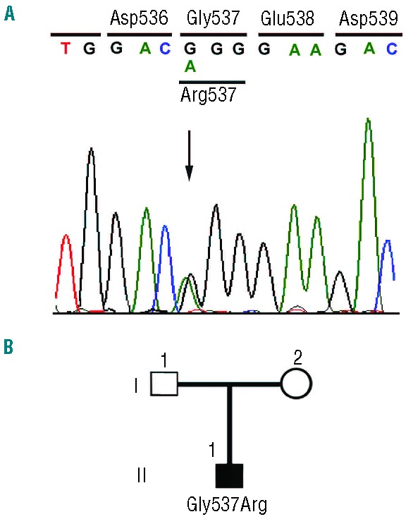
Hypoxia-inducible factor 2α (HIF-2α) plays a pivotal role in the balancing of oxygen requirements throughout the body. The protein is a transcription factor that modulates the expression of a wide array of genes and, in turn, controls several key processes including energy metabolism, erythropoiesis and angiogenesis. We describe here the identification of two cases of familial erythrocytosis associated with heterozygous HIF2A missense mutations, namely Ile533Val and Gly537Arg. Ile533Val is a novel mutation and represents the genetic HIF2A change nearest to Pro-531, the primary hydroxyl acceptor residue, so far identified. The Gly537Arg missense mutation has already been described in familial erythrocytosis. However, our patient is the only described case of a de novo HIF2A mutation associated with the development of congenital polycythemia. Functional in vivo studies, based on exogenous expression of hybrid HIF-2α transcription factors, indicated that these genetic alterations lead to the stabilization of HIF-2α protein. All the identified polycythemic subjects with HIF2A mutations show serum erythropoietin in the normal range, independently of the hematocrit values and phlebotomy frequency. The erythroid precursors obtained from the peripheral blood of patients showed an altered phenotype, including an increased rate of growth and a modified expression of some HIF-2α target genes. These results suggest the novel proposal that polycythemia observed in subjects with HIF2A mutations might also be due to primary changes in hematopoietic cells and not only secondary to increased erythropoietin levels.


Figures

Figure 1.
Figure 1.
Identification of the Arg537Gly mutation in the HIF2A gene. (A) Detection of the c.1609 G>A (Gly537Arg) mutation by PCR-direct sequencing. PCR-direct sequencing was performed on total peripheral blood DNA using specific primers to amplify exon 12 of the HIF2A gene. Sequencing detected a heterozygous G to A change at base 1609 in patient 1, as indicated by the arrow, as compared to the wild-type sequence (upper panel). Nucleotides 1604–1617 are shown. Bases are as follows: G black; A green; T red; C, blue. (B) The pedigree of the family of patient 1 is shown. Squares represent male family members, circles represent female family members, and solid symbols represent family members with erythrocytosis; the genotype is shown under the symbol.
Figure 2.
Figure 2.
Identification of the Val533Ile mutation in the HIF2A gene. (A) Detection of the c.1597 A>G (Ile 533Val) mutation by PCR-direct sequencing. PCR-direct sequencing was performed on total peripheral blood DNA using specific primers to amplify exon 12. Sequencing detected a heterozygous A to G change at base 1597 in patient 2, as indicated by the arrow, as compared to the wild-type sequence (upper panel). Nucleotides 1589–1603 are shown. Bases are as follows: G, black; A, green; T, red; C, blue. (B) The pedigree of the family of patient 2 is shown. Squares represent male family members, circles represent female family members, and solid symbols represent family members with erythrocytosis; genotypes are shown under each symbol.
Figure 3.
Figure 3.
Identification of the Ile533Val mutation by BsmF1 digestion. Lymphocytic cDNA from a control (lane 2), patient 2 (lanes 1 and 4), his daughter (lane 3) and his affected son (lane 5) were amplified using primers localized in HIF2A exon 12. The amplified materials were not digested (lane 1) or digested with BmsF1 (lanes 2–5). The unrestricted PCR product is 868 bp long. BmsF1 digestion results in two fragments of 517 and 351 bp in the absence of the Val533Ile mutation (lanes 2 and 3). The presence of the c1597 A>G mutation creates a second BmsF1 restriction site in the 351 bp fragment and the formation of two further fragments of 206 and 145 bp each (lanes 4 and 5). MW, molecular weight.
Figure 4.
Figure 4.
HIF-2α peptide hydroxylation. (A) Sequences of HIF-1α and HIF-2α peptides employed in the VBC binding assay for proline hydroxylation. The arrow shows the hydroxyacceptor proline. (B) Different known percentage of hydroxyproline HIF-α peptides (depicted in panel A) were subjected to a VBC binding assay a described in the Methods section. (C) Wild-type and mutant HIF-1α and HIF-2α peptides (P564A and P531A, respectively) were subjected to in vitro hydroxylation by recombinant purified PHD isoforms. (D) Wild-type and mutant HIF-1α and HIF-2α peptides (Met531Ala mutation) were subjected to in vitro hydroxylation by recombinant purified PHD isoforms.
Figure 5.
Figure 5.
Functional effects of HIF-2α mutations. (A) Sequences of wild-type and mutant HIF-2α peptides employed. (B) HIF-2α peptides (depicted in panel A) and containing the indicated point mutations were used for a VBC binding assay in the absence of recombinant PHD isoforms. (C) The same HIF-2α peptides were used for the VBC binding assay following hydroxylation by the recombinant PHD as indicated. While wild-type peptides were efficiently hydroxylated by all PHD, the HIF-2α G456R mutation showed reduced hydroxylation specifically by PHD3 by approximately 40%. (D) Subconfluent Hek293 cultures were co-transfected with pGRE5xE1bluc, 250 ng of pM3-HIF-2α (amino acids 404–569) and 200 ng of the respective PHD expression construct or empty expression vector. Different mutants of pM3-HIF-2α were used, as reported in the figure, including: the wild-type (WT) form; a form mutated in both the hydroxylable proline residues (PP/AA) and three forms encoding G537W, G537R and I533V mutants. pRL-SV40 was used to normalize for transfection efficiency. Twenty-four hours post-transfection, cultures were equally distributed into 12-well plates and grown for an additional 24 h in 20% oxygen. Cells were subjected to a dual luciferase assay.
Figure 6.
Figure 6.
Effects of HIF-2α mutations on peripheral erythroid precursor phenotypes. (A) Results of real-time PCR assays. Total RNA was prepared from primary cultures of peripheral erythroid precursors. RNA was reverse transcribed and the concentrations of transcripts of VEGF the vascular endothelial growth factor gene ( ), adrenomedullin gene (ADM), N-myc downstream regulated gene 1 (NDRG1), and transferrin receptor gene (TFR) were determined by the real-time PCR. The values obtained were reported as fold-increase over the transcript concentrations from primary cultures of erythroid precursors from control subjects. Means from three separate experiments are shown. T bars indicate standard deviations. (B) Growth rate of primary cultures of peripheral erythroid precursors. Proliferation was determined by cell counting. The content of erythropoietin in the cell culture medium was 100 mU/mL. Means from three separate experiments are shown. T bars indicate standard deviations. (C) Content of vascular endothelial growth factor (VEGF) and soluble transferrin receptor (sTFR) in the growth media of primary cultures of peripheral erythroid precursors. The media of the first and second weeks of incubation were collected and VEGF and sTFR levels were determined by ELISA. Means from three separate experiments are shown. T bars indicate standard deviations.

References

    1. Abbrecht PH, Littell JK. Plasma erythropoietin in men and mice during acclimatization to different altitudes. J Appl Physiol. 1972;32(1):54–8 - PubMed
    1. Jelkmann W. Erythropoietin after a century of research: younger than ever. Eur J Haematol. 2007;78(3):183–205 - PubMed
    1. Maxwell PH, Osmond MK, Pugh CW, Heryet A, Nicholls LG, Tan CC, et al. Identification of the renal erythropoietin-producing cells using transgenic mice. Kidney Int. 1993;44(5):1149–62 - PubMed
    1. Wenger RH, Hoogewijs D. Regulated oxygen sensing by protein hydroxylation in renal erythropoietin-producing cells. Am J Physiol Renal Physiol. 2010; 298(6):F1287–96 - PubMed
    1. Obara N, Suzuki N, Kim K, Nagasawa T, Imagawa S, Yamamoto M. Repression via the GATA box is essential for tissue-specific erythropoietin gene expression. Blood. 2008;111(10):5223–32 - PubMed

Publication types

Substances

Supplementary concepts

LinkOut - more resources