. 2013 May 28;4(3):e00298-13.
doi: 10.1128/mBio.00298-13.

Human embryonic stem cell lines model experimental human cytomegalovirus latency

Affiliations

Human embryonic stem cell lines model experimental human cytomegalovirus latency

Rhiannon R Penkert et al. mBio. .

Abstract

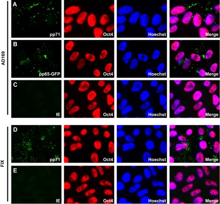
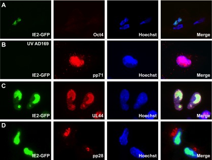
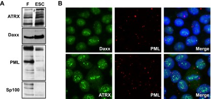
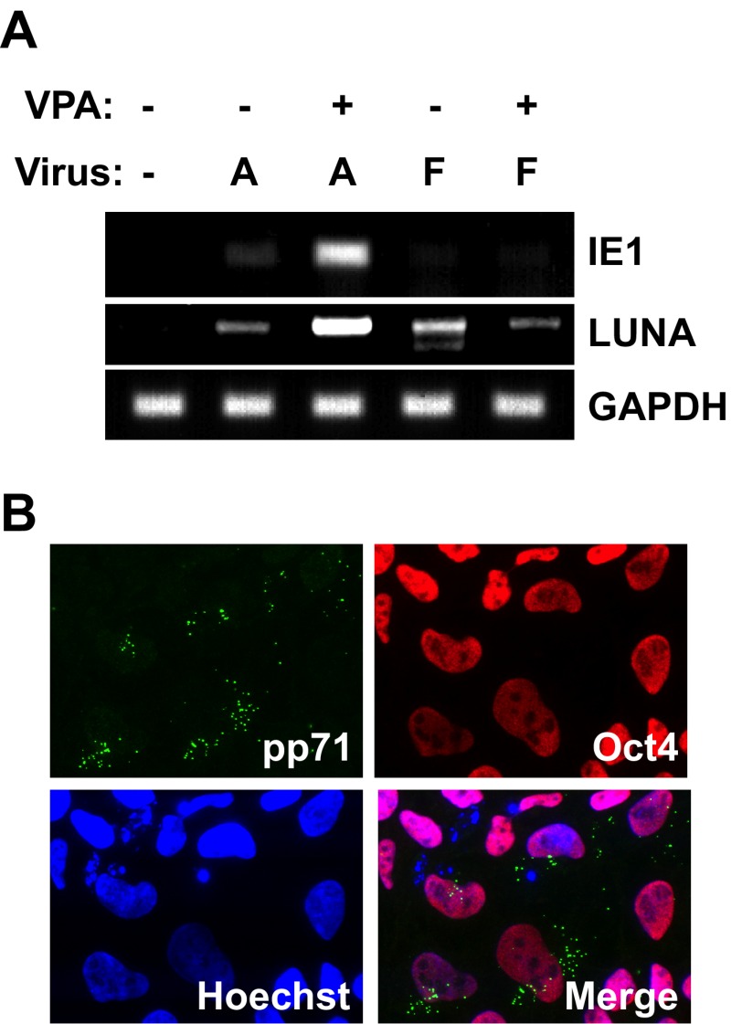
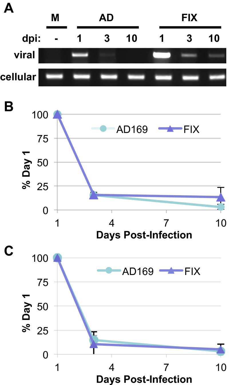
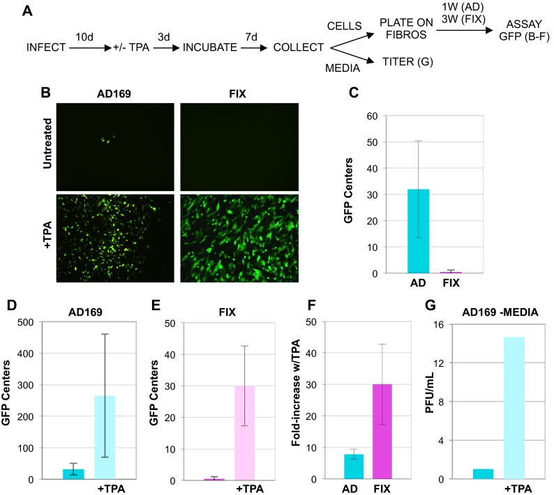
Herpesviruses are highly successful pathogens that persist for the lifetime of their hosts primarily because of their ability to establish and maintain latent infections from which the virus is capable of productively reactivating. Human cytomegalovirus (HCMV), a betaherpesvirus, establishes latency in CD34(+) hematopoietic progenitor cells during natural infections in the body. Experimental infection of CD34(+) cells ex vivo has demonstrated that expression of the viral gene products that drive productive infection is silenced by an intrinsic immune defense mediated by Daxx and histone deacetylases through heterochromatinization of the viral genome during the establishment of latency. Additional mechanistic details about the establishment, let alone maintenance and reactivation, of HCMV latency remain scarce. This is partly due to the technical challenges of CD34(+) cell culture, most notably, the difficulty in preventing spontaneous differentiation that drives reactivation and renders them permissive for productive infection. Here we demonstrate that HCMV can establish, maintain, and reactivate in vitro from experimental latency in cultures of human embryonic stem cells (ESCs), for which spurious differentiation can be prevented or controlled. Furthermore, we show that known molecular aspects of HCMV latency are faithfully recapitulated in these cells. In total, we present ESCs as a novel, tractable model for studies of HCMV latency.


Figures

FIG 1
FIG 1
HCMV enters embryonic stem cells (ESCs) but does not initiate lytic infection. ESCs grown on coverslips were infected with HCMV strain AD169 (A and C), FIX (D and E), or AD169 pp65-GFP (B) at an MOI of 3. The cells growing on coverslips were harvested 24 h postinfection, and the indicated viral (pp71 and IE1) or cellular (Oct4) proteins were visualized by indirect immunofluorescence microscopy. Viral pp65 was detected as a GFP signal by fluorescence microscopy. The nuclei were counterstained with Hoechst stain.
FIG 2
FIG 2
Differentiated ESCs support lytic-phase gene expression. ESCs grown on coverslips were treated with TPA for 3 days and subsequently infected with HCMV strain AD169 IE2-GFP (A, C, and D) or UV-inactivated AD169 IE2-GFP (B) at an MOI of 1. The cells growing on coverslips were harvested at 4 h (A and B), 3 days (C), or 5 days (D) postinfection. The indicated viral (pp71, UL44, and pp28) or cellular (Oct4) proteins were visualized by indirect immunofluorescence microscopy. Viral IE2 was detected as a GFP signal by fluorescence microscopy. The nuclei were counterstained with Hoechst stain.
FIG 3
FIG 3
Differentiated ESCs support productive infection. ESCs treated with TPA for 3 days were subsequently infected with HCMV strain AD169 at an MOI of 1. Infectious virions accumulated in the medium at 2 or 8 days postinfection (dpi) were quantitated by plaque assay.
FIG 4
FIG 4
ESCs express PML-NB proteins but do not assemble canonical PML-NBs. (A) Equal amounts of protein lysates generated from uninfected fibroblasts (F) or uninfected ESCs were analyzed by Western blotting with the indicated antibodies. (B) Uninfected ESCs grown on coverslips were analyzed by indirect immunofluorescence microscopy for the indicated proteins. The merge panels show nuclei counterstained with Hoechst stain.
FIG 5
FIG 5
HCMV gene expression in ESCs is regulated in a manner indistinguishable from experimental latency in primary CD34+ cells. (A) ESCs not treated with VPA (−) or pretreated with VPA (+) for 1 h were infected with HCMV strain AD169 (A) or HCMV strain FIX (F) at an MOI of 3. RNA extracted at 24 hpi was subjected to RT-PCR to monitor the expression of the indicated viral (IE1 and LUNA) or cellular (GAPDH) gene. (B) ESCs grown on coverslips and pretreated with VPA for 1 h were infected with AD169 at an MOI of 3. The indicated proteins were imaged by indirect immunofluorescence microscopy.
FIG 6
FIG 6
Viral genomes are maintained in ESCs for at least 10 days. (A) ESCs were mock infected (M) or infected with HCMV strain AD169 (AD) or FIX at an MOI of 3. Total DNA isolated at the indicated day postinfection (dpi) was analyzed by PCR for the presence of viral (UL123) or cellular (GAPDH) DNA. (B and C) Viral genomes at the indicated dpi were quantitated by Li-Cor imaging for three independent biological replicates (B) or by real-time PCR for two biological replicates (C), normalized to cellular genome levels and are expressed as a percentage of the viral genome level detected on day 1 for each individual viral strain.
FIG 7
FIG 7
HCMV reactivates from latently infected ESCs upon their differentiation. (A) Reactivation assay flow chart. ESCs infected with HCMV strain AD169 (AD) or HCMV strain FIX for 10 days (10d) were treated with TPA (+) or not treated with TPA (−) for 3 days, cultured for an additional 7 days, and then separated into fractions consisting of either attached cells or clarified medium. Cells (top right) were coplated with fibroblasts (FIBROS) for the indicated number of weeks (1 week [1W] or 3 weeks [3W]), and then infectious centers were quantitated by counting GFP foci. Data from this portion of the assay are presented in panels B to F. The titers of virus in the medium fraction (bottom right) were determined directly by a plaque assay. Data from this portion of the assay are presented in panel G. (B) Representative images used to quantify GFP foci detected in untreated or TPA-treated cells infected with AD169 or FIX. (C) Quantitation of GFP foci detected in single wells of untreated, HCMV-infected ESCs. (D) Quantitation of GFP foci detected in single wells of untreated or TPA-treated ESCs infected with AD169. (E) Quantitation of GFP foci detected in single wells of untreated or TPA-treated ESCs infected with FIX. (F) Calculated fold increase for TPA-treated versus untreated cells infected with the indicated virus. (G) Infectious virions accumulated in the medium rescued from AD169-infected ESCs untreated or treated with TPA were quantitated by plaque assay. In all graphs, error bars represent standard deviations.

References

    1. Mocarski E, Shenk T, Pass R. 2007. Cytomegaloviruses, p 2701–2772 In Knipe D, Howley P, Griffin DE, Lamb RA, Martin MA, Roizman B, Straus SE, Fields virology. Lippincott Williams and Wilkins, Philadelphia, PA.
    1. Marschall M, Stamminger T. 2009. Molecular targets for antiviral therapy of cytomegalovirus infections. Future Microbiol. 4:731–742 - PubMed
    1. McGregor A, Choi KY. 2011. Cytomegalovirus antivirals and development of improved animal models. Expert Opin. Drug Metab. Toxicol. 7:1245–1265 - PMC - PubMed
    1. Goodrum F, Caviness K, Zagallo P. 2012. Human cytomegalovirus persistence. Cell. Microbiol. 14:644–655 - PMC - PubMed
    1. Reeves M, Sinclair J. 2008. Aspects of human cytomegalovirus latency and reactivation. Curr. Top. Microbiol. Immunol. 325:297–313 - PubMed

Publication types