. 2013 Aug;81(8):2899-908.
doi: 10.1128/IAI.00542-13. Epub 2013 May 28.

Borrelia hermsii acquisition order in superinfected ticks determines transmission efficiency

Affiliations

Borrelia hermsii acquisition order in superinfected ticks determines transmission efficiency

Paul F Policastro et al. Infect Immun. 2013 Aug.

Abstract

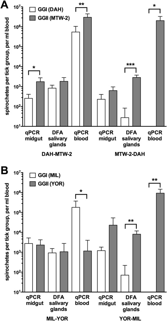
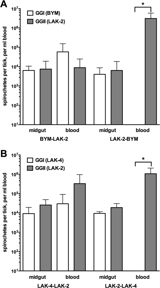
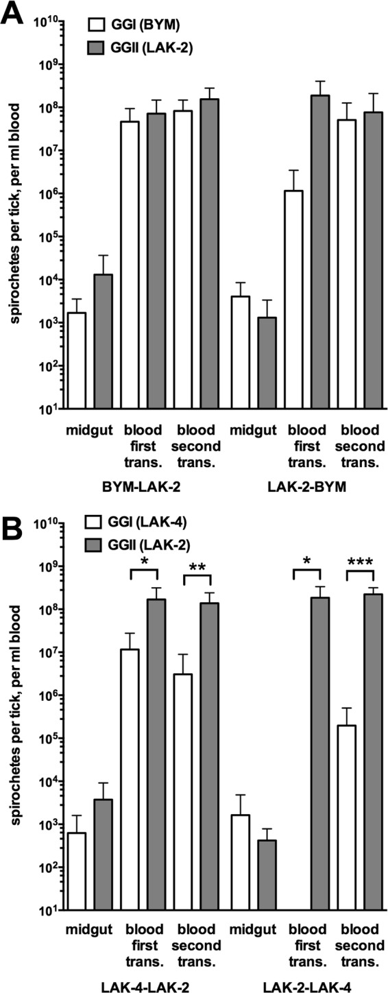
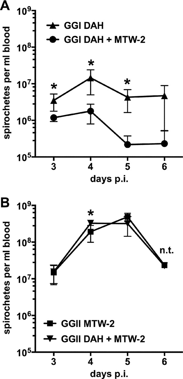
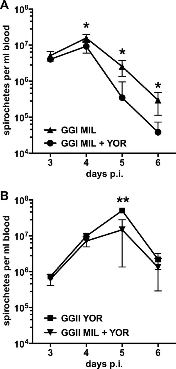
Multilocus sequence typing of Borrelia hermsii isolates reveals its divergence into two major genomic groups (GG), but no differences in transmission efficiency or host pathogenicity are associated with these genotypes. To compare GGI and GGII in the tick-host infection cycle, we first determined if spirochetes from the two groups could superinfect the tick vector Ornithodoros hermsi. We infected mice with isolates from each group and fed ticks sequentially on these mice. We then fed the infected ticks on naive mice and measured GGI and GGII spirochete densities in vector and host, using quantitative PCR of genotype-specific chromosomal DNA sequences. Sequential feedings resulted in dual tick infections, showing that GGI or GGII primary acquisition did not block superinfection by a secondary agent. On transmission to naive mice at short intervals after acquisition, ticks with primary GGI and secondary GGII spirochete infections caused mixed GGI and GGII infections in mice. However, ticks with primary GGII and secondary GGI spirochete infections caused only GGII infections with all isolate pairs examined. At longer intervals after acquisition, the exclusion of GGI by GGII spirochetes declined and cotransmission predominated. We then examined GGI and GGII spirochetemia in mice following single inoculation and coinoculation by needle and found that GGI spirochete densities were reduced on multiple days when coinoculated with GGII. These findings indicate that dual GGI-GGII spirochete infections can persist in ticks and that transmission to a vertebrate host is dependent on the order of tick acquisition and the interval between acquisition and transmission events.


Figures

Fig 1
Fig 1
GGI and GGII B. hermsii in tick midgut, salivary gland, and mouse blood following transmission by tick bite with three ticks per mouse. Ticks fed at primary and secondary acquisitions on infected mice were fed in groups of three per mouse on naive recipient mice at 5 months after the secondary-acquisition feed. The values represent qPCR-detected GGI and GGII B. hermsii in DNA from tick midguts dissected 2 weeks posttransmission and blood collected during initial spirochetemia after tick feeding (qPCR; means and standard deviations [SD]) and DFA-detected Vtp-6-positive GGI and Vtp-5-positive GGII B. hermsii in pairs of tick salivary glands that were dissected 2 weeks posttransmission (DFA; means and SD). Tick mean values were calculated from the total for each set of three midguts or salivary gland pairs per mouse. The numbers of tick-mouse samples per transmission assay were as follows: (A) DAH–MTW-2, n = 5, and MTW-2–DAH, n = 4; (B) MIL-YOR, n = 6, and YOR-MIL, n = 5. The brackets indicate significant differences between GGI and GGII values by unpaired t test: *, P = 0.05 to 0.01; **, P = 0.01 to 0.001; ***, P < 0.001.
Fig 2
Fig 2
DFA of B. hermsii in the salivary glands of infected O. hermsi. Fixed tissue was incubated with monoclonal antibodies H4825 to Vtp-6-Alexa 568 conjugate (red channel) and H3548 to Vtp-5-Alexa 488 conjugate (green channel) and viewed at ×200 magnification. Salivary gland duct and tracheal branches (blue channel) were detected by autofluorescence. (A) Gland from tick with primary GGI MIL (Vtp-6; red) and secondary GGII YOR (Vtp-5; green) infections. (B) Gland from tick with primary GGII YOR (Vtp-5; green) and secondary GGI MIL (Vtp-6; red) infections. Scale bar in panel B, 20 μm.
Fig 3
Fig 3
GGI and GGII B. hermsii in tick midgut and mouse blood following transmission by tick bite with one tick per mouse. Ticks fed at primary and secondary acquisitions on infected mice were fed individually on naive recipient mice at 6 to 10 months after the secondary-acquisition feed. The values represent means and SD for qPCR-detected GGI and GGII spirochetes in DNA from tick midguts dissected 2 weeks posttransmission and blood collected during initial spirochetemia after tick feeding. The numbers of tick-mouse samples per transmission assay were as follows: (A) BYM–LAK-2 and LAK-2–BYM, n = 5 for both groups (both isolates possess the vtp-1 allele). (B) LAK-4–LAK-2 and LAK-2–LAK-4, n = 5 for both groups (both isolates were from a single focus of endemicity). The brackets indicate significant differences between GGI and GGII values by unpaired t test: *, P = 0.01 to 0.05.
Fig 4
Fig 4
GGI and GGII B. hermsii in tick midgut and mouse blood following serial transmission to naive mice by tick bite with one tick per mouse. Ticks fed at primary and secondary acquisitions on infected mice were fed individually on naive recipient mice at 9 to 15 months after the secondary-acquisition feed, held for 3 months, and refed on naive mice. The values represent means and SD for qPCR-detected GGI and GGII spirochetes in DNA from tick midguts dissected 1 month after the second transmission (trans.) and from blood collected during initial spirochetemia posttransmission. The numbers of tick-mouse samples per transmission assay were as follows: (A) BYM–LAK-2, n = 7, and LAK-2–BYM, n = 4; (B) LAK-4–LAK-2, n = 7, and LAK-2–LAK-4, n = 6. The brackets indicate significant differences between GGI and GGII values by unpaired t test: *, P = 0.05 to 0.01; **, P = 0.01 to 0.001; ***, P < 0.001.
Fig 5
Fig 5
GGI and GGII B. hermsii in mouse blood during initial spirochetemia following single- and dual-needle inoculation with DAH and MTW-2. Cultured DAH or MTW-2 spirochetes (1 × 10³) were injected intravenously on day 0 postinoculation (p.i.) in a final volume of 0.1 ml for individual inoculations; coinoculations (DAH + MTW-2) consisted of 1 × 10³ DAH and 1 × 10³ MTW-2 spirochetes in 0.1 ml. The values represent means ± SD of qPCR-detected GGI (A) and GGII (B) spirochetes in DNA from blood collected during spirochetemia on days 3 to 6 postinoculation (n = 5 for each group). No GGII spirochetes were detected in mice inoculated with DAH, and no GGI spirochetes were detected in mice inoculated with MTW-2; thus, these data are not plotted. Three DAH–MTW-2 and four MTW-2 mice were euthanized after blood collection on day 5. Significant differences between single and coinoculated values by unpaired t test are indicated: *, P = 0.01 to 0.05. n.t., no t test on day 6 for a single MTW-2 mouse.
Fig 6
Fig 6
GGI and GGII B. hermsii in mouse blood during initial spirochetemia following single- and dual-needle inoculation with MIL and YOR. Cultured MIL or YOR spirochetes (1 × 10³) were injected intravenously on day 0 p.i. in a final volume of 0.1 ml for individual inoculations; coinoculations (MIL + YOR) consisted of 1 × 10³ MIL and 1 × 10³ YOR spirochetes in 0.1 ml. No GGII spirochetes were detected in mice inoculated with MIL, and no GGI spirochetes were detected in mice inoculated with YOR; thus, these data are not plotted. The values represent means ± SD of qPCR-detected GGI (A) and GGII (B) spirochetes in DNA from blood collected during spirochetemia on days 3 to 6 postinoculation (n = 5 for each group). Note that the density range differs from that in Fig. 5. Significant differences between single and coinoculated values by unpaired t test are indicated: *, P = 0.05 to 0.01; **, P = 0.01 to 0.001.

Similar articles

Cited by

References

    1. Dworkin MS, Schwan TG, Anderson DE, Jr, Borchardt SM. 2008. Tick-borne relapsing fever. Infect. Dis. Clin. North Am. 22:449–468, viii - PMC - PubMed
    1. Schwan TG, Raffel SJ, Schrumpf ME, Porcella SF. 2007. Diversity and distribution of Borrelia hermsii. Emerg. Infect. Dis. 13:436–442 - PMC - PubMed
    1. Bunikis J, Tsao J, Garpmo U, Berglund J, Fish D, Barbour AG. 2004. Typing of Borrelia relapsing fever group strains. Emerg. Infect. Dis. 10:1661–1664 - PMC - PubMed
    1. Porcella SF, Raffel SJ, Anderson DE, Gilk SD, Bono JL, Schrumpf ME, Schwan TG. 2005. Variable tick protein in two genomic groups of the relapsing fever spirochete Borrelia hermsii in western North America. Infect. Immun. 73:6647–6658 - PMC - PubMed
    1. Schwan TG, Policastro PF, Miller Z, Thompson RL, Damrow T, Keirans JE. 2003. Tick-borne relapsing fever caused by Borrelia hermsii, Montana. Emerg. Infect. Dis. 9:1151–1154 - PMC - PubMed

Publication types

LinkOut - more resources