

Peroxisome proliferator-activated receptor-β/δ regulates angiogenic cell behaviors and oxygen-induced retinopathy
- PMID: 23716627
- PMCID: PMC3687964
- DOI: 10.1167/iovs.13-11608
Peroxisome proliferator-activated receptor-β/δ regulates angiogenic cell behaviors and oxygen-induced retinopathy
Abstract
Purpose: To develop new therapies against ocular neovascularization (NV), we tested the effect of peroxisome proliferator-activated receptor-β/δ (PPAR-β/δ) agonism and antagonism on angiogenic behaviors and in human retinal microvascular endothelial cells (HRMEC) and on preretinal NV in rat oxygen-induced retinopathy (OIR).
Methods: HRMECs were treated with the PPAR-β/δ agonist GW0742 and the antagonist GSK0660. Messenger RNA levels of a PPAR-β/δ target gene, angiopoietin-like-4 (angptl4) were assayed by qRT-PCR. HRMEC proliferation and tube formation were assayed according to standard protocols. OIR was induced in newborn rats by exposing them to alternating 24-hour episodes of 50% and 10% oxygen for 14 days. OIR rats were treated with GW0742 or GSK0660. Angptl4 protein levels were assessed by ELISA and preretinal NV was quantified by adenosine diphosphatase staining.
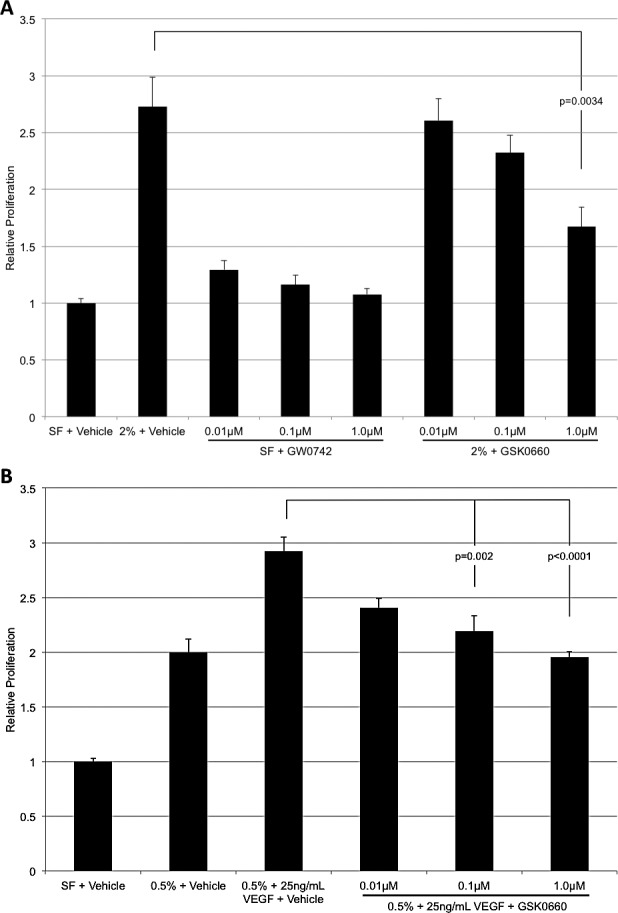
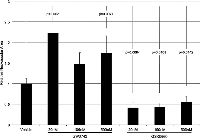
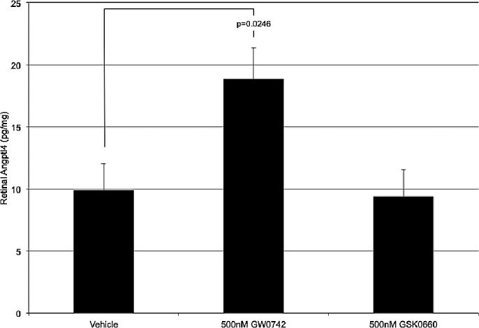
Results: GW0742 significantly increased angptl4 mRNA, and GSK0660 significantly decreased angptl4 mRNA. GW0742 had no effect on HRMEC proliferation, but caused a significant and dose-responsive increase in tube formation. GSK0660 significantly reduced serum-induced HRMEC proliferation and tube formation in a dose-dependent manner. Intravitreal injection of GW0742 significantly increased total retinal Angptl4 protein, but intravitreal injection of GSK0660 had no effect. Intravitreal injection of GW0742 significantly increased retinal NV, as did GW0742 administered by oral gavage. Conversely, both intravitreal injection and intraperitoneal injection of GSK0660 significantly reduced retinal NV.
Conclusions: PPAR-β/δ activation exacerbates, and its inhibition reduces, preretinal NV. PPAR-β/δ may regulate preretinal NV through a prodifferentiation/maturation mechanism that depends on Angptl4. Pharmacologic inhibition of PPAR-β/δ may provide a rational basis for therapeutic targeting of ocular NV.
Keywords: angiogenesis; nuclear transcription factor; peroxisome proliferator-activated receptor; retinopathy of prematurity; vascular endothelial growth factor.
Figures


References
-
- Klagsbrun M. Regulators of angiogenesis: stimulators, inhibitors, and extracellular matrix. J Cell Biochem. 1991; 47: 199–200 - PubMed
-
- Folkman J, Shing Y. Angiogenesis. J Biol Chem. 1992; 267: 10931–10934 - PubMed
-
- Folkman J, D'Amore PA. Blood vessel formation: what is its molecular basis? Cell. 1996; 87: 1153–1155 - PubMed
-
- Risau W. Mechanisms of angiogenesis. Nature. 1997; 386: 671–674 - PubMed
-
- Li J, Zhang YP, Kirsner RS. Angiogenesis in wound repair: angiogenic growth factors and the extracellular matrix. Microsc Res Tech. 2003; 60: 107–114 - PubMed
Publication types
MeSH terms
Substances
Grants and funding
LinkOut - more resources
Full Text Sources
Other Literature Sources
