. 2013 Aug;41(14):6952-9.
doi: 10.1093/nar/gkt409. Epub 2013 May 28.

UGA codon position-dependent incorporation of selenocysteine into mammalian selenoproteins

Affiliations

UGA codon position-dependent incorporation of selenocysteine into mammalian selenoproteins

Anton A Turanov et al. Nucleic Acids Res. 2013 Aug.

Abstract

It is thought that the SelenoCysteine Insertion Sequence (SECIS) element and UGA codon are sufficient for selenocysteine (Sec) insertion. However, we found that UGA supported Sec insertion only at its natural position or in its close proximity in mammalian thioredoxin reductase 1 (TR1). In contrast, Sec could be inserted at any tested position in mammalian TR3. Replacement of the 3'-UTR of TR3 with the corresponding segment of a Euplotes crassus TR restricted Sec insertion into the C-terminal region, whereas the 3'-UTR of TR3 conferred unrestricted Sec insertion into E. crassus TR, in which Sec insertion is normally limited to the C-terminal region. Exchanges of 3'-UTRs between mammalian TR1 and E. crassus TR had no effect, as both proteins restricted Sec insertion. We further found that these effects could be explained by the use of selenoprotein-specific SECIS elements. Examination of Sec insertion into other selenoproteins was consistent with this model. The data indicate that mammals evolved the ability to limit Sec insertion into natural positions within selenoproteins, but do so in a selenoprotein-specific manner, and that this process is controlled by the SECIS element in the 3'-UTR.


Figures
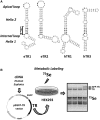
Figure 1.
Figure 1.
Experimental design of the study. (A) SECIS elements used in the study. SECIS element images were generated with SECISearch. The SECIS element core and apical loop are shown in bold, and the essential structural motifs are highlighted. Type II SECISes contain an additional stem in the apical loop. eTR1 and eTR2 designate Euplotes TR1 and TR2, respectively, and hTR1 and hTR3 designate human TR1 and TR3. (B) Design of the study. cDNAs corresponding to selenoproteins are cloned into the pEGFP-C3 vector (unless indicated otherwise). Site-directed mutagenesis is used to introduce mutations in the original sequences. HEK 293 cells are transfected with the resulting constructs, and 24 h after transfection, cells are labeled by supplementing the medium with 75Se for an additional 24 h. Proteins from each transfection are resolved by SDS–PAGE, and selenoprotein patterns are visualized. The EGFP-fusion of TRs is used to distinguish the expressed proteins (e.g. EGFP-TR; shown with an arrow) from endogenous selenoproteins (e.g. TR, shown with an arrow). Other detected bands represent endogenous selenoproteins, which serve as an internal control.
Figure 2.
Figure 2.
Position-dependent Sec insertion into hTR1 and mTGR. Cells were transfected with EGFP-hTR1 (A–C) or mTGR (D) constructs or with the indicated chimeric constructs containing Euplotes sequences. All expressed TRs had single in-frame UGA codons at indicated positions (natural or unnatural). Transfected HEK 293 cells were metabolically labeled with 75Se, and proteins separated by SDS–PAGE, transferred onto PVDF membranes, and selenoprotein patterns were visualized with a PhosphorImager. Left panels: Sec incorporation in TRs assayed by 75Se labeling. Numbers correspond to the UGA codon positions. Asterisk indicates natural positions of Sec-encoding UGA codons in TRs. Arrows show positions of full length Sec containing proteins. Middle panels: Schematic representation of constructs. Natural SECIS element sequences are shown in black, and when replaced with a foreign sequence, in gray. Right panels: This part of the figure shows ORF, 3′-UTR and SECIS element sequences that were used in indicated constructs. Hash mark indicates that the replaced sequence includes the C-terminal part of the coding sequence together with the 3′-UTR. (A) Expression of EGFP-hTR1. Sec insertion in hTR1 with its natural SECIS element. (B) Sec insertion in hTR1 with the 3′UTR from eTR1. (C) Sec insertion in hTR1 with the SECIS element from eTR1. (D) Expression of mTGR. Sec insertion in mTGR with its natural SECIS element.
Figure 3.
Figure 3.
Position-dependent Sec insertion in hTR3. (A) Expression of EGFP-hTR3 in HEK 293 cells. Sec insertion in hTR3 with its natural SECIS element. (B) Sec insertion in hTR3 with the 3′UTR from eTR2. (C) Sec insertion in hTR3 with the SECIS element from eTR2. The experiment was carried out as described in Figure 2.
Figure 4.
Figure 4.
Role of SECIS elements in position-dependent Sec insertion. (A) Expression of EGFP-eTR1 in HEK 293 cells. Sec insertion in eTR1 with its natural SECIS element. (B) Sec insertion in eTR1 with the SECIS element from hTR1. (C) Sec insertion in eTR1 with the SECIS element from hTR3. The experiment was carried out as described in Figure 2.
Figure 5.
Figure 5.
Role of the 3′-UTR and SECIS element in position-dependent Sec insertion. (A) Expression of EGFP-eTR2 in HEK 293 cells. Sec insertion in eTR2 with its natural SECIS element. (B) Sec insertion in eTR2 with the 3′-UTR from hTR3. (C) Sec insertion in eTR2 with the SECIS element from hTR3. (D) Sec insertion in eTR2 with a modified form of the natural SECIS element. (E) Sec insertion in eTR2 with the 3′-UTR from human selenoprotein P. The experiment was carried out as described in Figure 2. The 2 × eTR2 indicates duplication of the sequence between the natural Sec UGA codon and the SECIS core in eTR2 mRNA.
Figure 6.
Figure 6.
Role of the apical loop in hTR3 SECIS element in Sec insertion. (A) Sec insertion in eTR2 with the SECIS element from hTR3 with the 11 nt deletion in the apical loop. (B) Sec insertion in eTR2 with the SECIS element from hTR3 with the 8 nt deletion in the apical loop. (C) Natural and modified hTR3 SECIS elements used in the study. (D) Multiple sequence alignment of the natural and modified hTR3 SECIS elements. The experiment was carried out as described in Figure 2. Δ11 indicates deletion of 11 nt in the apical loop of the SECIS element. Δ8 indicates deletion of 8 nt in the apical loop of SECIS element.

References

    1. Kryukov GV, Castellano S, Novoselov SV, Lobanov AV, Zehtab O, Guigo R, Gladyshev VN. Characterization of mammalian selenoproteomes. Science. 2003;300:1439–1443. - PubMed
    1. Mariotti M, Ridge PG, Zhang Y, Lobanov AV, Pringle TH, Guigo R, Hatfield DL, Gladyshev VN. Composition and evolution of the vertebrate and mammalian selenoproteomes. PLoS One. 2012;7:e33066. - PMC - PubMed
    1. Arner ES. Selenoproteins-What unique properties can arise with selenocysteine in place of cysteine? Exp. Cell Res. 2010;316:1296–1303. - PubMed
    1. Donovan J, Copeland P. Threading the needle: getting selenocysteine into proteins. Antioxid. Redox Signal. 2010;12:881–892. - PMC - PubMed
    1. Squires JE, Berry MJ. Eukaryotic selenoprotein synthesis: mechanistic insight incorporating new factors and new functions for old factors. IUBMB Life. 2008;60:232–235. - PubMed

Publication types