. 2013 Jun 11;110(24):9891-6.
doi: 10.1073/pnas.1308336110. Epub 2013 May 28.

Genetic confirmation for a central role for TNFα in the direct action of thyroid stimulating hormone on the skeleton

Affiliations

Genetic confirmation for a central role for TNFα in the direct action of thyroid stimulating hormone on the skeleton

Li Sun et al. Proc Natl Acad Sci U S A. .

Erratum in

  • Proc Natl Acad Sci U S A. 2013 Jul 23;110(30):12498. Baliram, Ramkumari [corrected to Baliram, Ramkumarie]

Abstract

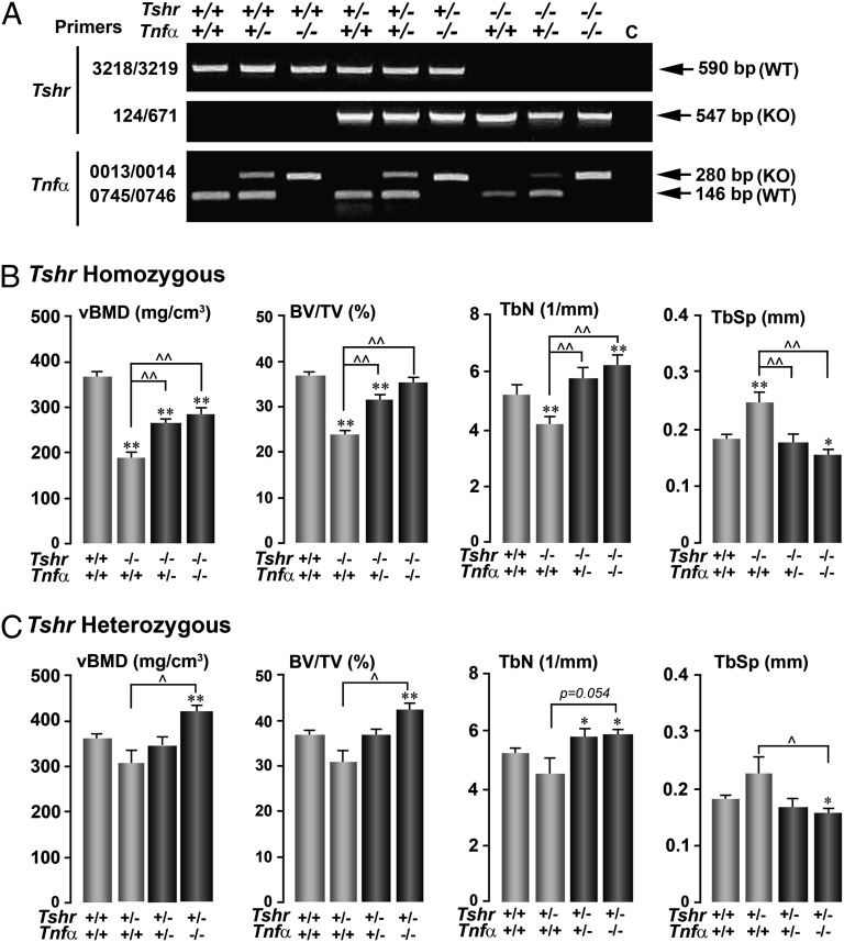
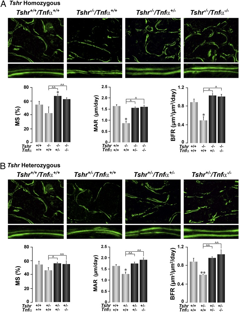
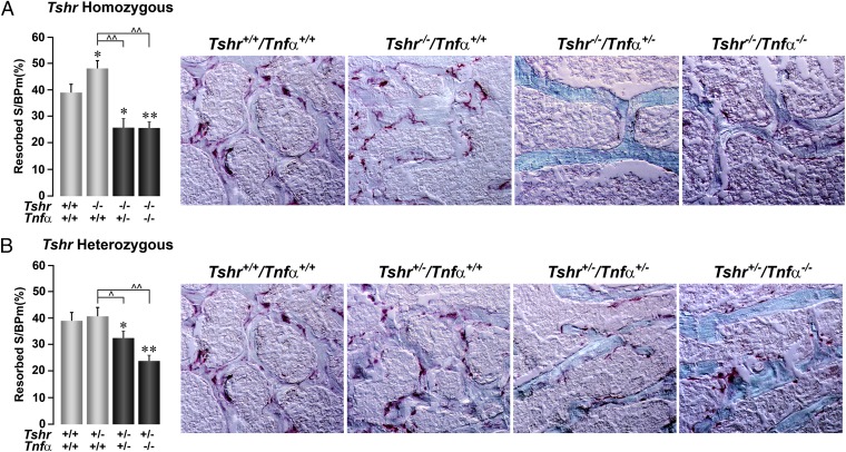
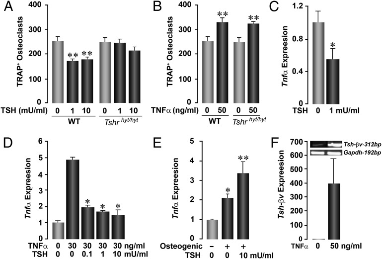
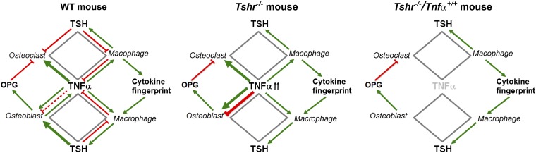
Clinical data showing correlations between low thyroid-stimulating hormone (TSH) levels and high bone turnover markers, low bone mineral density, and an increased risk of osteoporosis-related fractures are buttressed by mouse genetic and pharmacological studies identifying a direct action of TSH on the skeleton. Here we show that the skeletal actions of TSH deficiency are mediated, in part, through TNFα. Compound mouse mutants generated by genetically deleting the Tnfα gene on a Tshr(-/-) (homozygote) or Tshr(+/-) (heterozygote) background resulted in full rescue of the osteoporosis, low bone formation, and hyperresorption that accompany TSH deficiency. Studies using ex vivo bone marrow cell cultures showed that TSH inhibits and stimulates TNFα production from macrophages and osteoblasts, respectively. TNFα, in turn, stimulates osteoclastogenesis but also enhances the production in bone marrow of a variant TSHβ. This locally produced TSH suppresses osteoclast formation in a negative feedback loop. We speculate that TNFα elevations due to low TSH signaling in human hyperthyroidism contribute to the bone loss that has traditionally been attributed solely to high thyroid hormone levels.

Keywords: bone density; bone metabolism; thyroid disease.


Conflict of interest statement

Conflict of interest statement: M.Z. is a named inventor of a pending patent application related to osteoclastic bone resorption filed by the Mount Sinai School of Medicine.

Figures

Fig. 1.
Fig. 1.
Deletion of Tnfα rescues the osteoporosis owing to Tshr deficiency. Agarose gels showing genotype of representative compound mutants, using specific primer sets to detect Tshr and Tnfα gene expression. (A) The effect of deleting the Tnfα gene on a homozygote (Tshr−/−) (B) or heterozygote (Tshr+/−) (C) background on microtomography-based parameters of trabecular architecture, namely vBMD, BV/TV, TbN, and TbSp. Statistics: Student t test with Bonferroni’s correction; comparisons as shown, *P ≤ 0.05; **P ≤ 0.01, compared with Tshr+/+/Tnfα+/+ mice; ^P ≤ 0.05; ^^P ≤ 0.01, comparisons with Tshr−/−/Tnfα+/+ (B) or Tshr+/−/Tnfα+/+ (C) mice; mean ± SEM is shown, up to eight mice per group.
Fig. 2.
Fig. 2.
Deletion of Tnfα rescues the low bone formation owing to Tshr deficiency. The effect of deleting the Tnfα gene on a homozygote (Tshr−/−) (A) or heterozygote (Tshr+/−) (B) background on bone formation, shown either as double-labeled surfaces (10×, Lower: magnified digitally to show differences in interlabel distances) or as quantitative estimates of MS, MAR, and BFR (units as shown). Statistics by Student t test with Bonferroni’s correction; comparisons as shown, *P ≤ 0.05; **P ≤ 0.01, compared with Tshr+/+/Tnfα+/+ mice; ^P ≤ 0.05; ^^P ≤ 0.01, comparisons with Tshr−/−/Tnfα+/+ (A) or Tshr+/−/Tnfα+/+ (B) mice; mean ± SEM is shown, 9–57 sections from up to six mice per group.
Fig. 3.
Fig. 3.
Deletion of Tnfα suppresses resorption. The effect of deleting the Tnfα gene on a homozygote (Tshr−/−) (A) or heterozygote (Tshr+/−) (B) background on bone resorption, shown either as TRAP-positive surfaces (10×; dark pink) or as quantitative estimates of resorption surfaces (resorbed S/BPm). Statistics by Student t test with Bonferroni’s correction; comparisons as shown, *P ≤ 0.05; **P ≤ 0.01, compared with Tshr+/+/Tnfα+/+ mice; ^P ≤ 0.05; ^^P ≤ 0.01, compared with Tshr−/−/Tnfα+/+ (A) or Tshr+/−/Tnfα+/+ (B) mice; mean ± SEM is shown, 10–134 sections from up to 12 mice per group.
Fig. 4.
Fig. 4.
Interactions between Tsh and Tnfα. Effect of Tsh (A) and Tnfα (B) on TRAP-positive osteoclast formation in ex vivo bone marrow cell cultures derived from WT mice or Tshrhyt/hyt mice, which harbor a mutated, signaling-deficient Tshr. Whereas TSH inhibits osteoclast formation in WT cultures, the effect is lost in cultures from Tshrhyt/hyt mice (A). In contrast, Tnfα stimulates osteoclastogenesis in both WT and Tshrhyt/hyt mice (B). TSH inhibits both basal (C) and Tnfα-induced (D) Tnfα expression in osteoclasts, whereas it stimulates Tnfα expression by osteoblasts differentiating in osteogenic media (10) (E) (by quantitative PCR). Tnfα stimulates the expression of Tsh-βv in RAW264.7 macrophages, shown as 312-bp product on agarose gel and quantitated by quantitative PCR (F). Statistics by Student t test with Bonferroni’s correction; all comparisons with zero dose, *P ≤ 0.05; **P ≤ 0.01.
Fig. 5.
Fig. 5.
A central role for Tnfα in hyperthyroid bone loss. Elevated Tnfα in Tshr−/− mice increases osteoclastogenesis by acting directly on the osteoclast precursor and by creating a footprint for the release of other pro-osteoclastogenic cytokines, such as IL-1 and IL-6. Excessive Tnfα also stimulates Tsh-βv production from macrophages, but in Tshr−/− mice the released molecule fails to inhibit osteoclastic bone resorption or to stimulate the production of osteoprotegerin, a Rank-l inhibitor. The Tsh-βv also fails to stimulate osteoblastic bone formation in Tshr−/− mice. Hence, profound hyperresorption, reduced bone formation, and bone loss ensue in Tsh, signaling deficiency. When Tnfα is removed from the Tshr−/− background, full recovery occurs.

References

    1. Abe E, et al. TSH is a negative regulator of skeletal remodeling. Cell. 2003;115(2):151–162. - PubMed
    1. Sun L, et al. Intermittent recombinant TSH injections prevent ovariectomy-induced bone loss. Proc Natl Acad Sci USA. 2008;105(11):4289–4294. - PMC - PubMed
    1. Baliram R, et al. Hyperthyroid-associated osteoporosis is exacerbated by the loss of TSH signaling. J Clin Invest. 2012;122(10):3737–3741. - PMC - PubMed
    1. Vincent BH, et al. Bone marrow cells produce a novel TSHbeta splice variant that is upregulated in the thyroid following systemic virus infection. Genes Immun. 2009;10(1):18–26. - PMC - PubMed
    1. Grimnes G, Emaus N, Joakimsen RM, Figenschau Y, Jorde R. The relationship between serum TSH and bone mineral density in men and postmenopausal women: The Tromsø study. Thyroid. 2008;18(11):1147–1155. - PubMed

Publication types

MeSH terms