. 2013 Jun 11;110(24):9856-61.
doi: 10.1073/pnas.1220864110. Epub 2013 May 28.

ZNF408 is mutated in familial exudative vitreoretinopathy and is crucial for the development of zebrafish retinal vasculature

Affiliations

ZNF408 is mutated in familial exudative vitreoretinopathy and is crucial for the development of zebrafish retinal vasculature

Rob W J Collin et al. Proc Natl Acad Sci U S A. .

Abstract

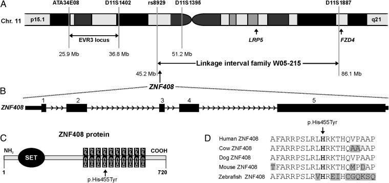
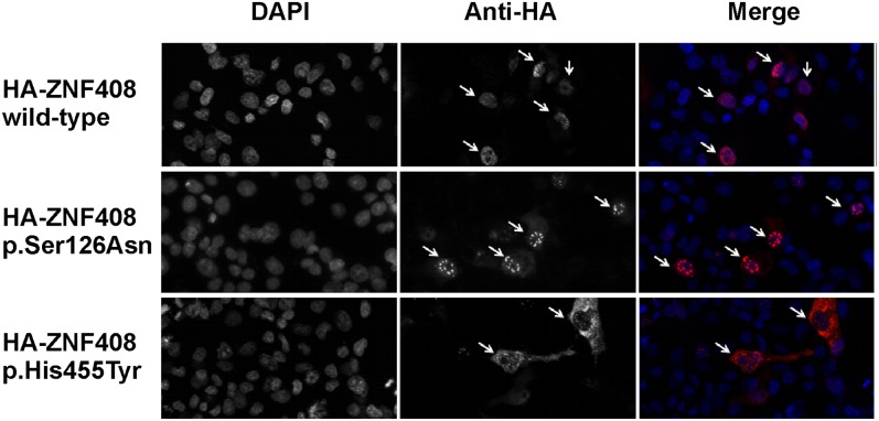
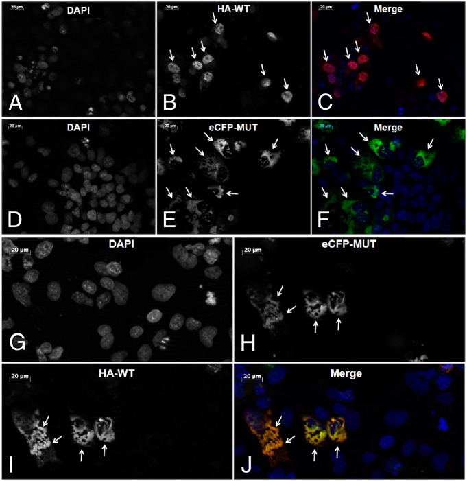
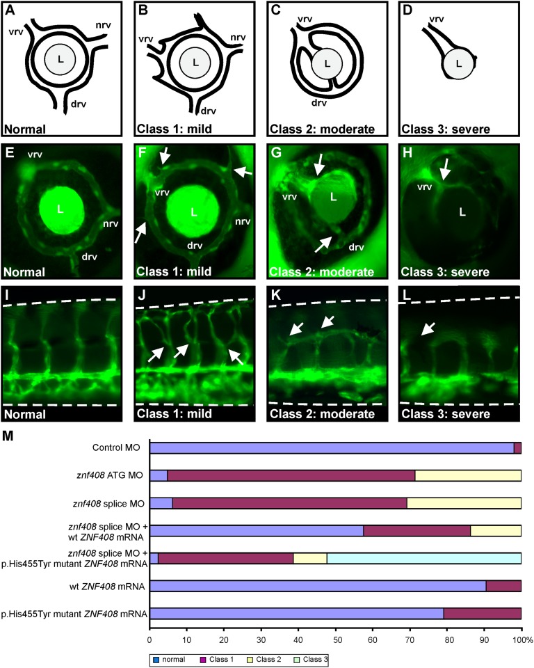
Familial exudative vitreoretinopathy (FEVR) is a genetically heterogeneous disorder characterized by abnormal vascularization of the peripheral retina, which can result in retinal detachment and severe visual impairment. In a large Dutch FEVR family, we performed linkage analysis, exome sequencing, and segregation analysis of DNA variants. We identified putative disease-causing DNA variants in proline-alanine-rich ste20-related kinase (c.791dup; p.Ser265ValfsX64) and zinc finger protein 408 (ZNF408) (c.1363C>T; p.His455Tyr), the latter of which was also present in an additional Dutch FEVR family that subsequently appeared to share a common ancestor with the original family. Sequence analysis of ZNF408 in 132 additional individuals with FEVR revealed another potentially pathogenic missense variant, p.Ser126Asn, in a Japanese family. Immunolocalization studies in COS-1 cells transfected with constructs encoding the WT and mutant ZNF408 proteins, revealed that the WT and the p.Ser126Asn mutant protein show complete nuclear localization, whereas the p.His455Tyr mutant protein was localized almost exclusively in the cytoplasm. Moreover, in a cotransfection assay, the p.His455Tyr mutant protein retains the WT ZNF408 protein in the cytoplasm, suggesting that this mutation acts in a dominant-negative fashion. Finally, morpholino-induced knockdown of znf408 in zebrafish revealed defects in developing retinal and trunk vasculature, that could be rescued by coinjection of RNA encoding human WT ZNF408 but not p.His455Tyr mutant ZNF408. Together, our data strongly suggest that mutant ZNF408 results in abnormal retinal vasculogenesis in humans and is associated with FEVR.


Conflict of interest statement

The authors declare no conflict of interest.

Figures

Fig. 1.
Fig. 1.
ZNF408 chromosomal location, genomic structure, and ZNF408 protein characteristics. (A) Part of chromosome 11 showing the W05-215 linkage interval. The locations of the FEVR genes FZD4 and LRP5, as well as the boundaries of the EVR3 locus (16), are depicted. (B) Exon-intron structure of the ZNF408 gene. (C) Schematic overview of ZNF408, a protein of 720 amino acids that is predicted to contain one putative SET and 10 C2H2-type zinc-finger domains. The position of the histidine residue that is mutated to a tyrosine is indicated. (D) Sequence comparison of ZNF408 proteins from several vertebrate species. The alignment contains the histidine residue that is mutated (in bold type), along with 10 flanking amino acids on each side. Identical residues are black on a white background, whereas nonidentical residues are black on a gray background.
Fig. 2.
Fig. 2.
Immunocytochemical analysis of COS-1 cells transiently transfected with constructs encoding HA-tagged WT and mutant (p.Ser126Asn, p.His455Tyr) ZNF408 proteins. DAPI stains cell nuclei, whereas the anti-HA antibody stains the ZNF408 fusion proteins (indicated by arrows). (Right) Merged pictures (DAPI in blue, and HA-tagged ZNF408 proteins in red). Representative examples are shown for each transfection, indicating full nuclear localization for the WT and p.Ser126Asn ZNF408 proteins, but with the p.His455Tyr mutant ZNF408 confined mainly to the cytoplasm (each indicated by arrows).
Fig. 3.
Fig. 3.
Cotransfection analysis of WT and p.His455Tyr mutant ZNF408. (A–C) Immunocytochemical analysis of transiently transfected HA-tagged WT ZNF408 proteins. Representative examples show WT ZNF408 expression inside the cell nuclei (arrows). Images are presented as DAPI (staining cell nuclei) (A), anti-HA (detecting HA-tagged ZNF408 proteins) (B), and merged pictures (DAPI in blue, HA-tagged ZNF408 in red) (C). (D–F) Fluorescence analysis of transiently transfected eCFP-p.His455Tyr mutant ZNF408 proteins. Mutant ZNF408 is located mainly outside the cell nuclei (indicated by arrows). Images are presented as DAPI (staining cell nuclei) (D), eCFP (detecting eCFP-ZNF408 mutant proteins) (E), and merged pictures (DAPI in blue, eCFP-tagged p.His455Tyr ZNF408 in green) (F). (G–J) Combined analysis of COS-1 cells cotransfected with HA-tagged WT ZNF408 as well as eCFP-p.His455Tyr mutant ZNF408 proteins. In cells that express both the mutant and the WT ZNF408, the WT ZNF408 is also retained in the cytoplasm (I and J). Images are presented as DAPI (staining cell nuclei) (G), eCFP (detecting eCFP-p.His455Tyr ZNF408 mutant proteins (H), anti-HA (detecting HA-tagged WT ZNF408 proteins) (I), and merged pictures (DAPI in blue, HA-tagged WT ZNF408 in red, eCFP-p.His455Tyr ZNF408 in green) (J).
Fig. 4.
Fig. 4.
Functional characterization of znf408 in zebrafish. (A–D) Schematic overview of zebrafish eye vasculature phenotypes observed on MO-induced knockdown, or expression of WT or mutant ZNF408. Embryos were divided into four subclasses: normal, mildly affected (class 1), moderately affected (class 2), and severely affected (class 3). In the normal situation, blood enters through the nasal vessel (nrv) and exits via the intraocular ring vessel through the dorsal (drv) and ventral (vrv) vessels. L, lens. (E–H) Representative images of GFP expression in the eyes of fli1:EGFP transgenic zebrafish larvae. Abnormal sprouting of the vessels in class 1 embryos is indicated by white arrows (F). In class 2 (G), the nrv is absent, the vrv is enlarged, and both vrv and drv are directly attached to the lens. In class 3 (H), the complete intraocular ring vessel is absent. (I–L) Lateral views of the trunk vasculature (dorsal side is up) in fli1:EGFP transgenic larvae. Intersegmental vessels project from the dorsal aorta (bottom horizontal vessel) toward the dorsal longitudinal vessel at the top. (J) In class 1 embryos, intersegmental vessel branching is disrupted (indicated by white arrows). (K and L) In class 2 and class 3 embryos, the intersegmental vessels failed to reach the dorsal longitudinal vessel. (M) Phenotypic classification of zebrafish larvae injected with control MO (10 ng), znf408-ATG MO (6 ng) or znf408-splice MO (6 ng), mRNA encoding human WT or p.His455Tyr mutant ZNF408, or combinations of splice MO and mRNAs. At least 50 larvae per condition were evaluated.

References

    1. Criswick VG, Schepens CL. Familial exudative vitreoretinopathy. Am J Ophthalmol. 1969;68(4):578–594. - PubMed
    1. Canny CLB, Oliver GL. Fluorescein angiographic findings in familial exudative vitreoretinopathy. Arch Ophthalmol. 1976;94(7):1114–1120. - PubMed
    1. Boonstra FN, et al. Clinical and molecular evaluation of probands and family members with familial exudative vitreoretinopathy. Invest Ophthalmol Vis Sci. 2009;50(9):4379–4385. - PubMed
    1. Chen ZY, et al. A mutation in the Norrie disease gene (NDP) associated with X-linked familial exudative vitreoretinopathy. Nat Genet. 1993;5(2):180–183. - PubMed
    1. Gow J, Oliver GL. Familial exudative vitreoretinopathy: An expanded view. Arch Ophthalmol. 1971;86(2):150–155. - PubMed

Publication types

MeSH terms

LinkOut - more resources