. 2013 Jun 11;110(24):E2209-18.
doi: 10.1073/pnas.1303573110. Epub 2013 May 28.

Antibody to a conserved antigenic target is protective against diverse prokaryotic and eukaryotic pathogens

Affiliations

Antibody to a conserved antigenic target is protective against diverse prokaryotic and eukaryotic pathogens

Colette Cywes-Bentley et al. Proc Natl Acad Sci U S A. .

Abstract

Microbial capsular antigens are effective vaccines but are chemically and immunologically diverse, resulting in a major barrier to their use against multiple pathogens. A β-(1→6)-linked poly-N-acetyl-d-glucosamine (PNAG) surface capsule is synthesized by four proteins encoded in genetic loci designated intercellular adhesion in Staphylococcus aureus or polyglucosamine in selected Gram-negative bacterial pathogens. We report that many microbial pathogens lacking an identifiable intercellular adhesion or polyglucosamine locus produce PNAG, including Gram-positive, Gram-negative, and fungal pathogens, as well as protozoa, e.g., Trichomonas vaginalis, Plasmodium berghei, and sporozoites and blood-stage forms of Plasmodium falciparum. Natural antibody to PNAG is common in humans and animals and binds primarily to the highly acetylated glycoform of PNAG but is not protective against infection due to lack of deposition of complement opsonins. Polyclonal animal antibody raised to deacetylated glycoforms of PNAG and a fully human IgG1 monoclonal antibody that both bind to native and deacetylated glycoforms of PNAG mediated complement-dependent opsonic or bactericidal killing and protected mice against local and/or systemic infections by Streptococcus pyogenes, Streptococcus pneumoniae, Listeria monocytogenes, Neisseria meningitidis serogroup B, Candida albicans, and P. berghei ANKA, and against colonic pathology in a model of infectious colitis. PNAG is also a capsular polysaccharide for Neisseria gonorrhoeae and nontypable Hemophilus influenzae, and protects cells from environmental stress. Vaccination targeting PNAG could contribute to immunity against serious and diverse prokaryotic and eukaryotic pathogens, and the conserved production of PNAG suggests that it is a critical factor in microbial biology.

Keywords: animal models; carbohydrates; immunotherapy; infectious diseases; malaria.


Conflict of interest statement

Conflict of interest statement: G.B.P., T.M.-L., M.L.G., Y.E.T., and N.E.N. are inventors of intellectual properties [human monoclonal antibody to PNAG (G.B.P. and T.M.-L.) and PNAG vaccines (G.B.P., T.M.-L., M.L.G., Y.E.T., and N.E.N.], which are licensed by Brigham and Women's Hospital to Alopexx Vaccine, LLC, and Alopexx Pharmaceuticals, LLC, entities in which G.B.P. also holds equity. As inventors of intellectual properties, these authors also have the right to receive a share of licensing-related income (royalties, fees) through Brigham and Women's Hospital from Alopexx Pharmaceuticals, LLC, and Alopexx Vaccine, LLC. G.B.P. and T.M.-L.'s interests were reviewed and are managed by the Brigham and Women's Hospital and Partners Healthcare in accordance with their conflict of interest policies. A.R. and C.P. are employees of Sanofi, Inc., which has licensed the technology for use of mAb F598 (designated as SAR279356) for treatment and prevention of human infections.

Figures
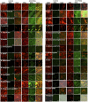
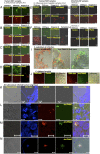
Fig. 1.
Fig. 1.
Expression of PNAG among diverse microbial pathogens. Cells from cultures of the indicated microbes were fixed with paraformaldehyde, placed onto slides, exposed to cold methanol, and then reacted with either control mAb F429 conjugated to AF488 or mAb F598 to PNAG also conjugated to AF488 (green fluorescence) as well as with SYTO 83 to visualize DNA (red fluorescence) and photographed using a scanning confocal laser microscopy. The label “dual” indicates overlay of red and green channels. (Scale bars: 10 µm; note that some micrographs lack bars due to cropping but are at same magnification as other micrographs of same strain.)
Fig. 2.
Fig. 2.
Demonstration that PNAG is a surface capsular polysaccharide for N. gonorrhoeae and nontypable H. influenzae and is also surface expressed on N. meningitidis serogroups B and A. Indicated organism (A–I) was reacted with control mAb F429 to P. aeruginosa alginate or mAb F598 to PNAG followed by protein A conjugated to 15-nM gold particles. (J) N. meningitidis serogroup A strain Z2087 cells were first reacted with either control mAb F429 or mAb F598 to PNAG then with 15-nm protein A gold particles followed by 1% glutaraldehyde to cross-link the human IgG1 mAbs and the 15-nm protein A gold label and to block further binding of protein A. Next, a mouse IgG mAb to the serogroup A capsule was applied to the grids, followed by a bridging rabbit antibody to mouse IgG then by 10-nm protein A gold particles. Control + anti-serogroup A panels show binding only of the antibody to serogroup A, whereas panels labeled anti-PNAG (598) + anti-serogroup A show binding of both antibodies. High magnification of indicated surface area (purple arrow) shows intercalation of selected examples of both 10-nm (purple circles) and 15-nm (green circles) gold particles on the bacterial surface.
Fig. 3.
Fig. 3.
In vivo expression of PNAG by various microbial pathogens. Samples of infected middle ear fluid (MEF) from humans with either S. pneumoniae (A–D) or nontypable H. influenzae (E and F) otitis media (OM) or chinchillas with OM due to S. pneumoniae serotype 19A (G and H) were treated with chitinase and reacted with control mAb F429-AF488 to P. aeruginosa alginate or treated with chitinase, dispersin B, or periodate in the case of nontypable H. influenzae (periodate destroyed S. pneumoniae cells) and reacted with mAb F598-AF488 to PNAG. (I) Colonic sections from mice with C. rodentium infection were stained with either control mAb F429-AF488 or mAb F598-AF488 to PNAG (green) plus SYTO 83 (red) to visualize DNA. Two different sections stained for both PNAG and DNA shown. (J) Sections from the cornea of a mouse with C. albicans keratitis stained with either control mAb F429-AF488 or mAb F598-AF488 to PNAG (green) plus SYTO 83 (red) to visualize DNA. Sections of a human lung infected with M. tuberculosis (K–N) were stained with SYTO 62 to visualize DNA (blue), rabbit antibody to M. tuberculosis followed by anti-rabbit IgG secondary antibody (red), or control mAb F429-AF488 or anti PNAG-mAb F598-AF488 (green). (Scale bars: white, 10 µm; red, 20 µM; some micrographs lack bars due to cropping but are at the same magnification as other micrographs of the same strain.)
Fig. 4.
Fig. 4.
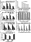
Opsonic or bactericidal killing of selected microbial pathogens by antibody to PNAG (anti-9GlcNH2-TT). (A and B) Opsonic killing of four strains of S. pneumoniae mediated by polyclonal rabbit (A) or mAb (B) to PNAG in the presence of HL60 phagocytes and complement (C′). Control for mAb F598 is irrelevant mAb F429. (C) Opsonic killing of three strains of E. faecalis mediated by polyclonal antibody to PNAG. (D) Opsonic killing of two strains (003 and 771) of S. pyogenes mediated by mAb F598 as well as a deletion mutant of strain 771 lacking the ability to produce the self-antigenic hyaluronic acid GAS capsule (strain 188). (E) Opsonic killing of C. albicans by indicated amount of mAb F598 in the presence of polymorphonuclear neutrophils (PMNs) and complement. (F and G) Bactericidal killing of six strains of N. gonorrhoeae (F) and five strains of N. meningitidis serogroup B (G) mediated by polyclonal rabbit antibody to PNAG and its indigenous C′. Killing calculated to subtract out any reductions in cfu counts by a comparable dilution of normal rabbit serum (NRS). Heat-inactivated complement (HI C′) controls for all N. gonorrhoeae strains had greater cfu counts surviving than in control NRS, and are not shown on figure. Bars represent means of triplicates to quadruplicates determined in the same assay; bars below the zero line indicate samples with colony counts greater than the control.
Fig. 5.
Fig. 5.
Protective efficacy of antibody to PNAG against experimental mouse infections caused by various pathogens. (A) Protection against lethality due to S. pyogenes (group A Streptococcus) infection initiated from an s.c. injection. (B) Protection against lethal sepsis in 3-d-old neonatal mice following i.p. injection of L. monocytogenes. (C) Protection against lethal, systemic infection in CBA/N mice infected intranasally with S. pneumoniae serotype 2 strain D39. P values for A–C determined by log-rank tests. (D) Reductions in bacterial burden 24 h after infection into the lungs of FVB mice given indicated dose of mAb F598 i.v. 4 h before intranasal infection with S. pneumoniae serotype 9V strain. Comparison group given the antibiotic cefotaxime (15 mg/kg) 1 and 4 h postinfection. P values determined by ANOVA (overall P < 0.001) and post hoc, pairwise comparisons made to the control. An irrelevant human IgG1 mAb had no effect on S. pneumoniae clearance in this model (not depicted). (E and F) Levels of N. meningitidis serogroup B strain B16B6 in the brains of 3-d-old mice given 50 µL of indicated antiserum 24 h before i.p. infection with two different doses of bacteria. Brain levels determined 24 h postinfection. P values determined by nonparametric t test. (G) Cumulative histopathologic scores in TRUC mice at 8 wk of age following treatment commenced in week 1 with either control human IgG1 mAb F105 or mAb F598 to PNAG. (H) Cumulative histopathologic scores in WT mice cross-fostered on TRUC females that transmit infectious colitis to the nursed animals by 8 wk of age. Treatment commenced in week 4 with either control human IgG1 mAb F105 or mAb F598 to PNAG. P values for G and H determined by nonparametric t test. (E–H) Symbols represent individual mice; lines represent median for the group.
Fig. 6.
Fig. 6.
Protective efficacy of mAb or polyclonal sera to PNAG against eukaryotic pathogens. (A) Reductions in cfu counts recovered from the cornea of C57BL/6 mice infected with C. albicans during keratitis. Mice treated with 200 µg i.p. of either control IgG1 mAb or F598 24 h before eye infection followed by topical application of 5 µg mAb per eye 24 and 32 h postinfection. Corneas recovered and processed after 48 h of infection. P value by nonparametric t test. (B) Reductions in cfu counts recovered from the cornea of mice infected with C. albicans during keratitis. Mice treated with either control IgG1 mAb or F598 by topical application of 5 µg per eye 4, 8, and 24 h postinfection with corneas recovered for processing 32 h postinfection. P value by nonparametric t test. Corneal pathology scores associated with these treatments are in Fig. S5. Symbols represent individual mice; lines represent the group medians. (C) Survival of C57BL/6 mice from systemic PNAG-positive P. berghei ANKA infection given i.p. injections of either 0.2 mL of normal goat serum or 0.2 mL of goat antibody raised to 9GlcNH2-TT on days −1, +2, +5, +8, +11, +14, +17, and +20. P value by log-rank test. (D) Lack of survival of C57BL/6 mice from systemic PNAG-negative, GFP-positive P. berghei ANKA infection given i.p. injections of either 0.2 mL of normal goat serum or 0.2 mL of goat antibody raised to 9GlcNH2-TT on days −1, +2, +5, and +8.

Comment in

References

    1. Centers for Disease Control and Prevention (CDC) Control of infectious diseases. MMWR Morb Mortal Wkly Rep. 1999;48(29):621–629. - PubMed
    1. Kwong PD, Mascola JR. Human antibodies that neutralize HIV-1: Identification, structures, and B cell ontogenies. Immunity. 2012;37(3):412–425. - PMC - PubMed
    1. Lingwood D, et al. Structural and genetic basis for development of broadly neutralizing influenza antibodies. Nature. 2012;489(7417):566–570. - PMC - PubMed
    1. Toltzis P, Jacobs MR. The epidemiology of childhood pneumococcal disease in the United States in the era of conjugate vaccine use. Infect Dis Clin North Am. 2005;19(3):629–645. - PubMed
    1. Ladhani SN. Two decades of experience with the Haemophilus influenzae serotype b conjugate vaccine in the United Kingdom. Clin Ther. 2012;34(2):385–399. - PubMed

Publication types

MeSH terms