. 2013 Jun 11;110(24):9710-5.
doi: 10.1073/pnas.1217042110. Epub 2013 May 28.

Structures of ABCB10, a human ATP-binding cassette transporter in apo- and nucleotide-bound states

Affiliations

Structures of ABCB10, a human ATP-binding cassette transporter in apo- and nucleotide-bound states

Chitra A Shintre et al. Proc Natl Acad Sci U S A. .

Abstract

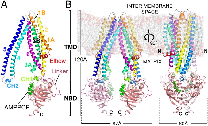
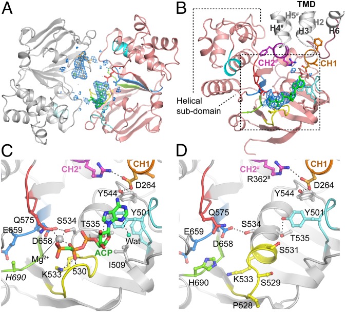
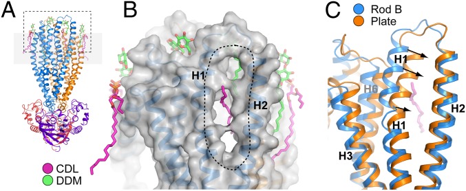
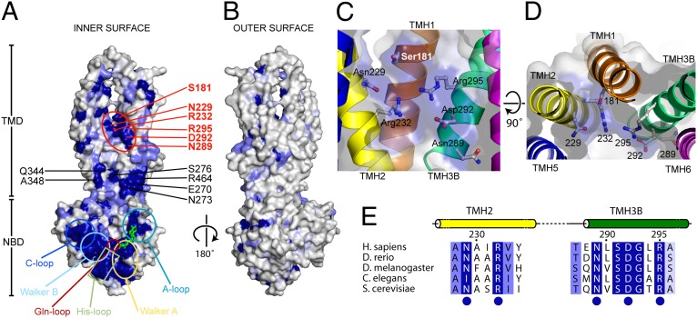
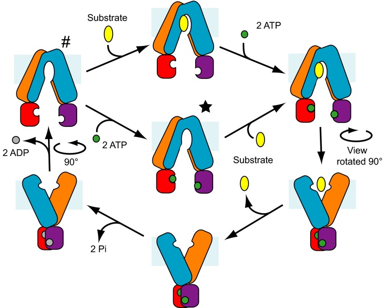
ABCB10 is one of the three ATP-binding cassette (ABC) transporters found in the inner membrane of mitochondria. In mammals ABCB10 is essential for erythropoiesis, and for protection of mitochondria against oxidative stress. ABCB10 is therefore a potential therapeutic target for diseases in which increased mitochondrial reactive oxygen species production and oxidative stress play a major role. The crystal structure of apo-ABCB10 shows a classic exporter fold ABC transporter structure, in an open-inwards conformation, ready to bind the substrate or nucleotide from the inner mitochondrial matrix or membrane. Unexpectedly, however, ABCB10 adopts an open-inwards conformation when complexed with nonhydrolysable ATP analogs, in contrast to other transporter structures which adopt an open-outwards conformation in complex with ATP. The three complexes of ABCB10/ATP analogs reported here showed varying degrees of opening of the transport substrate binding site, indicating that in this conformation there is some flexibility between the two halves of the protein. These structures suggest that the observed plasticity, together with a portal between two helices in the transmembrane region of ABCB10, assist transport substrate entry into the substrate binding cavity. These structures indicate that ABC transporters may exist in an open-inwards conformation when nucleotide is bound. We discuss ways in which this observation can be aligned with the current views on mechanisms of ABC transporters.

Keywords: ABC mitochondrial erythroid; X-ray crystallography; cardiolipin; human membrane protein structure; nucleotide complex.


Conflict of interest statement

The authors declare no conflict of interest.

Figures

Fig. 1.
Fig. 1.
Structure of ABCB10 in complex with the nonhydrolysable nucleotide analog AMPPCP, showing that ABCB10 is in an open conformation, even when it is bound to nucleotide analogs. Cartoon representations of the ABCB10/AMPPCP complex monomer (A) and homodimer (B) as seen in the rod-form B crystal structure. The structures have a single monomer in the asymmetric unit, the dimer is generated by a crystallographic twofold.
Fig. 2.
Fig. 2.
Comparison of the structures of the ABCB10 homodimer in the absence (apo) and presence of bound nucleotide analogs, with the structures of ceP-gp in the open-inwards conformation [PDB ID code: 4F4C (10)] and Sav1866 [PDB ID code 2HYD (11)] in the open-outwards conformation. Transporters are viewed perpendicular to their (pseudo) twofold symmetry axes. (B) Alignment of NBDs of the structures shown in A. viewed looking toward the membrane. The ABCB10 monomers (blue/purple and orange/red respectively), nucleotides (green), and the NBD’s C-loop (cyan) are highlighted. The black lines/circles below each NBD pair indicate the translation required to bring the NBDs in the closed conformation for catalysis [the distance is the separation between the nucleotide γ-phosphate (green) and the C-α of the first glycine in the catalytic C loop of the adjacent NBD (cyan)]. An additional rotational component is also required for proper alignment of the NBDs in the closed state. Where trinucleotide is not present in the structure, the position of the γ-phosphate has been inferred by superposition of the AMPPCP complex. The C-terminal extension in the Sav1866 structure has been omitted for clarity.
Fig. 3.
Fig. 3.
Interactions of nucleotides with the ABCB10 inward-facing conformation. (A) Schematic representation of NBDs of ABCB10 homodimer in the highest-resolution structure (rod form A) viewed looking from the TMD. AMPPCP (green/orange) are bound to each NBD but do not make inter-NBD interaction with the ABC transporter consensus sequence LSGGQ C-loop motif (cyan). (B) Individual NBD viewed looking from adjacent NBD. NBD (pink) with ABC Transporter nucleotide binding signature motifs colored as in C. TMD (gray) and coupling helices (CH1, orange; CH2#, purple) are highlighted. Omit FoFc density for AMPPCP/Mg2+ (blue mesh) is shown contoured at 3 σ. Dotted box indicates zoomed region in C and D. Detailed view of nucleotide binding site in rod form A/AMPPCP complex (C) and nucleotide-free form (D). Oxygen atoms are colored red, nitrogen atoms blue, phosphate atoms orange and carbon atoms are colored according to the location of the atom: The AMPPCP carbon atoms are shown in green. The conserved NBD sequence motifs are colored Walker A (yellow), Walker B (light blue), A-loop (pale cyan), Gln-loop (red), His-loop (light green), coupling helix 1 (CH1, orange), and coupling helix 2 (CH2, magenta). The C-loop (cyan) is not visible as it is more than 16 Å away from the nucleotide binding site in this conformation. Residues/secondary structure elements marked with a pound symbol (#) denote regions contributed by homodimer partner. The side-chain of His690 is disordered and has not been modeled in rod form A.
Fig. 4.
Fig. 4.
ABCB10 has cardiolipin and detergent bound to the transmembrane helices and a portal between helices TMH1 and TMH2, which is open in the rod crystal form and closed in the plate-form crystals. (A) Overview of the ABCB10 structure showing the location of lipid (magenta) and detergent (green) binding sites. (B) Molecular surface representation of the TMD in rod form A crystals, with lipid and detergent molecules shown in magenta and green and the portal between TMH1 and TMH2 indicated with a dotted line. In the rod-form structures TMH1 and TMH2 are loosely packed revealing a 7 Å wide × 30 Å long portal connecting the central cavity of the TMDs with the membrane environment. The portal is occupied by a CDL alkyl chain (magenta). (C) In the plate form crystals TMH1 and TMH2 are packed closer together, with the portal closed. Structures are viewed in the same orientation as in A.
Fig. 5.
Fig. 5.
Sequence conservation of residues in ABCB10. Conservation is mapped onto a molecular surface representation of the concave inner cavity (A) and convex outer surface (B) of ABCB10. The molecular surface shown is a composite representing one-half “leaflet” of the transporter dimer, comprising residues from TMH1–TMH3 and TMH6-NBD in one monomer and TMH4/5 from the second monomer (residues 311–424). Residues are colored according to sequence conservation, invariant (dark blue), highly conserved (slate; 2–4 related amino acids), and moderately conserved (pale blue; 3–8 related amino acids). Sequence conservation was calculated based on an alignment of eighty ABCB10 homologs (human to yeast) using the CONSURF server (38). The conserved patch in the TMD between TMH2/TMH3B adjacent to the portal region (indicated in red) defines a unique ABCB10 signature sequence. (C and D) Perpendicular views of residues defined by ABCB10 signature sequence (Asn229, Arg232, Asn289, Asp295, and Arg295). All of the side-chains project toward the lumen of the transporter. Side-chains are shown along with a semitransparent molecular surface. (E) Conserved signature sequence defined by residues located in central portion of TMH2 and at the N-terminal end of TMH3B.
Fig. 6.
Fig. 6.
Overview of the steps proposed for the transport cycle of ABCB10 and other ABC transporters of the exporter family. The TMDs are colored blue and orange, with their associated NBDs colored purple and red. ATP is shown in green, ADP in gray and the transport substrate in yellow. An asterisk (*) and pound symbol (#) indicate the conformations observed for nucleotide-bound and nucleotide-free ABCB10, respectively.

References

    1. Locher KP. Review. Structure and mechanism of ATP-binding cassette transporters. Philos Trans R Soc Lond B Biol Sci. 2009;364(1514):239–245. - PMC - PubMed
    1. Zolnerciks JK, Andress EJ, Nicolaou M, Linton KJ. Structure of ABC transporters. Essays Biochem. 2011;50(1):43–61. - PubMed
    1. Aittoniemi J, et al. Review. SUR1: A unique ATP-binding cassette protein that functions as an ion channel regulator. Philos Trans R Soc Lond B Biol Sci. 2009;364(1514):257–267. - PMC - PubMed
    1. Sheppard DN, Welsh MJ. Structure and function of the CFTR chloride channel. Physiol Rev. 1999;79(1) Suppl:S23–S45. - PubMed
    1. Gottesman MM. Mechanisms of cancer drug resistance. Annu Rev Med. 2002;53:615–627. - PubMed

Publication types

MeSH terms