. 2013 Jun 11;110(24):9879-84.
doi: 10.1073/pnas.1303524110. Epub 2013 May 28.

HHLA2 is a member of the B7 family and inhibits human CD4 and CD8 T-cell function

Affiliations

HHLA2 is a member of the B7 family and inhibits human CD4 and CD8 T-cell function

Ruihua Zhao et al. Proc Natl Acad Sci U S A. .

Abstract

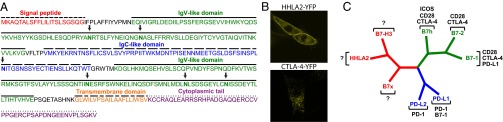
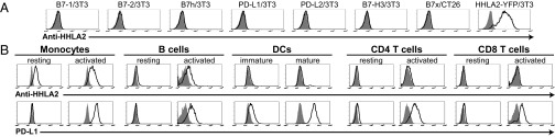
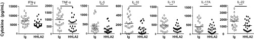
T-cell costimulation and coinhibition generated by engagement of the B7 family and their receptor CD28 family are of central importance in regulating the T-cell response, making these pathways very attractive therapeutic targets. Here we describe HERV-H LTR-associating protein 2 (HHLA2) as a member of the B7 family that shares 10-18% amino acid identity and 23-33% similarity to other human B7 proteins and phylogenetically forms a subfamily with B7x and B7-H3 within the family. HHLA2 is expressed in humans but not in mice, which is unique within the B7 and CD28 families. HHLA2 protein is constitutively expressed on the surface of human monocytes and is induced on B cells after stimulation with LPS and IFN-γ. HHLA2 does not interact with other known members of the CD28 family or the B7 family, but does bind a putative receptor that is constitutively expressed not only on resting and activated CD4 and CD8 T cells but also on antigen-presenting cells. HHLA2 inhibits proliferation of both CD4 and CD8 T cells in the presence of T-cell receptor signaling. In addition, HHLA2 significantly reduces cytokine production by T cells including IFN-γ, TNF-α, IL-5, IL-10, IL-13, IL-17A, and IL-22. Thus, we have identified a unique B7 pathway that is able to inhibit human CD4 and CD8 T-cell proliferation and cytokine production. This unique human T-cell coinhibitory pathway may afford unique strategies for the treatment of human cancers, autoimmune disorders, infection, and transplant rejection and may help to design better vaccines.


Conflict of interest statement

The authors declare no conflict of interest.

Figures

Fig. 1.
Fig. 1.
HHLA2 is a member of the B7 family and forms a subfamily with B7x and B7-H3. (A) Predicted signal peptide, IgV-like and IgC-like domains, transmembrane region, and cytoplasmic tail of human HHLA2 protein were indicated. The potential N-glycosylation sites were arrowed. (B) Confocal microscopy showed that human HHLA2-YFP protein was predominantly expressed on cell membranes of the 3T3 cells, whereas human CTLA-4-YFP fusion protein was mainly localized intracellularly in the 3T3 cells. (C) Phylogenetic tree of the human B7 family. The phylogenetic comparison of human B7 molecules was generated by PAUP version 4.0b10. The family was divided into three groups: HHLA2, B7x, and B7-H3 for group III; PD-L1 and PD-L2 for group II; and B7-1, B7-2, and B7h for group I. Receptors for human B7 molecules were also indicated.
Fig. 2.
Fig. 2.
Analysis of endogenous HHLA2 protein expression by flow cytometry with specific mAb. (A) 3T3 or CT26 cells were transfected with MSCV vectors to stably express cell surface human B7-1, B7-2, B7h, PD-L1, PD-L2, B7-H3, B7x, and HHLA2-YFP. Transfectants were stained with an anti-HHLA2 mAb clone 566.1 (open histograms) or isotype control (shaded histograms) for FACS. (B) Human PBMCs were stained with biotin–anti-HHLA2 mAb/APC-streptavidin, and PE-, FITC-, or Percp-Cy5.5–conjugated anti-CD14 (monocytes), anti-CD19 (B cells), anti-CD4, anti-CD8, and anti–PD-L1. Monocytes and B cells were activated with LPS/IFN-γ for 3 d, whereas T cells were activated with anti-CD3 for 3 d. Immature DCs were generated from blood monocytes incubated with GM-CSF/IL-4 and were induced with LPS/IFN-γ to become mature DCs. Endogenous HHLA2 protein was highly detected on monocytes and induced on B cells, whereas PD-L1 was induced on activated immune cells. Anti-HHLA2 mAb (open histograms) and isotype control (shaded histograms) are shown. Results represent at least seven experiments.
Fig. 3.
Fig. 3.
HHLA2 does not bind other known members of the CD28 and B7 families. (A) 3T3 or CT26 cells were transfected with MSCV vectors to stably express cell surface human CD28, CTLA-4 without cytoplasmic tail, ICOS, PD-1, B7-1, B7-2, B7h, PD-L1, PD-L2, B7-H3, and B7x. All transfectants were stained with specific mAbs (open histograms) or control Abs (shaded histograms). (B) Transfectants were stained with HHLA2-Ig fusion protein (open histograms) or control fusion proteins Ig or B7x-Ig (shaded histograms) and then stained with a PE-conjugate antihuman IgG Fc. (C) As positive controls, 3T3 cells expressing PD-L1 or PD-L2 were stained with PD-1–Ig (open histograms) or control Ig (shaded histograms).
Fig. 4.
Fig. 4.
T cells and other immune cells constitutively express a putative receptor for HHLA2. (A) T cells, B cells, and monocytes from PBMCs and DCs derived from blood monocytes were stained with HHLA2-Ig fusion protein (open histograms) or control Ig (shaded histograms) and then stained with a PE-conjugate antihuman IgG Fc. CD4 and CD8 T cells were stimulated with anti-CD3 for 3 d, whereas B cells and monocytes were stimulated with LPS/IFN-γ for 3 d. Immature DCs were generated from blood monocytes and were induced with LPS/IFN-γ to be mature DCs. HHLA2 bound T cells, B cells, monocytes, and DCs. ICOS was induced on activated CD4 and CD8 T cells, whereas PD-L1 was induced on APCs. (B) In contrast to these immune cells, HHLA2 bound neither human HeLa cells nor mouse 3T3 cells. HHLA2-Ig fusion protein (open histograms) and control Ig (shaded histograms) are shown. Results represent at least five experiments.
Fig. 5.
Fig. 5.
Coinhibition of HHLA2 on TCR-mediated CD4 and CD8 T-cell proliferation. (A) T cells purified from PBMCs were activated with a combination of plate-bound anti-CD3 and either plate-bound HHLA2-Ig (4 μg/mL), control Ig (4 μg/mL), or B7x-Ig (4 μg/mL) for 3 d. Metabolic activity was then determined by MTT assay. (B and C) CFSE-labeled T cells were stimulated with a combination of plate-bound anti-CD3 and either plate-bound HHLA2-Ig (10 μg/mL), control Ig (10 μg/mL), or B7x-Ig (10 μg/mL) for 5 d. T cells were then stained with anti-CD4 and anti-CD8 and analyzed by flow cytometry. Representative FACS plots showed CFSE dilution among CD4 and CD8 T cells (B). The percentages of proliferating CD4 and CD8 T cells were calculated by CFSE dilution (C). n = 9–12, *P < 0.05; **P < 0.01, ***P < 0.001.
Fig. 6.
Fig. 6.
Inhibition of HHLA2 on cytokine production from T cells. Purified T cells were stimulated with a combination of plate-bound anti-CD3 and either plate-bound HHLA2-Ig (4 μg/mL) or control Ig (4 μg/mL) for 3 d. The cytokine levels of the supernatants were measured using Th1/Th2/Th9/Th17/Th22 flowcytomix. HHLA2 significantly reduced production of seven cytokines from T cells including IFN-γ, TNF-α, IL-5, IL-10, IL-13, IL-17A, and IL-22. n = 24, **P < 0.01, ***P < 0.001.

References

    1. Chen L. Co-inhibitory molecules of the B7-CD28 family in the control of T-cell immunity. Nat Rev Immunol. 2004;4(5):336–347. - PubMed
    1. Greenwald RJ, Freeman GJ, Sharpe AH. The B7 family revisited. Annu Rev Immunol. 2005;23:515–548. - PubMed
    1. Zang X, Allison JP. The B7 family and cancer therapy: Costimulation and coinhibition. Clin Cancer Res. 2007;13(18 Pt 1):5271–5279. - PubMed
    1. Swallow MM, Wallin JJ, Sha WC. B7h, a novel costimulatory homolog of B7.1 and B7.2, is induced by TNFalpha. Immunity. 1999;11(4):423–432. - PubMed
    1. Yoshinaga SK, et al. T-cell co-stimulation through B7RP-1 and ICOS. Nature. 1999;402(6763):827–832. - PubMed

Publication types

MeSH terms