. 2013 Jun 11;110(24):10022-7.
doi: 10.1073/pnas.1303673110. Epub 2013 May 28.

Crystal structure of the FRP and identification of the active site for modulation of OCP-mediated photoprotection in cyanobacteria

Affiliations

Crystal structure of the FRP and identification of the active site for modulation of OCP-mediated photoprotection in cyanobacteria

Markus Sutter et al. Proc Natl Acad Sci U S A. .

Abstract

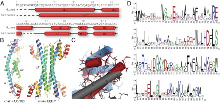
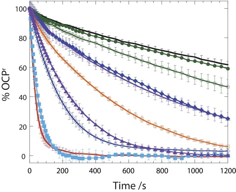
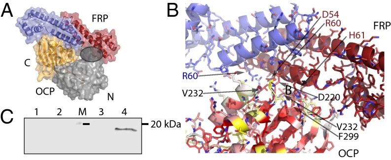
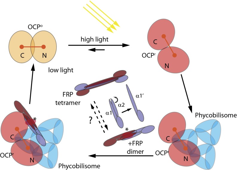
Photosynthetic reaction centers are sensitive to high light conditions, which can cause damage because of the formation of reactive oxygen species. To prevent high-light induced damage, cyanobacteria have developed photoprotective mechanisms. One involves a photoactive carotenoid protein that decreases the transfer of excess energy to the reaction centers. This protein, the orange carotenoid protein (OCP), is present in most cyanobacterial strains; it is activated by high light conditions and able to dissipate excess energy at the site of the light-harvesting antennae, the phycobilisomes. Restoration of normal antenna capacity involves the fluorescence recovery protein (FRP). The FRP acts to dissociate the OCP from the phycobilisomes by accelerating the conversion of the active red OCP to the inactive orange form. We have determined the 3D crystal structure of the FRP at 2.5 Å resolution. Remarkably, the FRP is found in two very different conformational and oligomeric states in the same crystal. Based on amino acid conservation analysis, activity assays of FRP mutants, FRP:OCP docking simulations, and coimmunoprecipitation experiments, we conclude that the dimer is the active form. The second form, a tetramer, may be an inactive form of FRP. In addition, we have identified a surface patch of highly conserved residues and shown that those residues are essential to FRP activity.

Keywords: Synechocystis; nonphotochemical quenching.


Conflict of interest statement

The authors declare no conflict of interest.

Figures

Fig. 1.
Fig. 1.
Structural overview of the FRP. (A) Primary and secondary structure of the Synechocystis sp. PCC 6803 FRP dimer and tetramer forms. Red tubes indicate α-helices, and the dashed line shows residues disordered in the structure. (B) Cartoon representation of the dimer and tetramer forms of the FRP observed in the crystals. Rainbow coloring from N to C termini from blue to red. (C) Alignment of the head domains of the dimer (red, chain B) and tetramer (blue, chain F; gray, chain E) illustrating the conservation of interactions of the head and stalk domain between the two forms. (D) Sequence conservation logo of the FRP with numbering corresponding to the Synechocystis sp. PCC 6803 protein.
Fig. 2.
Fig. 2.
Amino acid conservation mapping onto the structure of the FRP. (A) FRP dimer and tetramer structures shown in surface representation are colored according to conservation (red, high; white, medium; yellow, low). (B) Close-up view of the proposed active site with stick representation of side chains. Side chain oxygen and nitrogen atoms are colored red and blue, respectively. The location of the twofold symmetry axis is indicated with a black ellipse.
Fig. 3.
Fig. 3.
Kinetics of dark reconversion of OCPr to OCPo at 8 °C in the absence and presence of WT and mutant FRPs with a ratio of OCP to FRP of 2:1: without FRP (black), with WT (red), and with W9L (closed squares; sky blue), H53L (open squares; orange), W50F (closed triangles; violet), W50L (open triangles; violet), R60L (closed circles,; green), R60K (open circles; green), D54E (open rhomboids; blue), or D54L (closed rhomboids; blue). Average of three independent experiments. Error bars represent SD.
Fig. 4.
Fig. 4.
Analysis of the FRP–OCP interaction. (A) Docking solution of the FRP dimer (active site region) to the C-terminal domain of the OCP. Overview of the best solution shown as the surface with the residues involved in the docking as a cartoon and the N-terminal domain in gray, the C-terminal domain in orange. The gray circle indicates the clash between the docked FRP and the N-terminal domain of OCP. (B) Details of the docking solution interaction between OCP and FRP. Amino acids in the OCP involved in the interaction are marked in black, and amino acids of the FRP are marked in blue and red. The surface of the OCP C-terminal domain is colored according to conservation (red, high; white, medium; yellow, low). (C) Anti-OCP immunoblot of the N- (lane 2) and C-terminal domains (lane 4) after coimmunoprecipitation of each domain with the OCP. Control experiments (without FRP) are also shown: N- (lane 1) and C-terminal controls (lane 3). Molecular mass marker (20 kDa; lane M) is denoted with a black line).
Fig. 5.
Fig. 5.
Model of the interplay between the FRP, OCP, and phycobilisome under high and low light conditions. The OCPo is converted to OCPr under high light conditions, undergoes a conformational change that exposes part of the carotenoid and R155, and binds to the phycobilisomes to quench excess energy. The FRP can bind to unattached or phycobilisome-attached OCPr, converting the OCPr into OCPo and dissociating it from the phycobilisomes. Under low light conditions, the decrease of OCPr concentration leads to a situation in which all of the phycobilisomes will be free of OCP and unquenched. The dimer FRP is the active form. The state of the FRP could be regulated by exterior factors that convert it to an inactive tetramer form under high light conditions by rearranging helix α2 to form an extended α1′. The FRP dissociates from the OCPo after conversion. The red ellipse and asterisk indicate the position of the FRP active site.

References

    1. Niyogi KK. Photoprotection revisited: Genetic and molecular approaches. Annu Rev Plant Physiol Plant Mol Biol. 1999;50:333–359. - PubMed
    1. Pascal AA, et al. Molecular basis of photoprotection and control of photosynthetic light-harvesting. Nature. 2005;436(7047):134–137. - PubMed
    1. Kerfeld CA. Structure and function of the water-soluble carotenoid-binding proteins of cyanobacteria. Photosynth Res. 2004;81(3):215–225. - PubMed
    1. Kerfeld CA. Water-soluble carotenoid proteins of cyanobacteria. Arch Biochem Biophys. 2004;430(1):2–9. - PubMed
    1. Kerfeld CA, et al. The crystal structure of a cyanobacterial water-soluble carotenoid binding protein. Structure. 2003;11(1):55–65. - PubMed

Publication types

MeSH terms

LinkOut - more resources