. 2013 Jun 11;110(24):9914-9.
doi: 10.1073/pnas.1304046110. Epub 2013 May 28.

Eosinophils secrete IL-4 to facilitate liver regeneration

Affiliations

Eosinophils secrete IL-4 to facilitate liver regeneration

Y P Sharon Goh et al. Proc Natl Acad Sci U S A. .

Abstract

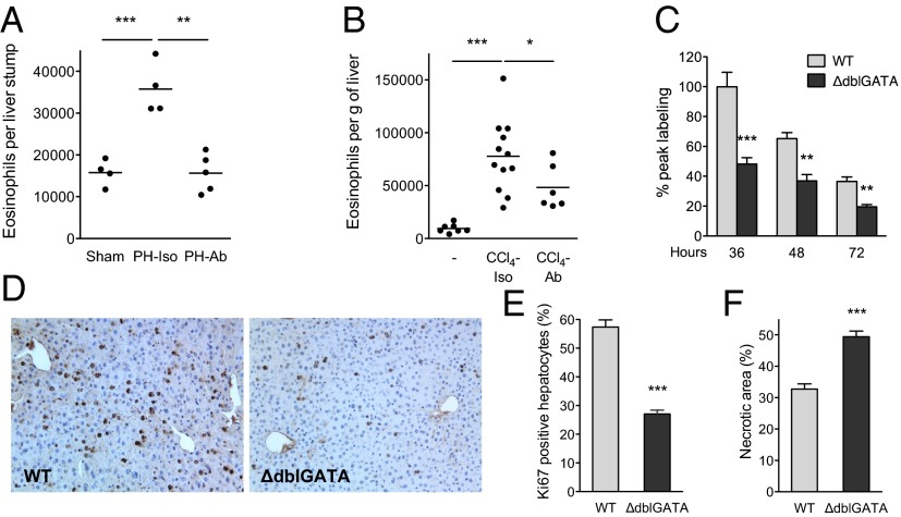
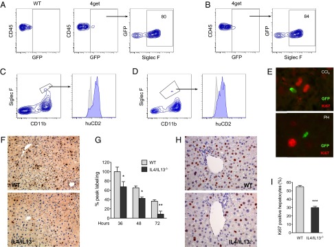
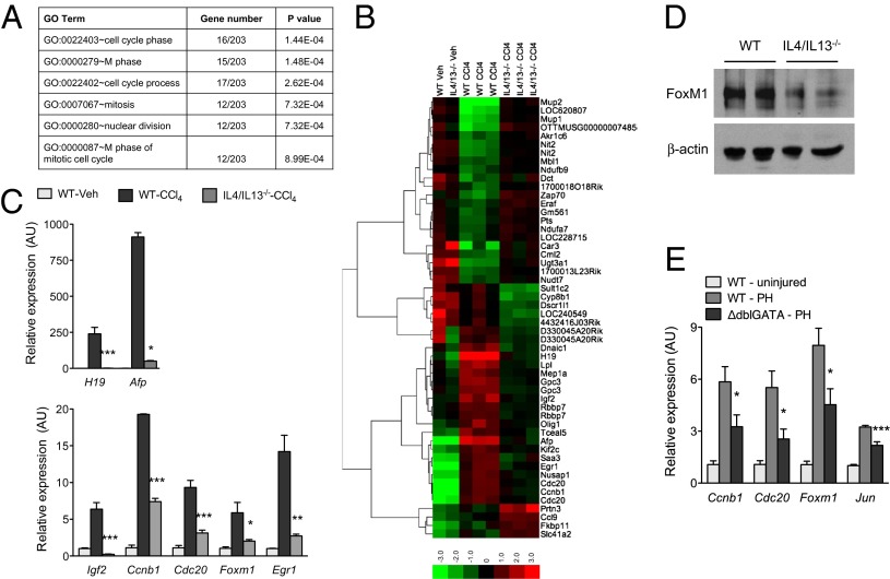
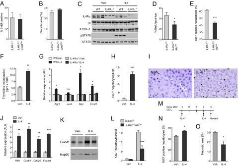
The liver is a central organ for the synthesis and storage of nutrients, production of serum proteins and hormones, and breakdown of toxins and metabolites. Because the liver is susceptible to toxin- or pathogen-mediated injury, it maintains a remarkable capacity to regenerate by compensatory growth. Specifically, in response to injury, quiescent hepatocytes enter the cell cycle and undergo DNA replication to promote liver regrowth. Despite the elucidation of a number of regenerative factors, the mechanisms by which liver injury triggers hepatocyte proliferation are incompletely understood. We demonstrate here that eosinophils stimulate liver regeneration after partial hepatectomy and toxin-mediated injury. Liver injury results in rapid recruitment of eosinophils, which secrete IL-4 to promote the proliferation of quiescent hepatocytes. Surprisingly, signaling via the IL-4Rα in macrophages, which have been implicated in tissue repair, is dispensable for hepatocyte proliferation and liver regrowth after injury. Instead, IL-4 exerts its proliferative actions via IL-4Rα in hepatocytes. Our findings thus provide a unique mechanism by which eosinophil-derived IL-4 stimulates hepatocyte proliferation in regenerating liver.

Keywords: inflammation; parasites; tissue injury and repair; type 2 immunity.


Conflict of interest statement

The authors declare no conflict of interest.

Figures

Fig. 1.
Fig. 1.
Impaired liver regeneration in mice lacking eosinophils. (A and B) Eosinophil numbers in livers of mice subjected to partial hepatectomy (PH) or toxin-induced injury (CCl4) were enumerated using flow cytometry. Migration of eosinophils into injured livers is integrin-dependent and can be blocked by antibodies against α4 and αL integrins (Ab). Isotype (Iso) immunoglobulins (IgG2a and IgG2b) were administered to control mice (n = 4–12 mice per group). (C) PH was performed in WT and ΔdblGATA mice, which lack eosinophils, and BrdU incorporation was examined at the indicated times to assess hepatocyte proliferation (n = 4–7 mice per genotype per time). (D) Remnant liver sections were stained for BrdU 36 h after partial hepatectomy. Representative images of WT and ΔdblGATA are shown. (E) Percentage of Ki67+ hepatocytes in WT and ΔdblGATA mice 2 d after administration of CCl4 (n = 3–5 mice per genotype). (F) Quantification of necrotic area in WT and ΔdblGATA mice 2 d after administration of CCl4 (n = 4–5 mice per genotype). *P < 0.05, **P < 0.01, ***P < 0.001. All data are presented as mean ± SEM.
Fig. 2.
Fig. 2.
IL-4/IL-13 promote liver regeneration. (A and B) Identification of eosinophils as the major cell population competent for IL-4 secretion after CCl4-mediated injury (A) and PH (B). Two days after administration of CCl4 or PH in the IL-4 reporter mice (4get mice), the nonhepatocyte fraction was isolated and analyzed for expression of GFP and markers of eosinophils. Representative data from 3 independent experiments are shown in A and B. (C and D) Eosinophils produce IL-4 in injured livers. Expression of huCD2 on eosinophils (Siglec F+CD11b+) 2 d after PH (C) or CCl4-induced injury (D) in KN2 mice. Isotype, gray histogram; huCD2, blue histogram. (E) GFP+ cells localize in proximity to proliferating hepatocytes (Ki67+) 2 d after injury with CCl4 (Upper) or PH (Lower). (F) Liver sections stained for BrdU 36 h after partial hepatectomy. Representative images of WT and IL-4/IL-13−/− are shown. (G) Liver regeneration in WT and IL-4/IL-13−/− mice was assessed by BrdU incorporation at indicated times after PH (n = 4–9 mice per genotype per time). (H) Representative liver sections from WT and IL-4/IL-13−/− mice were stained for Ki67 2 d after administration of CCl4. (I) Percentage of Ki67+ hepatocytes in WT and IL-4/IL-13−/− mice 2 d after CCl4 treatment (n = 5 mice per genotype). *P < 0.05, **P < 0.01, ***P < 0.001. All data are presented as mean ± SEM.
Fig. 3.
Fig. 3.
IL-4/IL-13 signaling promotes cell cycle progression after liver injury. (A) Global gene expression analysis of WT and IL-4/IL-13−/− livers was performed 2 d after injury with CCl4. Gene ontology (GO) terms associated with cell cycle and mitosis are enriched among the differentially expressed genes. (B) Heat map presentation of differentially expressed genes in livers of WT and IL-4/IL-13−/− mice 2 d after injury with CCl4 (red, high; green, low). (C) Quantitative RT-PCR analysis of genes associated with hepatocyte proliferation and liver regeneration. mRNAs from WT and IL-4/IL-13−/− mice were analyzed 2 d after injury with CCl4 (n = 3–5 per genotype and treatment). (D) Western blot analysis of FoxM1 in WT and IL-4/IL-13−/− livers. (E) Quantitative RT-PCR analysis of genes associated with hepatocyte proliferation in WT and ΔdblGATA mice was performed 2 d after PH (n = 5–8 per genotype and treatment). *P < 0.05, **P < 0.01, ***P < 0.001. All data are presented as mean ± SEM.
Fig. 4.
Fig. 4.
IL-4 stimulates hepatocyte proliferation in a cell-autonomous manner. (A and B) IL4Rα signaling in myeloid cells is not required for liver regeneration. (A) Liver regeneration in IL4RαL/L and IL4RαL/LLysMCre mice was assessed by BrdU incorporation at 36 h after PH (n = 3–5 mice per genotype). (B) Quantification of necrotic area in IL4RαL/L and IL4RαL/LLysMCre mice 2 d after administration of CCl4 (n = 4–5 per genotype). (C) Immunoblot analyses of IL-4/IL-13 signaling receptors in primary hepatocytes. (D and E) IL4Rα signaling in hepatocytes is required for liver regeneration after injury. (D) BrdU incorporation in IL4RαL/L and IL4RαL/LAlbCre mice was assessed 36 h after PH (n = 6–7 mice per genotype). (E) Quantification of Ki67+ hepatocytes in IL4RαL/L and IL4RαL/LAlbCre mice 2 d after administration of CCl4 (n = 5–7 mice per genotype). (FK) Stimulation with IL-4 promotes hepatocyte proliferation in vitro and in vivo. (F) 3H-thymidine incorporation in primary hepatocytes after in vitro stimulation with IL-4. Data are representative of 2 independent experiments. (G) Quantitative RT-PCR analysis of genes associated with hepatocyte proliferation in primary hepatocytes stimulated with Veh or IL-4 for 48 h (n = 3 per genotype and treatment). (H) WT C57BL/6J mice were injected with Veh or IL-4 for 5 d, and hepatocyte proliferation was quantified by Ki67 positivity (n = 5–6 per treatment group). (I) Representative sections showing staining for Ki67 in livers of mice treated with Veh or IL-4. (J) Quantitative RT-PCR analysis of genes associated with hepatocyte proliferation and liver growth in WT mice treated with Veh and IL-4. (K) Immunoblot analysis of FoxM1 expression in livers of mice treated with Veh or IL-4. (L) Mitogenic actions of IL-4 in IL4RαL/L and IL4RαL/LAlbCre mice. Ki67+ hepatocytes were quantified in Veh or IL-4-treated mice (n = 5 mice per genotype and treatment). (MO) Treatment with IL-4 protects mice from CCl4-induced liver injury. (M) Schematic for dosing of IL-4 to mice administered CCl4. (N) Percentage of Ki67+ hepatocytes in Veh and IL-4-treated mice 3 d after CCl4 administration (n = 4 per treatment group). (O) Quantification of necrotic area in Veh and IL-4-treated mice 3 d after CCl4-induced injury (n = 4–5 mice per treatment group). *P < 0.05, **P < 0.01, ***P < 0.001. All data are presented as mean ± SEM.

References

    1. Michalopoulos GK. Liver regeneration after partial hepatectomy: critical analysis of mechanistic dilemmas. Am J Pathol. 2010;176(1):2–13. - PMC - PubMed
    1. Taub R. Liver regeneration: From myth to mechanism. Nat Rev Mol Cell Biol. 2004;5(10):836–847. - PubMed
    1. Higgins GM, Anderson RM. Experimental pathology of the liver. I. Restoration of the liver of the white rat following partial surgical removal. Arch Pathol (Chic) 1931;12:186–202.
    1. Michalopoulos GK, DeFrances MC. Liver regeneration. Science. 1997;276(5309):60–66. - PubMed
    1. Huang W, et al. Nuclear receptor-dependent bile acid signaling is required for normal liver regeneration. Science. 2006;312(5771):233–236. - PubMed

Publication types

MeSH terms

Substances

Associated data