Conditional deletion of the human ortholog gene Dicer1 in Pax2-Cre expression domain impairs orofacial development
- PMID: 23716939
- PMCID: PMC3656520
- DOI: 10.4103/0971-6866.107984
Conditional deletion of the human ortholog gene Dicer1 in Pax2-Cre expression domain impairs orofacial development
Abstract
Background: Orofacial clefts are common worldwide and result from insufficient growth and/or fusion during the genesis of the derivatives of the first pharyngeal arch and the frontonasal prominence. Recent studies in mice carrying conditional and tissue-specific deletions of the human ortholog Dicer1, an RNAse III family member, have highlighted its importance in cell survival, differentiation, proliferation, and morphogenesis. Nevertheless, information regarding Dicer1 and its dependent microRNAs (miRNAs) in mammalian palatogenesis and orofacial development is limited.
Aims: To describe the craniofacial phenotype, gain insight into potential mechanisms underlying the orofacial defects in the Pax2-Cre/Dicer1 CKO mouse, and shed light on the role of Dicer1 in mammalian palatogenesis.
Materials and methods: Histological and molecular assays of wild type (WT) and Pax2-Cre/Dicer1(loxP/loxP) (Dicer1 CKO) mice dissected tissues have been performed to characterize and analyze the orofacial dysmorphism in Pax2-Cre/Dicer1(loxP/loxP) mouse.
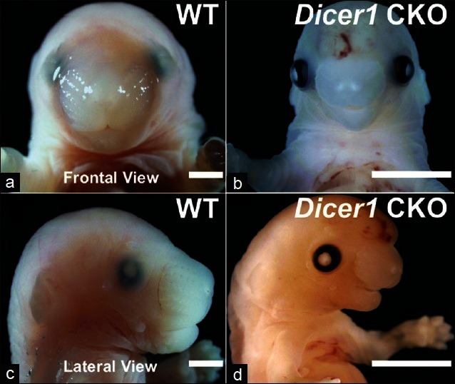
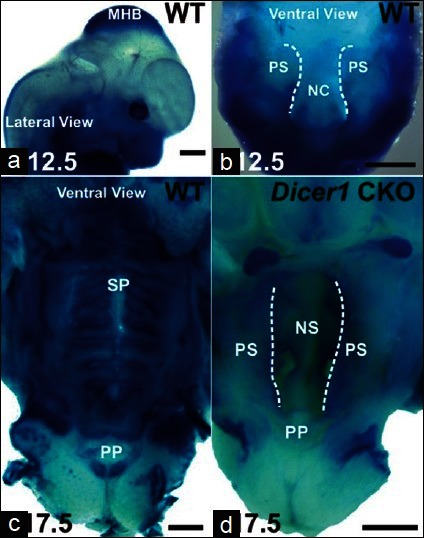
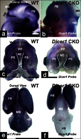
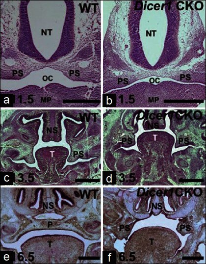
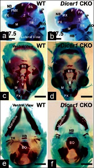
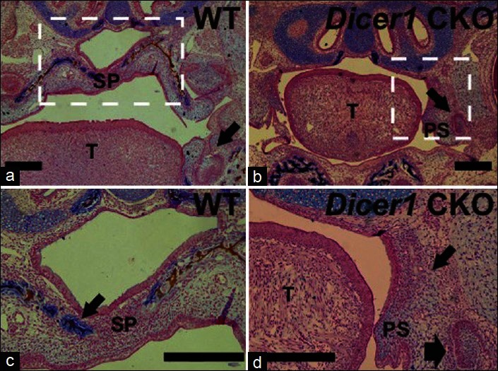
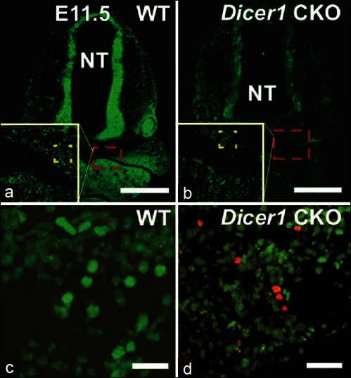
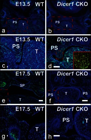
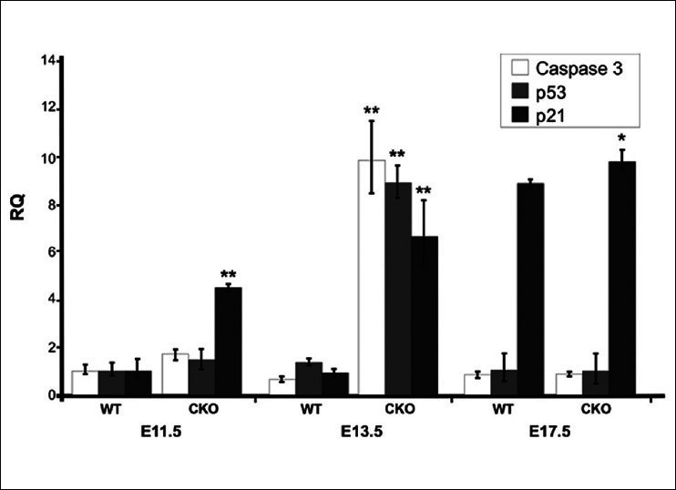
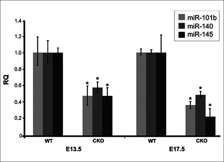
Results: Dicer1 CKO mice exhibit late embryonic lethality and severe craniofacial dysmorphism, including a secondary palatal cleft. Further analysis suggest that Dicer1 deletion neither impacts primary palatal development nor the initial stages of secondary palatal formation. Instead, Dicer1 is implicated in growth, differentiation, mineralization, and survival of cells in the lateral palatal shelves. Histological and molecular analysis demonstrates that secondary palatal development becomes morphologically arrested prior to mineralization around E13.5 with a significant increase in the expression levels of apoptotic markers (P < 0.01).
Conclusions: Pax2-Cre-mediated Dicer1 deletion disrupts lateral palatal outgrowth and bone mineralization during palatal shelf development, therefore providing a mammalian model for investigating the role of miRNA-mediated signaling pathways during palatogenesis.
Keywords: Cleft palate; Dicer1; cranial neural crest cells; microRNAs; palatogenesis.
Conflict of interest statement
Figures
References
-
- Ferguson MW. Palate development. Development. 1988;103:41–60. - PubMed
-
- Chai Y, Maxson RE., Jr Recent advances in craniofacial morphogenesis. Dev Dyn. 2006;235:2353–75. - PubMed
-
- Minoux M, Rijli FM. Molecular mechanisms of cranial neural crest cell migration and patterning in craniofacial development. Development. 2010;137:2605–21. - PubMed
-
- Jugessur A, Farlie PG, Kilpatrick N. The genetics of isolated orofacial clefts: From genotypes to subphenotypes. Oral Dis. 2009;15:437–53. - PubMed
Grants and funding
LinkOut - more resources
Full Text Sources
Molecular Biology Databases