Clinical Trial
. 2013;9(5):e1003366.
doi: 10.1371/journal.ppat.1003366. Epub 2013 May 23.

Cis and trans acting factors involved in human cytomegalovirus experimental and natural latent infection of CD14 (+) monocytes and CD34 (+) cells

Affiliations
Clinical Trial

Cis and trans acting factors involved in human cytomegalovirus experimental and natural latent infection of CD14 (+) monocytes and CD34 (+) cells

Cyprian C Rossetto et al. PLoS Pathog. 2013.

Abstract

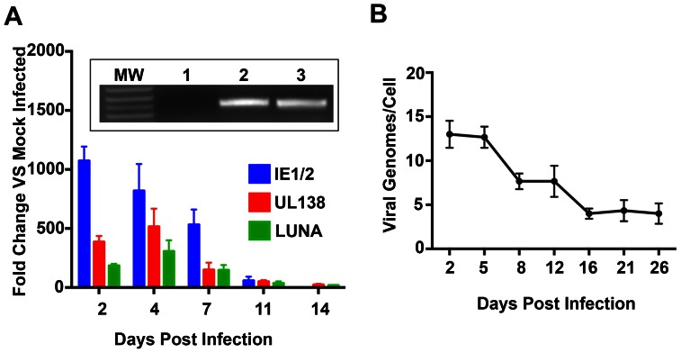
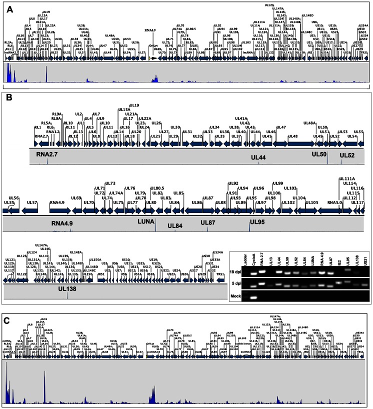
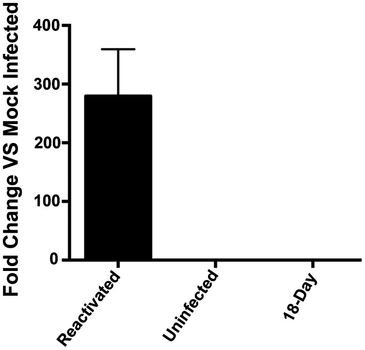
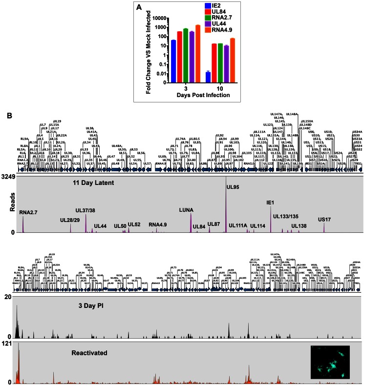
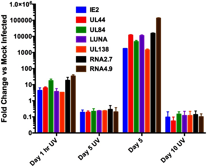
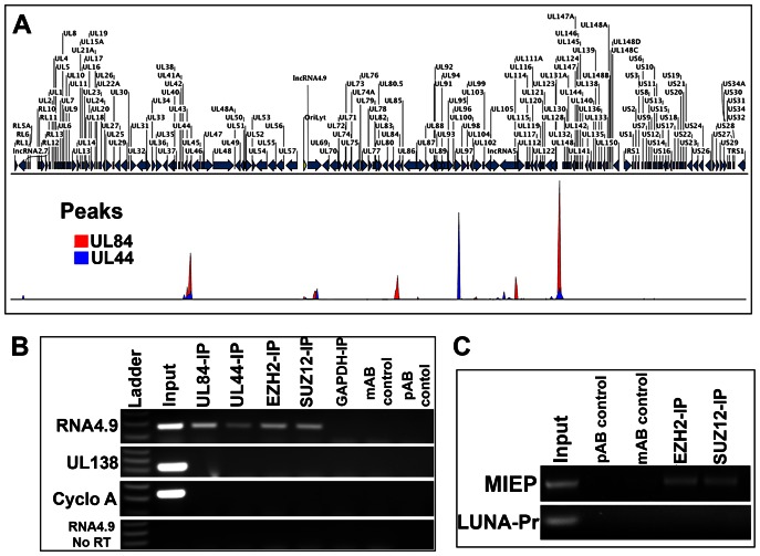
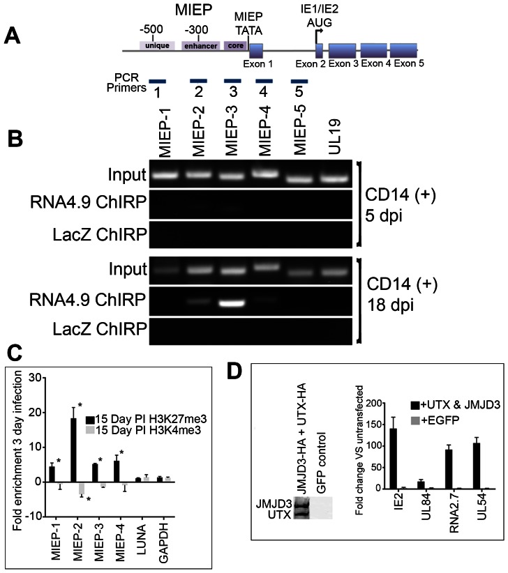
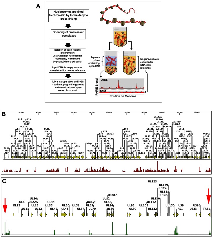
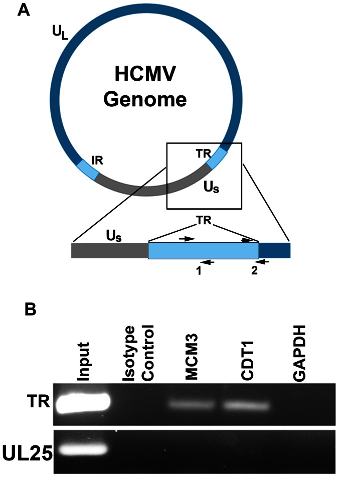
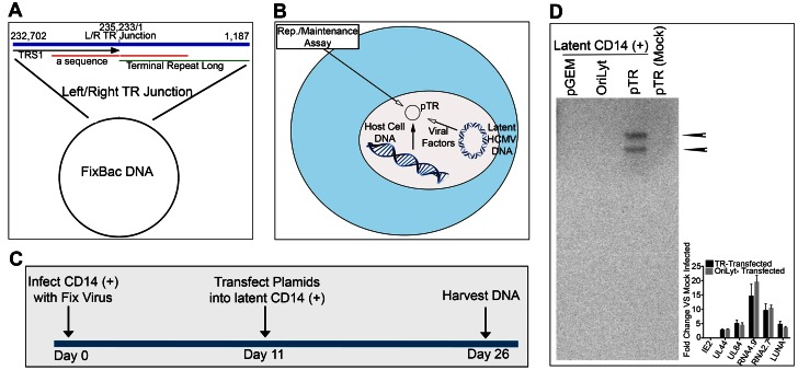
The parameters involved in human cytomegalovirus (HCMV) latent infection in CD14 (+) and CD34 (+) cells remain poorly identified. Using next generation sequencing we deduced the transcriptome of HCMV latently infected CD14 (+) and CD34 (+) cells in experimental as well as natural latency settings. The gene expression profile from natural infection in HCMV seropositive donors closely matched experimental latency models, and included two long non-coding RNAs (lncRNAs), RNA4.9 and RNA2.7 as well as the mRNAs encoding replication factors UL84 and UL44. Chromatin immunoprecipitation assays on experimentally infected CD14 (+) monocytes followed by next generation sequencing (ChIP-Seq) were employed to demonstrate both UL84 and UL44 proteins interacted with the latent viral genome and overlapped at 5 of the 8 loci identified. RNA4.9 interacts with components of the polycomb repression complex (PRC) as well as with the MIE promoter region where the enrichment of the repressive H3K27me3 mark suggests that this lncRNA represses transcription. Formaldehyde Assisted Isolation of Regulatory Elements (FAIRE), which identifies nucleosome-depleted viral DNA, was used to confirm that latent mRNAs were associated with actively transcribed, FAIRE analysis also showed that the terminal repeat (TR) region of the latent viral genome is depleted of nucleosomes suggesting that this region may contain an element mediating viral genome maintenance. ChIP assays show that the viral TR region interacts with factors associated with the pre replication complex and a plasmid subclone containing the HCMV TR element persisted in latently infected CD14 (+) monocytes, strongly suggesting that the TR region mediates viral chromosome maintenance.


Conflict of interest statement

The authors have declared that no competing interests exist.

Figures

Figure 1
Figure 1. Infected CD14 (+) cells express latency associated transcripts and stable DNA copy number after 14 days in culture.
(A) RT-qPCR analysis for the expression of IE1/IE2, LUNA and UL138 mRNA in infected CD14 (+) monocytes. CD14 (+) cells were infected with FIX BAC virus and total cellular RNA was harvested various days post infection and subjected to RT-qPCR using TaqMan primers and probes specific for IE1 and IE2 mRNA. Inset figure: Detection of viral genomic DNA in infected CD14 (+) monocytes by PCR. Lanes: 1, uninfected; 2, 5-day post infection; 3, 18 day post infection. Primers used were specific for the TR region of the genome. (B) CD14 (+) monocytes infected cell viral genome copy number. Cells were harvested at various days post infection and subjected to qPCR using primers and probes specific for the HCMV viral chromosome. Absolute viral genome copy number was determined by comparing to a standard curve and calculated on a per cell basis. Error bars are the standard deviation of the mean for 4 separate wells.
Figure 2
Figure 2. RNA-Seq analysis of HCMV infected CD14+ cells.
(A) HCMV transcriptome 5 days post infection. (B) HCMV transcriptome 18 days post infection. Transcript analysis was performed using CLC Bio Genomics Workbench software. Peaks were calculated and determined based on 4 independent experiments and using a minimum of 20 read cut off and pValue of <0.05 was used to determine peaks shown. Peaks identified from 18 day CD14 (+) latent infection were: UL138, UL95, UL87, UL84, UL52, UL50, UL44, LUNA, RNA4.9 and RNA2.7. Peak heights are indicative of relative transcript accumulation in infected cells. Arrows indicate direction of transcripts. Inset: RT-PCR evaluation of identified ORFs during lytic or latent infection. Negative controls for latent infection: IE2, UL13 and US21. CycloA = cyclophillin A. (C) HCMV transcriptome upon reactivation of latently infected CD14 (+) monocytes with treatment of IL-6 and culturing cells on a surface that allows for cell attachment.
Figure 3
Figure 3. Detection of supernatant virus from reactivated CD14 (+) monocytes.
Supernatants were collected from CD14 (+) monocytes infected with HCMV after 18 days post infection as well as from cell reactivated after 18 days in culture. Uninfected supernatants were also collected. qPCR was performed using primers and probes specific for the UL54 and UL95 loci. A total of 6 wells (3 for each probe) for each sample were collected and error bars are the standard deviation from the mean from 6 replicates.
Figure 4
Figure 4. RNA-Seq analysis of HCMV infected CD34 (+) cells.
Latent transcriptome from CD34 (+) cells shares core transcripts with those observed from CD14 (+) latent infection. (A) qPCR evaluation of specific gene expression from CD34 (+) cells infected with FIX BAC virus at 3 and 10 days post infection. (B) RNA-Seq detection of transcripts from infected CD34 (+) cells during latent (11 days post infection), 3 days post infection and from cells reactivated after 11 days post infection. Peaks were calculated and determined based on 3 independent experiments and using a minimum of 20 read cut off and pValue of <0.05 was used to determine peaks shown. Peak maps are shown where the number of transcript reads is shown to the left of the graphs. CD34 (+) cells latency-associated transcripts identified were: RNA2.7, UL28/29, UL37/38, UL44, UL50, UL52, RNA4.9, LUNA, UL84, UL87, UL95, UL111A, UL114, IE1, UL133, UL135, UL138, US17.
Figure 5
Figure 5. UV inactivation of virus abrogates accumulation of IE2 and latency associated transcripts in CD14 (+) monocytes.
CD14 (+) monocytes were infected with either wt FIX BAC virus or UV inactivated virus. Total cellular RNA was harvested at 1 hr, 5 and 10 days post infection and qPCR was performed to detect transcripts IE2, UL44, UL84, LUNA, UL138, lncRNA 2.7 and lncRNA4.9. Transcript fold increase was determined using mock infected as reference. 3 separate experiments were preformed and error bars are the SD of the mean.
Figure 6
Figure 6. UL44 and UL84 interact with the HCMV latent viral genome in CD14 (+) monocytes.
(A) ChIP-Seq analysis peak map of UL44 and UL84 in HCMV latently infected CD14 (+) monocytes. UL44 or UL84 specific antibodies were used to immunoprecipitate protein-DNA complexes followed by next generation sequencing. Shown is the HCMV (Fix strain) virus genome and the location of DNA sequence reads, Red peaks = UL84 protein binding, Blue peaks = UL44 protein binding. (B) RNA4.9 interacts with components for the polycomb repressive complex. HCMV latently infected monocytes were fixed and RNA-protein complexes were immunoprecipitated with antibodies specific for EZH2, SUZ12 or UL84. RNA reverse-transcribed and cDNA was detected by PCR using primers specific for RNA4.9 or Cyclophilin A. Control immunoprecipitations were performed using an isotype control antibodies, mAb control (isotype control for SUZ12 and UL84), pAb control (isotype control for EZH2 and UL44). (C) SUZ12 and EZH2 interact with the MIEP region during latent infection. ChIP assays were performed from latently infected CD14 (+) cells using antibodies specific for SUZ12 or EZH2. Primers specific for the promoter of the MIE gene locus were used as well as control primers specific for the LUNA promoter.
Figure 7
Figure 7. RNA4.9 physically interacts with the HCMV latent viral chromosome.
(A) Schematic of the HCMV MIE gene and promoter/enhancer region. Regions amplified by PCR primers are shown that are specific for the unique, enhancer, core promoter, exon 1 and the first intron of IE2. (B) ChIRP analysis of RNA4.9 binding to regions of the MIE promoter and gene locus. Infected CD14 (+) cells were harvested at 5 and 18 days post infection and ChIRP was performed. Control ChIRP was performed using biotinylated hybridization primers specific for LacZ. Amplification of the UL19 region was used as a control for ChIRP PCR amplification. (C) Increase in the enrichment of the repressive H3K27me3 mark at the MIEP during latency is consistent with the binding of RNA4.9. CD14 (+) cells infected with HCMV were harvested at 3-days post infection or during latency at 15 days post infection and ChIP assays were performed using antibodies specific for H3K27me3, H3K4me3 or IgG control antibody. Fold enrichment of H3K27me3 or H3K4me3 was calculated by IgG subtracted % input of each locus divided by the IgG subtracted % input of the control gene GAPDH. Each sample was evaluated in triplicate and the error bars are the SD of the mean. Data is shown as fold enrichment compared to tri methylation state at 3 days PI. Statistical analysis was done using multiple t-test, *P Value<0.001. (D) Increase in IE mRNA accumulation in cells transfected UTX and JMJD3 expression plasmids. Latently infected CD14 (+) cells were transfected with plasmids expressing UTX and JMJD3. Expression of UTX and JMJD3 was confirmed by Western blot, left panel. Graph shows the increase in fold accumulation of specific mRNAs. Error bars are the standard deviation of the average of three experiments.
Figure 8
Figure 8. Analysis of nucleosome depletion in HCMV infected CD14 (+) monocytes.
(A) Schematic of FAIRE method. FAIRE-Seq was performed using 4 or 18 day infected CD14 (+) monocytes. (B) FAIRE-Seq analysis of 4 day post infection of CD14 (+) monocytes. (C) FAIRE-Seq analysis of 18 day latently infected CD14 (+) monocytes. Sequencing reads were mapped to VR1814. Red Arrows indicate nucleosome depletion at the TR region of the HCMV genome.
Figure 9
Figure 9. Interaction of MCM3 and CDT1 with HCMV TR DNA sequences in latently infected CD14 (+) monocytes.
Latently infected cells were treated with formaldehyde and subjected to ChIP using antibodies specific for MCM3 or CDT1. (A) Schematic of the HCMV genome showing the terminal repeat (TR) region. Also shown are primer sets specific for the TR segment of the genome (1 and 2). (B) ChIP assay showing an interaction of the TR region of the genome with MCM3 and CDT1 in latently infected cells. Control immunoprecipitations were the use of an isotype antibody control and an antibody specific for GAPDH. Control PCR primers were specific for the HCMV UL25 ORF.
Figure 10
Figure 10. A plasmid clone containing the circularized TR region of HCMV genome is maintained in latently infected CD14 (+) monocytes.
(A) Schematic showing the circularized region of the HCMV genome subcloned into the plasmid vector pGEM7zf(−). (B) Schematic showing the development of a latent replication/maintenance assay in CD14 (+) monocytes. (C) Infection/transfection protocol used to evaluate plasmid maintenance in latently infected CD14 (+) monocytes. (D) Southern blot of a Gardella gel containing samples from latently infected CD14 (+) monocytes transfected with plasmids pGEM7zf(−), pOriLyt, pTR or pTR from uninfected CD14 (+) cells 26 days post transfection. Arrows indicate the presence of two bands in the pTR lane at 15 days post transfection, 25 days post infection.

References

    1. Landolfo S, Gariglio M, Gribaudo G, Lembo D (2003) The human cytomegalovirus. Pharmacol Ther 98: 269–297. - PubMed
    1. Mocarski ESaC, T. C. (2001) Cytomegaloviruses and Their Replication. In: Knipe DMaH, P. M., editor. Virology. 4 ed. Philadelphia: Lippincott Williams & Wilkins. pp. 2629–2674.
    1. Pari GS, Anders DG (1993) Eleven loci encoding trans-acting factors are required for transient complementation of human cytomegalovirus oriLyt-dependent DNA replication. J Virol 67: 6979–6988. - PMC - PubMed
    1. Pari GS, Kacica MA, Anders DG (1993) Open reading frames UL44, IRS1/TRS1, and UL36–38 are required for transient complementation of human cytomegalovirus oriLyt-dependent DNA synthesis. J Virol 67: 2575–2582. - PMC - PubMed
    1. Pari GS (2008) Nuts and bolts of human cytomegalovirus lytic DNA replication. Curr Top Microbiol Immunol 325: 153–166. - PubMed

Publication types

MeSH terms