. 2013 May 13:4:136.
doi: 10.3389/fpls.2013.00136. eCollection 2013.

Vitamin D in plants: a review of occurrence, analysis, and biosynthesis

Affiliations

Vitamin D in plants: a review of occurrence, analysis, and biosynthesis

Rie B Jäpelt et al. Front Plant Sci. .

Abstract

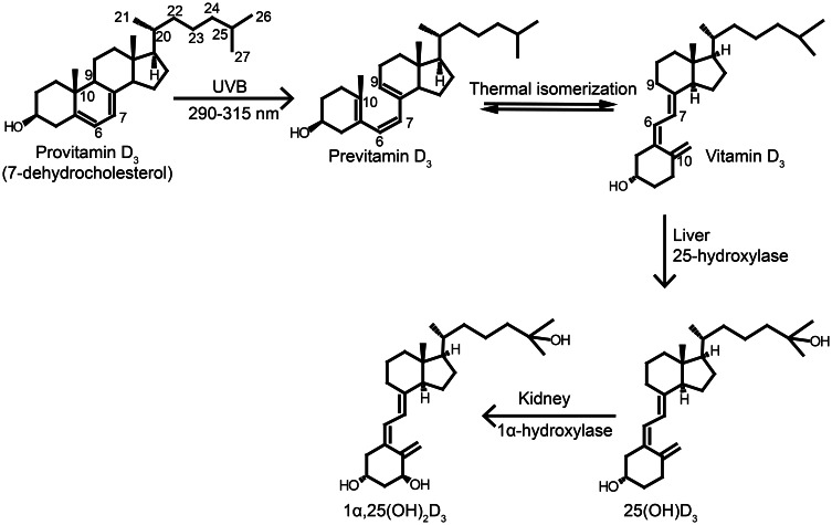
The major function of vitamin D in vertebrates is maintenance of calcium homeostasis, but vitamin D insufficiency has also been linked to an increased risk of hypertension, autoimmune diseases, diabetes, and cancer. Therefore, there is a growing awareness about vitamin D as a requirement for optimal health. Vitamin D3 is synthesized in the skin by a photochemical conversion of provitamin D3, but the necessary rays are only emitted all year round in places that lie below a 35° latitude. Unfortunately, very few food sources naturally contain vitamin D and the general population as a results fail to meet the requirements. Fish have the highest natural content of vitamin D expected to derive from an accumulation in the food chain originating from microalgae. Microalgae contain both vitamin D3 and provitamin D3, which suggests that vitamin D3 exist in the plant kingdom and vitamin D3 has also been identified in several plant species as a surprise to many. The term vitamin D also includes vitamin D2 that is produced in fungi and yeasts by UVB-exposure of provitamin D2. Small amounts can be found in plants contaminated with fungi and traditionally only vitamin D2 has been considered present in plants. This review summarizes the current knowledge on sterol biosynthesis leading to provitamin D. It also addresses the occurrence of vitamin D and its hydroxylated metabolites in higher plants and in algae and discusses limitations and advantages of analytical methods used in studies of vitamin D and related compounds including recent advances in analytical technologies. Finally, perspectives for a future production of vitamin D biofortified fruits, vegetables, and fish will be presented.

Keywords: 1; 25-dihydroxy vitamin D; 25-hydroxy vitamin D; algae; biosynthesis; detection; plants; sterols; vitamin D.


Figures

Figure 1
Figure 1
Synthesis and activation of vitamin D. Vitamin D3 is synthesized in the skin upon UVB exposure. The UVB exposure of provitamin D3 (7-dehydrocholesterol) in the skin breaks the B-ring to form previtamin D3, which undergoes thermally induced rearrangement to vitamin D3. Vitamin D3 is transported to the liver where it is hydroxylated at C-25 by the enzyme 25-hydroxylase producing 25OHD3, which is the major circulating form in vertebrates. The 25OHD3 is hydroxylated a second time at C-1 in the kidneys to the active metabolite 1,25(OH)2D3. Figure adapted from Jäpelt et al. (2012).
Figure 2
Figure 2
The core structure of sterols is a fused four ring (A,B,C, and D ring). Various groups are added to the core structure to generate a variety of sterols. Numbering of the carbon atoms is according to the 1989 IUPAC-IUB recommendations.
Figure 3
Figure 3
Basic structures of free sterol and its conjugates. The side chain R varies between various sterols. Figure Adapted from Toivo et al. (2001).
Figure 4
Figure 4
First part of the biosynthetic pathway of sterols. Solid arrows mean one reaction and doted arrow multiple steps. HMG-CoA, 3-hydroxymethyl-3-glutaryl coenzyme A; HMGR, HMG-CoA reductase; IPP, Isopentyl pyrophosphate; DMAPP, Dimethylalkyl pyrophosphate; FPP, Farnesyl pyrophosphate; SQS, squalene synthase; SQE, squalene epoxidase.
Figure 5
Figure 5
Cyclization of 2,3-oxidosqualene forms either lanosterol or cycloartenol via a series of enzymatic cyclizations leading to sterols in plants, fungi and animals. CAS, cycloartenol synthase; LAS, lanosterol synthase.
Figure 6
Figure 6
Simplified cholesterol biosynthesis. Lanosterol is converted to cholesterol in a series of enzyme reactions. Dashed arrows indicate more than one biosynthetic step. Solid arrows indicate a biosynthetic step regulated by: (1), sterol-Δ24-reductase; (2), lathosterol 5-desaturase; (3), 7-dehydrocholesterol reductase.
Figure 7
Figure 7
The last five steps of the ergosterol biosynthetic pathway in Saccharomyces cerevis. ERG6, S-adenosylmethionine sterol methyltransferase; ERG2, C8-C7 sterol isomerase; ERG3, Δ5-desaturase; ERG5, Δ22-desaturase; ERG4, Δ24-reductase. Figure adapted from Lees et al. (1995).
Figure 8
Figure 8
The figure represents the biosynthetic pathways to sterols downstream from cycloartenol. Hypothetical pathway for 24-desmethylsterols (left) marked with dashed box. Simplified biosynthetic pathways for 24-demethylsterols (middle) and 24-ethylsterols (right). Dashed arrows indicate more than one biosynthetic step. Solid arrows indicate a biosynthetic step regulated by: (A) SMT1, (B) SMT2, (C) DIM/DWARF1, (D) DWARF7/STE1, (E) C4-demethylase and (F) DWARF5.

References

    1. Aburjai T., Al-Khalil S., Abuirjeie M. (1998). Vitamin D3 and its metabolites in tomato, potato, egg plant and zucchini leaves. Phytochemistry 49, 2497–2499
    1. Aburjai T., Bernasconi S., Manzocchi L. A., Pelizzoni F. (1997). Effect of calcium and cell immobilization on the production of cholecalciferol and its derivatives by Solanum malacoxylon cell cultures. Phytochemistry 46, 1015–1018
    1. Aburjai T., Bernasconi S., Manzocchi L., Pelizzoni F. (1996). Isolation of 7-dehydrocholesterol from cell cultures of Solanum malacoxylon. Phytochemistry 43, 773–776
    1. Andrews J. W., Murai T., Page J. W. (1980). Effects of dietary cholecalciferol and ergocalciferol on catfish. Aquaculture 19, 49–54
    1. Armas L. A. G., Hollis B. W., Heaney R. P. (2004). Vitamin D2 is much less effective than vitamin D3 in humans. J. Clin. Endocrinol. Metab. 89, 5387–5391 10.1210/jc.2004-0360 - DOI - PubMed