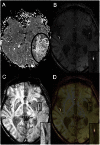
Comparison of susceptibility weighted imaging and TOF-angiography for the detection of Thrombi in acute stroke
- PMID: 23717426
- PMCID: PMC3662691
- DOI: 10.1371/journal.pone.0063459
Comparison of susceptibility weighted imaging and TOF-angiography for the detection of Thrombi in acute stroke
Abstract
Background and purpose: Time-of-flight (TOF) angiography detects embolic occlusion of arteries in patients with acute ischemic stroke due to the absence of blood flow in the occluded vessel. In contrast, susceptibility weighted imaging (SWI) directly enables intravascular clot visualization due to hypointense susceptibility vessel signs (SVS) in the occluded vessel. The aim of this study was to compare the diagnostic accuracy of both methods to determine vessel occlusion in patients with acute stroke.
Methods: 94 patients were included who presented with clinical symptoms for acute stroke and displayed a delay on the time-to-peak perfusion map in the territory of the anterior (ACA), middle (M1, M1/M2, M2/M3) or posterior (PCA) cerebral artery. The frequency of SVS on SWI and vessel occlusion or stenosis on TOF-angiography was compared using the McNemar-Test.
Results: 87 of 94 patients displayed a clearly definable SVS on SWI. In 72 patients the SVS was associated with occlusion or stenosis on TOF-angiography. Fifteen patients exclusively displayed SVS on SWI (14 M2/M3, 1 M1), whereas no patient revealed exclusively occlusion or stenosis on TOF-angiography. Sensitivity for detection of embolic occlusion within major vessel segments (M1, M1/M2, ACA, and PCA) did not show any significant difference between both techniques (97% for SWI versus 96% for TOF-angiography) while the sensitivity for detection of embolic occlusion within M2/M3 was significantly different (84% for SWI versus 39% for TOF-angiography, p<0.00012).
Conclusions: SWI and TOF-angiography provide similar sensitivity for central thrombi while SWI is superior for the detection of peripheral thrombi in small arterial vessel segments.
Conflict of interest statement
Figures


References
-
- Hacke W, Donnan G, Fieschi C, Kaste M, von Kummer R, et al. (2004) Association of outcome with early stroke treatment: pooled analysis of ATLANTIS, ECASS, and NINDS rt-PA stroke trials. Lancet 363: 768–774. - PubMed
-
- Furlan A, Higashida R, Wechsler L, Gent M, Rowley H, et al. (1999) Intra-arterial prourokinase for acute ischemic stroke. The PROACT II study: a randomized controlled trial. Prolyse in Acute Cerebral Thromboembolism. JAMA 282: 2003–2011. - PubMed
-
- Rovira A, Orellana P, Alvarez-Sabin J, Arenillas JF, Aymerich X, et al. (2004) Hyperacute ischemic stroke: middle cerebral artery susceptibility sign at echo-planar gradient-echo MR imaging. Radiology 232: 466–473. - PubMed
-
- Huang P, Chen CH, Lin WC, Lin RT, Khor GT, et al. (2012) Clinical applications of susceptibility weighted imaging in patients with major stroke. J Neurol 259: 1426–1432. - PubMed
-
- Sehgal V, Delproposto Z, Haacke EM, Tong KA, Wycliffe N, et al. (2005) Clinical applications of neuroimaging with susceptibility-weighted imaging. J Magn Reson Imaging 22: 439–450. - PubMed
Publication types
MeSH terms
LinkOut - more resources
Full Text Sources
Other Literature Sources

