. 2013 May 23;8(5):e64196.
doi: 10.1371/journal.pone.0064196. Print 2013.

Retroviral restriction factor APOBEC3G delays the initiation of DNA synthesis by HIV-1 reverse transcriptase

Affiliations

Retroviral restriction factor APOBEC3G delays the initiation of DNA synthesis by HIV-1 reverse transcriptase

Madison B Adolph et al. PLoS One. .

Abstract

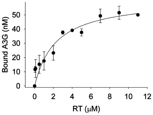
It is well established that the cytosine deaminase APOBEC3G can restrict HIV-1 virions in the absence of the virion infectivity factor (Vif) by inducing genome mutagenesis through deamination of cytosine to uracil in single-stranded HIV-1 (-)DNA. However, whether APOBEC3G is able to restrict HIV-1 using a deamination-independent mode remains an open question. In this report we use in vitro primer extension assays on primer/templates that model (-)DNA synthesis by reverse transcriptase from the primer binding site (PBS) and within the protease gene of HIV-1. We find that APOBEC3G is able to decrease the initiation of DNA synthesis by reverse transcriptase approximately 2-fold under conditions where reverse transcriptase is in excess to APOBEC3G, as found in HIV-1 virions. However, the delay in the initiation of DNA synthesis on RNA templates up to 120 nt did not decrease the total amount of primer extended after extended incubation unless the concentration of reverse transcriptase was equal to or less than that of APOBEC3G. By determining apparent Kd values of reverse transcriptase and APOBEC3G for the primer/templates and of reverse transcriptase binding to APOBEC3G we conclude that APOBEC3G is able to decrease the efficiency of reverse transcriptase-mediated DNA synthesis by binding to the RNA template, rather than by physically interacting with reverse transcriptase. All together the data support a model in which this deamination-independent mode of APOBEC3G would play a minor role in restricting HIV-1. We propose that the deamination-independent inhibition of reverse transcriptase we observed can be a mechanism used by APOBEC3G to slow down proviral DNA formation and increase the time in which single-stranded (-)DNA is available for deamination by APOBEC3G, rather than a direct mechanism used by APOBEC3G for HIV-1 restriction.


Conflict of interest statement

Competing Interests: The authors have declared that no competing interests exist.

Figures

Figure 1
Figure 1. RT-mediated primer extension from the HIV-1 PBS in the presence of A3G.
An 18 nt 32P-labeled RNA primer containing a sequence complementary to the HIV-1 PBS was annealed to a 106 nt RNA containing the PBS (sketch). Complete extension of the primer results in a product of 82 nt (sketch). The p/t was used at a concentration of 10 nM. (A) Primer extension by RT (480 nM) in the absence (0∶1) or presence (4∶1, 8∶1, 16∶1, 32∶1, 48∶1) of increasing amounts of A3G relative to the p/t concentration. Reactions were sampled at 5 and 60 min. (B) Quantification of primer extension (%) from gels shown in panel A for 5 min (black bars) and 60 min (grey bars). (C) Primer extension reactions carried out as described for (A), but in the absence of the NC protein. (D) Quantification of primer extension (%) from gel shown in panel C for 5 min (black bars) and 60 min (grey bars). (E) Primer extension by RT (120 nM) in the absence (0∶1) or presence (4∶1, 48∶1) of increasing amounts of A3G relative to the p/t concentration. Reactions were sampled at 5 and 60 min. (F) Quantification of primer extension (%) from gel shown in panel E for 5 min (black bars) and 60 min (grey bars). Primer extension (%) values are averaged from three independent trials. Designations for significant difference from 0∶1 values were p≤0.001 (***), p≤0.01(**), or p≤0.05 (*).
Figure 2
Figure 2. Decreased A3G-mediated inhibition of RT polymerization from a partially extended RNA primer.
A 24 nt 32P-labeled RNA/DNA primer containing the RNA sequence complementary to the HIV-1 PBS and 6 DNA bases to mimic partial extension by RT was annealed to a 106 nt RNA containing the PBS (sketch). Complete extension of the primer results in a product of 82 nt (sketch). The p/t was used at a concentration of 10 nM. (A) Primer extension by RT (480 nM) in the absence (0∶1) or presence (4∶1, 8∶1, 16∶1, 32∶1, 48∶1) of increasing amounts of A3G relative to the p/t concentration. Reactions were sampled at 2.5 and 60 min. (B) Quantification of primer extension (%) from gels shown in panel A for 2.5 min (black bars) and 60 min (grey bars). (C) Primer extension by RT (120 nM) in the absence (0∶1) or presence (4∶1, 48∶1) of increasing amounts of A3G relative to the p/t concentration. Reactions were sampled at 2.5 and 60 min. (D) Quantification of primer extension (%) from gel shown in panel C for 2.5 min (black bars) and 60 min (grey bars). Primer extension (%) values are averaged from three independent trials. Designations for significant difference from 0∶1 values were p≤0.001 (***), p≤0.01(**), or p≤0.05 (*).
Figure 3
Figure 3. RT polymerization from a DNA primer is less susceptible to A3G-mediated inhibition.
A 20 nt 32P-labeled DNA primer was annealed to a 120 nt RNA containing part of the HIV-1 protease gene (sketch). Complete extension of the primer results in a DNA of 120 nt (sketch). The p/t was used at a concentration of 10 nM. (A) Primer extension by RT (480 nM) in the absence (0∶1) or presence (4∶1, 8∶1, 16∶1, 32∶1, 48∶1) of increasing amounts of A3G relative to the p/t concentration. Reactions were sampled at 2.5 and 60 min. (B) Quantification of primer extension (%) from gels shown in panel A for 2.5 min (black bars) and 60 min (grey bars). Primer extension (%) values are averaged from three independent trials. Designations for significant difference from 0∶1 values were p≤0.001 (***), p≤0.01(**), or p≤0.05 (*).
Figure 4
Figure 4. Analysis of the interaction between RT and fluorescently labeled A3G using steady-state fluorescence depolarization.
The apparent dissociation constant (Kd) was determined by titrating increasing concentrations (x-axis) of RT to 50 nM of fluorescein (F)-labeled A3G. RT binds F-A3G with an apparent Kd of 2.3±0.8 µM. Fraction of F-A3G bound is calculated from anisotropy values averaged from three independent trials.
Figure 5
Figure 5. A3G E259Q inhibition of RT-mediated primer extension from the HIV-1 PBS.
An 18 nt 32P-labeled RNA primer containing a sequence complementary to the HIV-1 PBS was annealed to a 106 nt RNA containing the PBS (sketch). Complete extension of the primer results in a product of 82 nt (sketch). The p/t was used at a concentration of 10 nM. (A) Primer extension by RT (480 nM) in the absence (0∶1) or presence (4∶1, 48∶1) of A3G E259Q relative to the p/t concentration. Reactions were sampled at 5 and 60 min. (B) Quantification of primer extension (%) from gel shown in panel A for 5 min (black bars) and 60 min (grey bars). Primer extension (%) values are averaged from three independent trials. Designations for significant difference from 0∶1 values were p≤0.001 (***), p≤0.01(**), or p≤0.05 (*).
Figure 6
Figure 6. A3G inhibits the rate of primer extension but not the processivity of RT.
The p/t was used at a concentration of 10 nM. (A) A 24 nt 32P-labeled RNA/DNA primer containing the RNA sequence complementary to the HIV-1 PBS and 6 DNA bases to mimic partial extension by RT was annealed to a 106 nt RNA containing the PBS (sketch on left). Complete extension of the primer results in a product of 82 nt (sketch on left). Primer extension by RT (480 nM) in the presence of a heparin trap and the absence (0∶1) or presence (4∶1) of A3G, indicated relative to p/t concentration. Reactions were sampled at 1 min and 5 min. (B) Quantification of primer extension (%) after 1 min from gel shown in panel A for A3G∶p/t ratios of 0∶1 (black bar) and 4∶1 (grey bar). (C) A 20 nt 32P-labeled DNA primer was annealed to a 120 nt RNA containing part of the HIV-1 protease gene (sketch on right). Complete extension of the primer results in a DNA of 120 nt (sketch on right). Primer extension by RT (480 nM) in the presence of a heparin trap and the absence (0∶1) or presence (4∶1) of A3G, indicated relative to p/t concentration. Reactions were sampled at 0.5 min and 2.5 min. (D) Quantification of primer extension (%) from gels shown in panel C for 0.5 min (black bars) and 2.5 min (grey bars). (E) Primer extension by RT (40 nM) on the protease p/t (sketch on right) in the absence (0∶1) or presence (4∶1) of A3G, indicated as relative to the p/t concentration. Reactions were sampled at 1, 2.5 and 5 min. (F) Quantification of primer extension (%) from gels shown in panel E for RT in the absence (open circles) or presence of A3G (black circles). The primer extension rate of RT (4.6%/min) is decreased 3-fold in the presence of A3G (1.5%/min). (G) Comparison of RT-mediated primer extension reactions in the absence (0∶1) or presence (4∶1) of A3G where only 5% of the p/t was extended to ensure single-hit conditions. Under these conditions the size of the extension products can be used to determine the processivity of RT. Primer extension (%) values are averaged from three (A–D) or two (E–F) independent trials. Designations for significant difference from 0∶1 values were p≤0.001 (***), p≤0.01(**), or p≤0.05 (*).

References

    1. Sheehy AM, Gaddis NC, Choi JD, Malim MH (2002) Isolation of a human gene that inhibits HIV-1 infection and is suppressed by the viral Vif protein. Nature 418: 646–650. - PubMed
    1. Sheehy AM, Gaddis NC, Malim MH (2003) The antiretroviral enzyme APOBEC3G is degraded by the proteasome in response to HIV-1 Vif. Nat Med 9: 1404–1407. - PubMed
    1. Yu X, Yu Y, Liu B, Luo K, Kong W, et al. (2003) Induction of APOBEC3G ubiquitination and degradation by an HIV-1 Vif-Cul5-SCF complex. Science 302: 1056–1060. - PubMed
    1. Marin M, Rose KM, Kozak SL, Kabat D (2003) HIV-1 Vif protein binds the editing enzyme APOBEC3G and induces its degradation. Nat Med 9: 1398–1403. - PubMed
    1. Kao S, Khan MA, Miyagi E, Plishka R, Buckler-White A, et al. (2003) The human immunodeficiency virus type 1 Vif protein reduces intracellular expression and inhibits packaging of APOBEC3G (CEM15), a cellular inhibitor of virus infectivity. J Virol 77: 11398–11407. - PMC - PubMed

Publication types