. 2013 Jul 11;122(2):227-38.
doi: 10.1182/blood-2012-11-465039. Epub 2013 May 29.

Tyrosine kinase inhibitors impair B-cell immune responses in CML through off-target inhibition of kinases important for cell signaling

Affiliations

Tyrosine kinase inhibitors impair B-cell immune responses in CML through off-target inhibition of kinases important for cell signaling

Hugues de Lavallade et al. Blood. .

Abstract

Tyrosine kinase inhibitors (TKIs) have significant off-target multikinase inhibitory effects. We aimed to study the impact of TKIs on the in vivo B-cell response to vaccination. Cellular and humoral responses to influenza and pneumococcal vaccines were evaluated in 51 chronic phase chronic myeloid leukemia (CML) patients on imatinib, or second-line dasatinib and nilotinib, and 24 controls. Following vaccination, CML patients on TKI had significant impairment of IgM humoral response to pneumococcus compared with controls (IgM titer 79.0 vs 200 U/mL, P = .0006), associated with significantly lower frequencies of peripheral blood IgM memory B cells. To elucidate whether CML itself or treatment with TKI was responsible for the impaired humoral response, we assessed memory B-cell subsets in paired samples collected before and after imatinib therapy. Treatment with imatinib was associated with significant reductions in IgM memory B cells. In vitro coincubation of B cells with plasma from CML patients on TKI or with imatinib, dasatinib, or nilotinib induced significant and dose-dependent inhibition of Bruton's tyrosine kinase and indirectly its downstream substrate, phospholipase-C-γ2, both important in B-cell signaling and survival. These data indicate that TKIs, through off-target inhibition of kinases important in B-cell signaling, reduce memory B-cell frequencies and induce significant impairment of B-cell responses in CML.


Figures

Figure 1
Figure 1
T-cell responses to influenza A vaccination in patients with CML on TKI and healthy controls. PBMCs collected before and 2 to 3 months postvaccination were thawed and stimulated for 24 hours with or without seasonal influenza vaccine at a final concentration of 1.5 µg/mL of hemagglutinin antigens or with phorbol 12-myristate 13-acetate (50 ng/mL) and ionomycin (2 μg/mL; Sigma Aldrich, Gillingham, United Kingdom) (positive control) for 19 hours at 37°C. Brefeldin A (10 µg/mL) (Sigma Aldrich) was added alone or with monensin (0.7 µL/mL) (BD/Pharmingen, San Diego, CA) and the degranulation marker CD107a-FITC (BD/Pharmingen). PBMCs were washed and stained with anti-CD3 and anti-CD8 antibodies, fixed/permeabilized (all BD Biosciences, Oxford, United Kingdom), and stained with anti–IFN-γ, anti–TNF-α, and anti–IL-2 antibodies (all BD/Pharmingen). Data acquisition was performed using FACSCalibur (BD Biosciences, Oxford, United Kingdom), and a minimum of 300 000 events were acquired. The threshold of positivity for cytokines and CD107a was set in order to minimize nonspecific staining in nonstimulated cells (negative control). Following vaccination, a response was considered positive if there was a minimum of 0.10% flu-specific T cells producing TNF-α or INF-γ and the percentage of antigen-specific T cells producing TNF-α or INF-γ was twofold or higher compared with prevaccination level. (A) Examples of preexisting CD8+ and CD4+ T-cell responses to influenza before vaccination in patients on TKI and a healthy control. (B) Examples of T-cell responses to influenza A vaccination in patients on TKI using IC assay. (C) Detection of influenza-specific CD8+ T cells using an HLA-A2–restricted GILGFVFTL (FluMP) pentamer: the fluorescence-activated cell sorter plot from a CML patient on dasatinib showing a robust CD8+ T-cell response to influenza vaccination is presented. Unstimulated PBMCs from HLA-A0201 patients and healthy controls were stained with HLA-A0201/GILGFVFTL (FluMP) Pro5 MHC I Pentamer (ProImmune, Oxford, United Kingdom) conjugated to APC and costained with anti–CD8-FITC (ProImmune) and anti–CD3-PerCP (BD Biosciences).
Figure 2
Figure 2
Pneumococcal IgM response following vaccination. (A) Pneumococcal IgM titers are presented at 4 weeks following vaccination in healthy controls and CML patients on TKI. A positive IgM pneumococcal response was defined as a fourfold rise in serum IgM titers or an IgM titer >200 U/mL 4 weeks postimmunization irrespective of the preimmunization titer. (B) The pneumococcal IgM response is presented before and 4 weeks after vaccination in responders (black lines) and nonresponders (dashed lines) for healthy controls and CML patients on TKI. (C) The postimmunization pneumococcal IgM titers are presented for CML patients on imatinib, nilotinib, and dasatinib. Bars represent medians with interquartile range.
Figure 3
Figure 3
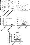
Relationship between memory B-cell subsets and pneumococcal humoral response. PBMCs were incubated with PE-cyanin7–conjugated anti-CD19 (Coulter Immunotech High Wycombe), PE-conjugated anti-human IgD (Southern Biotechnology Associates), APC-conjugated anti-human IgM (The Jackson Laboratory), and FITC-conjugated anti-CD27 (DakoCytomation). Cells were then washed and acquired on FACSCalibur (BD Biosciences, Oxford, United Kingdom). A minimum of 5000 events were acquired on the B-cell gate, and the results are expressed as a percentage of CD19 events. FlowJo software (TreeStar) was used for data analysis. Because IgM memory B cells express both IgD and IgM, coexpression of either IgD or IgM together with CD27 was used to define this subset. IgM memory B cells (CD19+ CD27+ IgMhigh IgD+/lo) and switched memory B cells (CD19+ CD27+ IgM IgD) were calculated using a modified Piqueras classification. (A) Patients who fail to mount a pneumococcal IgM response have significantly lower frequencies of IgM memory B cells compared with responders and healthy controls. (B) Scatter plot evaluating the association between pneumococcal IgM titers and IgM memory B-cell frequencies in CML patients. Samples were correlated using the Spearman rank correlation test. (C) Frequencies of class-switched memory B cells in the 33 patients who achieved a postimmunization IgG >200 U/mL compared with the 6 patients who failed to mount a positive pneumococcal IgM and IgG response; bars represent medians with interquartile range. A positive IgM Pneumovax II response was defined as a fourfold rise in serum IgM titers or an IgM titer >200 U/mL 4 weeks postimmunization irrespective of the preimmunization titer. A positive IgG response was defined as a twofold rise in serum IgG titer or an IgG titer >200 U/mL at 1 or 3 months. (D) IgM memory B-cell frequencies at diagnosis (prior to initiation of imatinib) and once CCyR was achieved on imatinib. (E) Class-switched memory B-cell frequencies at diagnosis (prior to initiation of imatinib) and once CCyR was achieved on imatinib. (F) B-cell phenotype of a CML patient who developed a positive pneumococcal IgM response (patient A) compared with a nonresponder (patient B).
Figure 4
Figure 4
Inhibition of Btk phosphorylation in CD19+ B cells from CML patients on TKI coincubated with autologous plasma. Cryopreserved PBMCs from CML patients on TKI (imatinib, n = 3; nilotinib, n = 3; and dasatinib, n = 3) were thawed, washed, and cocultured with autologous plasma or RPMI/10% fetal calf serum overnight. PBMCs were then stimulated with 5 mL of 50 mM of H2O2 for 15 minutes at 37°C. The stimulation was terminated by the addition of 5 mL prewarmed Cytofix Buffer (BD Biosciences, San Jose, CA) at 37°C for 12 minutes. Cells were fixed/permeabilized and stained with pBtk-PE (BD Biosciences) and APC-conjugated anti-CD19 (BD Biosciences). Data acquisition was performed on the FACSCalibur, and FlowJo software was used for analysis. MFI of Btk phosphorylation following incubation with autologous plasma in gated CD19+ B cells from representative CML patients on imatinib, dasatinib, or nilotinib is presented (right panel).
Figure 5
Figure 5
Btk and PLC-γ2 phosphorylation inhibition by imatinib, dasatinib, and nilotinib. (A) To assess the impact of TKI on normal B cells, PBMCs from healthy controls were isolated and cultured in the presence or absence of increasing concentrations of TKIs, namely, 1 to 50 μM of imatinib (LC Laboratories), 1 to 50 μM of nilotinib (LC Laboratories), or 1 to 100 nM of dasatinib (LC Laboratories) for 2 hours. PBMCs were then stimulated with goat anti-human IgG and IgM F(ab')2 (0.5 mg/mL solution) at a final concentration of 10 μg/mL for 20 minutes, and cells were stained with pBtk-PE or pPLC-γ2-PE, APC-conjugated anti-CD19 (BD Biosciences, San Jose, CA), PerCP-conjugated anti-human IgM (BD Biosciences, Oxford, United Kingdom), and FITC-conjugated anti-CD27 (DakoCytomation). Cells were gated on lymphocytes: the panels on the top depict the unstimulated negative control, and on the bottom anti-IgG and IgM-induced phosphorylation of Btk (left) and PLC-γ2 (right). (B) PBMCs were coincubated with imatinib (10 μM), nilotinib (10 μM), and dasatinib (100 nM) for 14 hours, and the viability of CD19+ B cells was assessed by staining with FITC-conjugated annexin and APC-conjugated anti-CD19 (both BD Biosciences, San Jose, CA). (C) Curve fit (linear regression) of TKI doses plotted against the percentage of Btk phosphorylation inhibition induced by each of the 3 TKIs, imatinib, nilotinib, and dasatinib (each experiment was performed a minimum of 3 times). The Y bar represents the percentage of gated population in which phosphorylated Btk or PLC-γ2 are detected.
Figure 6
Figure 6
Btk and PLC-γ2 phosphorylation inhibition in B-cell subsets. (A) Btk phosphorylation in B-cell subsets cultured in the presence or absence of 10 μM of imatinib, 100 nM of dasatinib, or 10 μM of nilotinib for 2 hours and stimulated with 10 μg/mL of anti-human IgG and IgM F(ab')2 for 20 minutes. Effect of the TKI on pBtk inhibition is shown in gated IgM memory B-cell, switched memory B-cell, and naive B-cell subsets. Each experiment was performed a minimum of 3 times. (B) MFI of BTK phosphorylation in gated CD19+ B cells from a representative healthy donor following incubation with 10 μM of imatinib, 100 nM of dasatinib, or 10 μM of nilotinib.

References

    1. de Lavallade H, Apperley JF, Khorashad JS, et al. Imatinib for newly diagnosed patients with chronic myeloid leukemia: incidence of sustained responses in an intention-to-treat analysis. J Clin Oncol. 2008;26(20):3358–3363. - PubMed
    1. Kantarjian H, Shah NP, Hochhaus A, et al. Dasatinib versus imatinib in newly diagnosed chronic-phase chronic myeloid leukemia. N Engl J Med. 2010;362(24):2260–2270. - PubMed
    1. Saglio G, Kim DW, Issaragrisil S, et al. ENESTnd Investigators. Nilotinib versus imatinib for newly diagnosed chronic myeloid leukemia. N Engl J Med. 2010;362(24):2251–2259. - PubMed
    1. Cwynarski K, Laylor R, Macchiarulo E, et al. Imatinib inhibits the activation and proliferation of normal T lymphocytes in vitro. Leukemia. 2004;18(8):1332–1339. - PubMed
    1. Mumprecht S, Matter M, Pavelic V, Ochsenbein AF. Imatinib mesylate selectively impairs expansion of memory cytotoxic T cells without affecting the control of primary viral infections. Blood. 2006;108(10):3406–3413. - PubMed

Publication types

MeSH terms