

Review
doi: 10.1016/j.clp.2013.02.002.
The biology of retinopathy of prematurity: how knowledge of pathogenesis guides treatment
Affiliations
- PMID: 23719305
- PMCID: PMC3673697
- DOI: 10.1016/j.clp.2013.02.002
Item in Clipboard
Review
The biology of retinopathy of prematurity: how knowledge of pathogenesis guides treatment
Clin Perinatol.
2013 Jun.
Abstract
Retinopathy of prematurity occurs because the retina of a preterm infant at birth is incompletely vascularized, and if the postnatal environment does not match the in utero environment that supported retinal development, the vessels and neural retina will not grow normally. Risk factors determined from many clinical studies and animal studies fall into 2 categories: prenatal factors and postnatal factors.
Copyright © 2013 Elsevier Inc. All rights reserved.
Figures
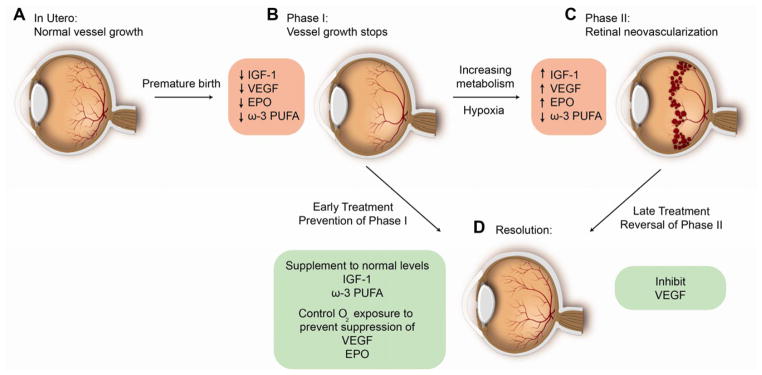
1. Laser photocoagulation of ROP: Hypoxic retina anterior to neovascularization in phase II of ROP which produces VEGF and Epo is destroyed to decrease pathological blood vessel formation (promoting C to D) 2. Anti-VEGF therapy: Direct suppression of neovascularization with suppression of VEGF (promoting C to D) 3. Increasing IGF-1 to in utero levels after birth: prevents vessel loss (phase I) (preventing A to B) to prevent phase II (C) 4. Control of oxygen after preterm birth: Prevents hyperoxia induced suppression of Hif regulated factors VEGF and Erythropoietin that are necessary for normal retinal vascular development thus preventing vessel loss (preventing A to B) 5. Maintaining adequate intake of the essential fatty acid DHA after preterm birth: Promotes normal vascularization and directly inhibits neovascularization (promote B to D and C to D) 6. Monitoring postnatal growth which is based on rate of increase of postnatal IGF-1 levels: Predicts the future development of neovascular ROP (C)
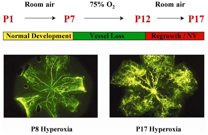
Neonatal mice are exposed to 75% oxygen from postnatal day 7 (P7) until P12 causing vessel loss and cessation of vascular growth to simulate Phase I of ROP. The central retinal microvessels are obliterated and radial vascular growth ceases. When the mice are returned to room air with incompletely vascularized retina, retinal neovascularization is seen, similar to phase II of ROP. Vessel proliferation is maximum at P17 then regresses, which also occurs in human ROP. These changes can be quantified in retinal flat mounts.
References
-
- Wikstrand MH, Hard AL, Niklasson A, Smith L, Lofqvist C, Hellstrom A. Maternal and neonatal factors associated with poor early weight gain and later retinopathy of prematurity. Acta Paediatr. 2011 - PubMed
-
- Singer D, Muhlfeld C. Perinatal adaptation in mammals: the impact of metabolic rate. Comp Biochem Physiol A Mol Integr Physiol. 2007;148(4):780–4. - PubMed
-
- Lofqvist C, Andersson E, Sigurdsson J, et al. Longitudinal postnatal weight and insulin-like growth factor I measurements in the prediction of retinopathy of prematurity. Arch Ophthalmol. 2006;124(12):1711–8. - PubMed
-
- Hellstrom A, Hard AL, Engstrom E, et al. Early weight gain predicts retinopathy in preterm infants: new, simple, efficient approach to screening. Pediatrics. 2009;123(4):e638–45. - PubMed
-
- Lofqvist C, Hansen-Pupp I, Andersson E, et al. Validation of a new retinopathy of prematurity screening method monitoring longitudinal postnatal weight and insulinlike growth factor I. Arch Ophthalmol. 2009;127(5):622–7. - PubMed
Publication types
MeSH terms
Substances
Grants and funding
LinkOut - more resources
Full Text Sources
Other Literature Sources
Medical
