. 2013 Jun 6;498(7452):118-22.
doi: 10.1038/nature12166. Epub 2013 May 29.

Control of angiogenesis by AIBP-mediated cholesterol efflux

Affiliations

Control of angiogenesis by AIBP-mediated cholesterol efflux

Longhou Fang et al. Nature. .

Abstract

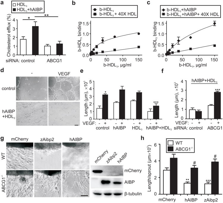
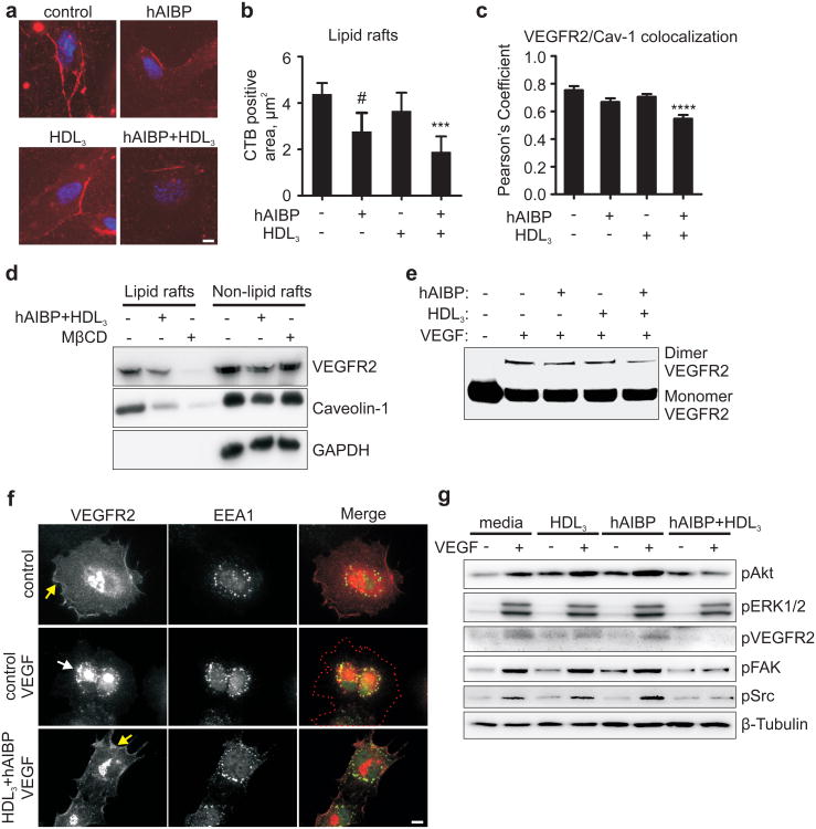
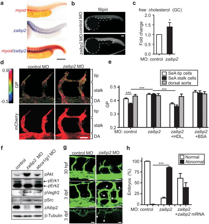
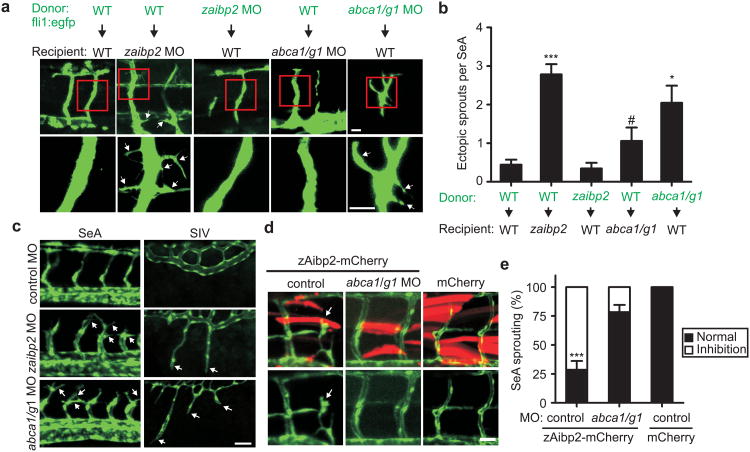
Cholesterol is a structural component of the cell and is indispensable for normal cellular function, although its excess often leads to abnormal proliferation, migration, inflammatory responses and/or cell death. To prevent cholesterol overload, ATP-binding cassette (ABC) transporters mediate cholesterol efflux from the cells to apolipoprotein A-I (apoA-I) and the apoA-I-containing high-density lipoprotein (HDL). Maintaining efficient cholesterol efflux is essential for normal cellular function. However, the role of cholesterol efflux in angiogenesis and the identity of its local regulators are poorly understood. Here we show that apoA-I binding protein (AIBP) accelerates cholesterol efflux from endothelial cells to HDL and thereby regulates angiogenesis. AIBP- and HDL-mediated cholesterol depletion reduces lipid rafts, interferes with VEGFR2 (also known as KDR) dimerization and signalling and inhibits vascular endothelial growth factor-induced angiogenesis in vitro and mouse aortic neovascularization ex vivo. Notably, Aibp, a zebrafish homologue of human AIBP, regulates the membrane lipid order in embryonic zebrafish vasculature and functions as a non-cell-autonomous regulator of angiogenesis. aibp knockdown results in dysregulated sprouting/branching angiogenesis, whereas forced Aibp expression inhibits angiogenesis. Dysregulated angiogenesis is phenocopied in Abca1 (also known as Abca1a) Abcg1-deficient embryos, and cholesterol levels are increased in Aibp-deficient and Abca1 Abcg1-deficient embryos. Our findings demonstrate that secreted AIBP positively regulates cholesterol efflux from endothelial cells and that effective cholesterol efflux is critical for proper angiogenesis.


Conflict of interest statement

The authors declare no conflicts of interests.

Figures

Figure 1
Figure 1. Role of AIBP in cholesterol efflux from EC and in vitro angiogenesis
a, hAIBP mediated-cholesterol efflux and effect of ABCG1 knockdown. HUVEC were transfected with control or ABCG1 siRNA, preloaded with 3H-cholesterol and incubated for 1 hour with 50 μg/ml HDL3 in the presence or absence of 0.2 μg/ml hAIBP. Efflux was measured as the 3H counts in the medium divided by the sum of 3H counts in the medium and the cells. Mean±SE; n=6. b and c, Effect of hAIBP on HDL3 binding to HUVEC. HUVEC were incubated on ice with the indicated concentration of biotinyated HDL3 (b-HDL3), in the presence or absence of hAIBP (at a 0.1:50 w/w hAIBP:HDL3 ratio) and 40× excess of unlabeled HDL. Each data point is Mean±SE from 3 to 7 independent experiments. The binding parameters for b-HDL3/HUVEC binding were calculated as Bmax = 0.8±0.1 and Kd = (0.33±0.10)×10−6 M in absence of hAIBP (panel b; R2=0.92, Sy.x=0.1), and Bmax=1.5±0.4 and Kd=(1.03±0.10)×10−6 M in the presence of hAIBP (panel c; R2=0.94, Sy.x=0.1). The differences in Bmax and Kd values were statistically significant (p<0.01 and p<0.05, respectively). d, Effect of hAIBP and HDL3 on EC tube formation. HUVEC were preincubated with or without 50 μg/ml HDL3 + 0.1 μg/ml hAIBP for 4 hours. Cells were then seeded on Matrigel, in the presence or absence of 20 ng/ml VEGF, and imaged following a 12-hour incubation. Scale, 100 μm. e, The length of EC tubes in the experiment illustrated in 1d and Supplementary Fig. 4. Mean±SE; n=5. f, Requirement for ABCG1 in hAIBP inhibition of angiogenesis. HUVEC were transfected with control or ABCG1 siRNA and assayed as in 1d. Mean±SE; n=6. g, Mouse aortic ring angiogenesis assay. Aortic rings from C57BL6 and Abcg1-/- mice were embedded in Matrigel. HEK293 cells transiently expressing mCherry, zAibp2 or hAIBP were inserted approximately 0.5 mm away from the aortic ring, and the plates were incubated with 10 ng/ml VEGF for 7 days. Images show the edge of the aortic rings facing the HEK293 cell clusters. Immunoblots show expression of hAIBP and zAibp2 (both detected with a Flag tag antibody) and mCherry in HEK293 cells. h, The length of aortic ring sprouts. Mean±SE; n=10. In all panels: #, not significant; *, p<0.05; **, p<0.01; ***, p<0.001.
Figure 2
Figure 2. Effect of AIBP on HUVEC lipid rafts, VEGFR2 localization, dimerization and signaling
a, Effect of hAIBP and HDL3 on lipid rafts. HUVEC were preincubated with 50 μg/ml HDL3, 0.1 μg/ml hAIBP, or 50 μg/ml HDL3 + 0.1 μg/ml hAIBP for 4 hours. Cells were stained for nuclei (blue, DAPI) and for lipid rafts (red, cholera toxin B (CTB) + anti-CTB antibody). Scale, 10 μm. b, The area of lipid rafts per cell. Mean±SE; n=10; **, p<0.01; #, p=0.08. c, Effect of hAIBP and HDL3 on caveolin-1 and VEGFR2 surface localization. HUVEC were incubated with hAIBP and/or HDL3 as in 2a, fixed and stained with antibodies to caveolin-1 and VEGFR2. Images were captured using TIRF microscopy (Supplementary Fig. 6) and Pearson's coefficient was calculated to assess surface colocalization of VEGFR2 with caveolin-1. Mean±SE; n=38-50; ***, p<0.001. d, VEGFR2 and caveolin-1 localization to lipid rafts. HUVEC were incubated with 20 μg/ml cholesterol-MβCD for six hours, followed by a 1 hour incubation with or without 50 μg/ml HDL3 + 0.1 μg/ml hAIBP, or a 30 min incubation with 10 mM MβCD. HUVEC lysates were separated into lipid rafts and non-lipid rafts fractions by ultracentrifugation, run on SDS-PAGE and blotted with VEGFR2 and caveolin-1 antibodies. e, Effect of hAIBP and HDL3 on VEGFR2 dimerization. HUVEC were preincubated with HDL3 and/or hAIBP as in 1a, followed by a 20 min stimulation with 50 ng/ml VEGF. Cells were treated with a crosslinking reagent, lysed and immunoprecipitated with a VEGFR2 antibody. Monomers and crosslinked dimers of VEGFR2 were visualized on western blot. f, Effect of hAIBP and HDL3 on VEGFR2 endocytosis. HUVEC were preincubated with or without 50 μg/ml HDL3 + 0.1 μg/ml hAIBP for 4 hours, then stimulated with 50 ng/ml VEGF for 20 min, fixed and stained with antibodies to VEGFR2 (red) and the early endosome marker EEA-1 (green). Yellow and white arrows point to the surface and endosomal localization of VEGFR2. Red dotted line traces cell contour. Scale, 10μm. g, Effect of hAIBP and HDL3 on VEGFR2 signaling. HUVEC were preincubated with HDL3 and/or hAIBP as in 2a, followed by a 20 min stimulation with 50 ng/ml VEGF. Total cell lysates were run on SDS-PAGE and probed as indicated.
Figure 3
Figure 3. Effect of Aibp deficiency on zebrafish cholesterol, membrane lipid order, Vegfr2 signaling and angiogenesis
a, Tissue distribution of zaibp2 mRNA in zebrafish embryos. Embryos at 24 hpf were fixed and WISH was performed with antisense myod and zaibp2 probes. Scale, 100 μm. b-c, Free cholesterol levels in zaibp2 morphants. b, Zebrafish embryos were injected with 8 ng of either control MO or zaibp2 MO. Twenty four hpf control and zaibp2 morphants were stained with filipin to detect free cholesterol in embryos. Note the yolks are artificially masked on the images. c, At 24 hpf, the trunk area (without yolk) was dissected, total lipids extracted, and free cholesterol levels determined by gas chromatography (GC). The cholesterol levels were normalized to the protein content and then to the values in control MO embryos. 50-70 embryos were pooled for each sample. Mean±SE; n=4; *, p<0.05. d, Effect of zaibp2 MO on SeA membrane lipid order. Tg(flk1:ras-cherry)s896 embryos were injected with control or zaibp2 MO as in 3b and at 24 hpf were stained with 5 μM Laurdan. In the same embryos, confocal images of mCherry fluorescence (bottom images) and the multiphoton images of Laurdan fluorescence (ex 800 nm, em 400-460 nm and 470-530 nm) were captured. The multiphoton results (top row images) are displayed as pseudocolored GP (generalized polarization, a measure of the membrane lipid order) images, cropped to show only the vasculature, i.e. mCherry-positive areas. Scale, 20 μm. e, The graph shows GP values in the areas corresponding to tip and stalk cells of growing SeA and the dorsal aorta (DA) as indicated in 3d. Some one-cell stage embryos were coinjected with 1 nl of 10 mg/ml human HDL3 or BSA. Note the Y-scale is from 0.2 to 0.5. Mean±SE; n=44-119 SeA in 25-49 embryos; ***, p<0.001. f, Phosphorylation of signaling proteins. Lysates of 24 hpf control (8 ng control MO), zaibp2 (8 ng zaibp2 MO) and abca1/g1 (4 ng abca1 MO + 4ng abcg1 MO) morphants were separated on SDS-PAGE and immunoblotted as indicated. g, Angiogenic defects in zaibp2 morphants. One-cell stage Tg(fli1:egfp)y1 zebrafish embryos were injected with 8 ng of either control or zaibp2 MO. The images are of SeA in 30 hpf embryos (top row), and SeA (middle row) and of SIV (bottom row) in 3 dpf embryos. Arrows point to dysregulated sprouts. Scale, 25 μm. h, Quantification of the number of embryos with normal and abnormal angiogenesis (SeA with ectopic branching). The abnormal angiogenesis was partially rescued by coinjection of 40 pg of zaibp2 mRNA lacking the MO targeting site. Mean±SE; n=100-149. ***, p<0.001.
Figure 4
Figure 4. Effect of Aibp and Abca1/Abcg1 deficiency on zebrafish angiogenesis
a, Mosaic expression analysis of EC branching in control, zaibp2 and abca1/abcg1 knockdown embryos. At 4 hpf, cells were isolated from donor embryos and transplanted into recipient embryos. Recipient embryos were analyzed at 3 dpf. Arrows point to aberrant ectopic branches/sprouts. Scale, 25 μm. b, Numbers of ectopic branches/filopodial projections per SeA. Mean±SE; n=8-16. #, not significant; *, p<0.05; ***, p<0.001. c, Angiogenic defects in abca1/abcg1 morphants. One-cell stage embryos were injected with 8 ng of control MO, 8 ng zaibp2 MO, or 4 ng abca1 MO + 4 ng abcg1 MO. Images of SeA (30 hpf) and SIV (3 dpf) are shown. Scale, 50 μm. d, Knockdown of abca1/g1 cancels the effect of zAibp2 overexpression. One-cell stage embryos were injected with 2 nl of 100 ng/μl myog:zaibp2-mCherry, myog:zaibp2-mCherry + abca1/g1 MO, or myog:mCherry. The arrow points to an aberrant SeA at the site of zAibp2-mCherry expression. Scale, 20μm. e, Abnormal SeA formation was quantified in 8-16 embryos per group. Mean±SE; *** p<0.001.

Comment in

  • Targeted cholesterol efflux.
    Fang L, Miller YI. Fang L, et al. Cell Cycle. 2013 Nov 1;12(21):3345-6. doi: 10.4161/cc.26401. Epub 2013 Sep 13. Cell Cycle. 2013. PMID: 24036659 Free PMC article. No abstract available.

References

    1. Bodzioch M, et al. The gene encoding ATP-binding cassette transporter 1 is mutated in Tangier disease. Nat Genet. 1999;22:347–351. - PubMed
    1. Rust S, et al. Tangier disease is caused by mutations in the gene encoding ATP-binding cassette transporter 1. Nat Genet. 1999;22:352–355. - PubMed
    1. Klucken J, et al. ABCG1 (ABC8), the human homolog of the Drosophila white gene, is a regulator of macrophage cholesterol and phospholipid transport. Proc Natl Acad Sci USA. 2000;97:817–822. - PMC - PubMed
    1. Yvan-Charvet L, et al. ATP-Binding Cassette Transporters and HDL Suppress Hematopoietic Stem Cell Proliferation. Science. 2010;328:1689–1693. - PMC - PubMed
    1. Armstrong AJ, Gebre AK, Parks JS, Hedrick CC. ATP-binding cassette transporter G1 negatively regulates thymocyte and peripheral lymphocyte proliferation. J Immunol. 2010;184:173–183. - PMC - PubMed

Publication types

MeSH terms