

E-cadherin is expressed by mono- and multinucleated histiocytes in cutaneous sarcoidal and foreign body granulomas
- PMID: 23719484
- PMCID: PMC3773293
- DOI: 10.1097/DAD.0b013e31828de7e0
E-cadherin is expressed by mono- and multinucleated histiocytes in cutaneous sarcoidal and foreign body granulomas
Abstract
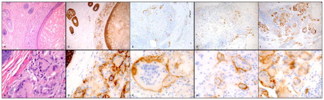
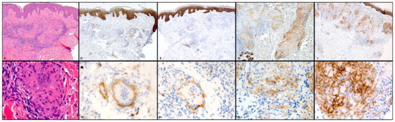
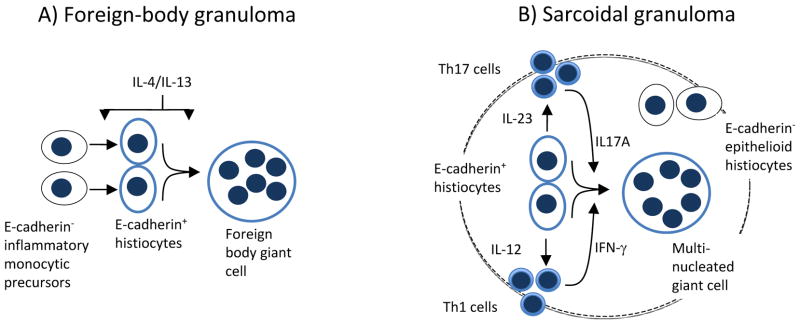
E-cadherin, a member of the cadherin family of transmembrane adhesion receptors, is critical for cutaneous barrier function, as it promotes keratinocyte and Langerhans cell adhesion in the epidermis. Recent murine models of chronic inflammation identified new E-cadherin expressing subsets of mononuclear phagocytes, including alternatively activated macrophages and selected inflammatory dendritic cells. It has been shown in vitro that expression of E-cadherin by murine macrophages promotes their homotypic aggregation and fusion to multinucleated giant cells (MNGCs), a signature cell type of granulomatous inflammation. The purpose of this study was to assess E-cadherin expression on histiocytes and giant cells in cutaneous granulomas in humans. E-cadherin expression was evaluated by immunohistochemistry of formalin-fixed paraffin-embedded skin biopsies of foreign body granulomas (n = 21) and sarcoidosis (n = 21). The results showed consistent membranous E-cadherin staining pattern on mononucleated histiocytes and MNGCs in both granuloma types. These E-cadherin expressing histiocytes are distinct from dermal Langerhans cells because they lacked CD1a expression. Our findings suggest that E-cadherin expressing mononuclear histiocytes are likely precursors for MNGCs in cutaneous granulomas and may play a critical role in disease pathogenesis.
Conflict of interest statement
Figures
References
-
- Boros DL. In: The Cellular Immunological Aspects of the Granulomatous Response. Boros DL, editor. Washington: ASM Press; 2003. pp. 1–28.
-
- Iannuzzi MC, Rybicki BA, Teirstein AS. Sarcoidosis. N Engl J Med. 2007;357(21):2153–65. Epub 2007/11/23. - PubMed
-
- Bergeron A, Bonay M, Kambouchner M, et al. Cytokine patterns in tuberculous and sarcoid granulomas: correlations with histopathologic features of the granulomatous response. Journal of immunology. 1997;159(6):3034–43. Epub 1997/09/23. - PubMed
-
- Ten Berge B, Paats MS, Bergen IM, et al. Increased IL-17A expression in granulomas and in circulating memory T cells in sarcoidosis. Rheumatology (Oxford) 2012;51(1):37–46. Epub 2011/11/15. - PubMed
-
- Kao WJ, McNally AK, Hiltner A, Anderson JM. Role for interleukin-4 in foreign-body giant cell formation on a poly(etherurethane urea) in vivo. J Biomed Mater Res. 1995;29(10):1267–75. Epub 1995/10/01. - PubMed
Publication types
MeSH terms
Substances
Grants and funding
LinkOut - more resources
Full Text Sources
Other Literature Sources
Medical
Miscellaneous
