. 2014 Jan;19(1):105-14.
doi: 10.1007/s12192-013-0438-7. Epub 2013 May 30.

Astragaloside IV attenuates glycated albumin-induced epithelial-to-mesenchymal transition by inhibiting oxidative stress in renal proximal tubular cells

Affiliations

Astragaloside IV attenuates glycated albumin-induced epithelial-to-mesenchymal transition by inhibiting oxidative stress in renal proximal tubular cells

Weiwei Qi et al. Cell Stress Chaperones. 2014 Jan.

Abstract

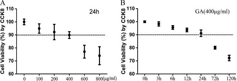
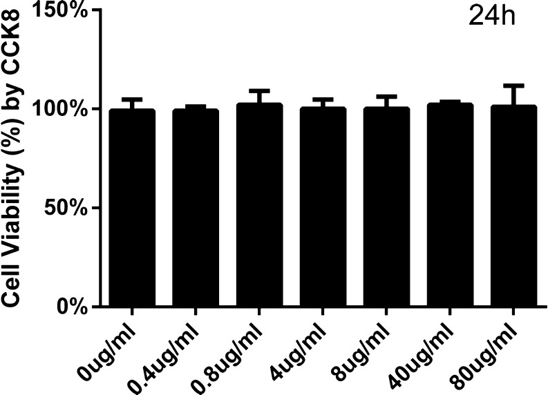
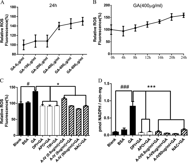
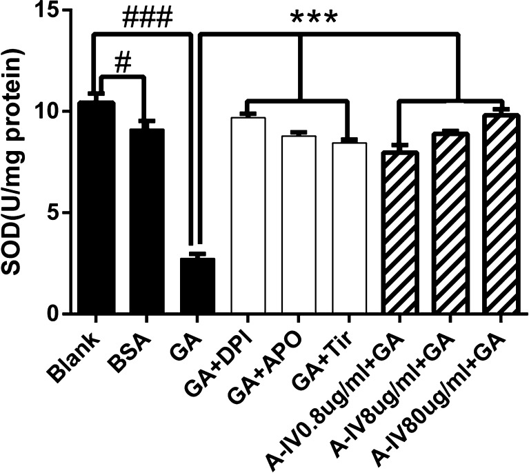
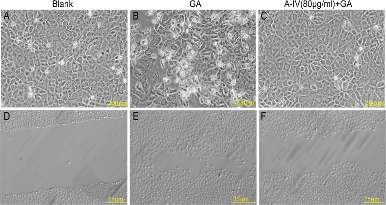
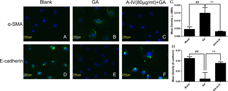
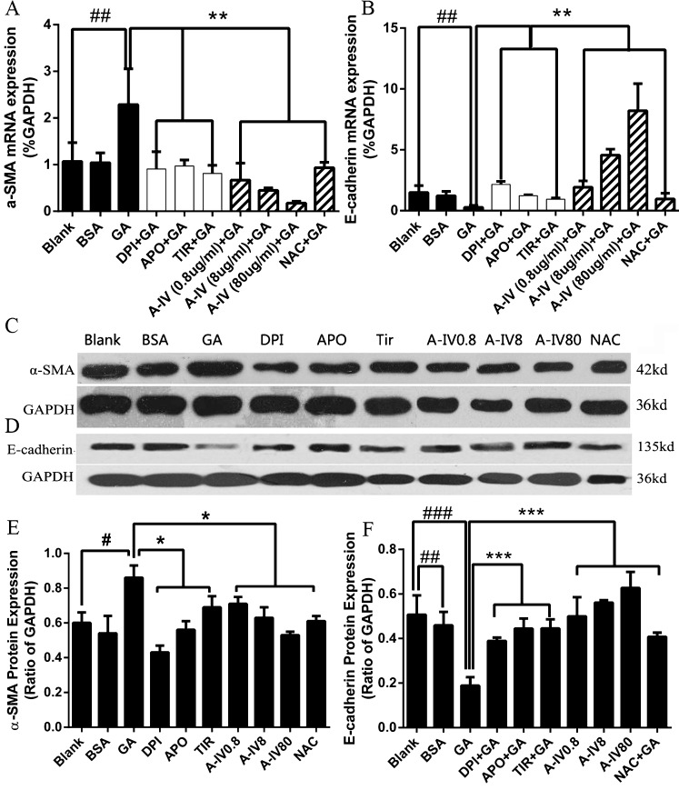
In diabetic kidney disease (DKD), epithelial-to-mesenchymal transition (EMT) is a classic pathological process in tubular damage. Oxidative stress is considered to play an important role in DKD. Astragaloside IV (A-IV), one of the main active ingredients of Astragalus membranaceus, exhibits a wide range of biological activities. However, the effect of A-IV on regulating EMT in tubular cells is unclear. This study aims to determine whether A-IV could attenuate glycated albumin (GA)-induced EMT in the NRK-52E cell line by inhibiting oxidative stress. GA and A-IV-induced cytotoxicity were assayed by CCK-8. The intercellular reactive oxygen species (ROS) level was detected by H2DCFDA. The activity of NADPH oxidase was assayed by adding exogenous NADPH oxidase, and the superoxide dismutase (SOD) units were observed by NBT. We used a microscope to examine the morphology of the NRK-52E cell line. We conducted a wound healing assay to measure cell mobility. To determine mRNA and protein expressions of α-SMA and E-cadherin, we used real-time polymerase chain reaction (real-time PCR), immunofluorescence, and western blot analysis. A-IV significantly attenuated GA-induced amplification of ROS, lowered the increased level of NADPH oxidase activity, and elevated the decreased level of SOD units. The GA-induced NRK-52E cell line showed increased expression of α-SMA and decreased expression of E-cadherin in mRNA and protein levels, whereas A-IV alleviated the expression of α-SMA and increased the expression of E-cadherin. Our data demonstrate that GA could induce NRK-52E cell line EMT through oxidative stress. This effect could be attenuated by A-IV via regulation of the impaired redox balance.


Figures

Fig. 1
Fig. 1
Effect of GA on cell viability (a, b). The viability of NRK-52E cells cultured with GA was assessed by performing a CCK-8 assay. a NRK-52E cells were stimulated with GA in different concentrations (0–800 μg/ml) for 24 h. b NRK-52E cells were incubated for 0–120 h with GA at the concentration of 400 μg/ml. The results are expressed as the mean ± SD values of three independent experiments
Fig. 2
Fig. 2
Effect of A-IV on cell viability. The viability of NRK-52E cells incubated with A-IV was tested by CCK-8 assay. NRK-52E cells were incubated with A-IV at the concentration from 0 to 80 μl/ml for 24 h. The results are expressed as the mean ± SD values of three independent experiments
Fig. 3
Fig. 3
GA-mediated ROS generation and NADPH oxidase activation were inhibited by A-IV in NRK-52E cells. a NRK-52E cells were treated with different concentrations of GA (0–800 μg/ml) for 24 h. b NRK-52E cells were incubated for 0–24 h with GA at the concentration of 400 μg/ml. c and d NRK-52E cells were stimulated by GA (400 μg/ml) for 24 h after pretreatment of NADPH oxidase inhibitors (DPI and Apo) or superoxide scavenger (Tiron) for 30 min and NRK-52E cells were pretreated with different concentrations of A-IV (0.8–80 μg/ml) or antioxidant NAC for 30 min before GA (400 μg/ml) treatment for 24 h. #P < 0.05 vs. blank; ###P < 0.001 vs. blank; *P < 0.05 vs. GA; ***P < 0.001 vs. GA. Values are expressed as the mean ± SD of three independent experiments
Fig. 4
Fig. 4
Effect of A-IV on GA induced the decrease of intracellular T-SOD level. NRK-52E cells were stimulated by GA (400 μg/ml) for 24 h after pretreatment of NADPH oxidase inhibitors (DPI and Apo) and superoxide scavenger (Tiron) for 30 min and NRK-52E cells were pretreated with different concentrations A-IV(0.8–80 μg/ml) or antioxidant NAC for 30 min before GA (400 μg/ml) treatment for 24 h. #P < 0.05 vs. blank, ###P < 0.001 vs. blank; ***P < 0.001 vs. GA. Data are presented as the mean ± SD of three independent experiments
Fig. 5
Fig. 5
Effect of A-IV on cell morphological transformation and cellular mobility. A typical epithelial shape of the NRK-52E cells cultured in the blank group (a) for 24 h was shown, with the characteristic cobblestone morphology. Exposure to 400 μg/ml GA (b) for 24 h induced a phenotypic transition. Pretreatment with 80 μg/ml A-IV for 30 min effectively weakened such changes induced by GA (c). Wound healing assay of NRK-52E cells was cultured in d the blank group, e 400 μg/ml GA group, and f GA (400 μg/ml) treating group with A-IV (80 μg/ml) for 24 h. A-IV attenuated the cell mobility caused by GA
Fig. 6
Fig. 6
A-IV inhibited GA-induced EMT in NRK-52E cells. NRK-52E cells were incubated in a, d the blank group, b, e 400 μg/ml GA group, c, f GA (400 μg/ml) treating group with A-IV (80 μg/ml) for 24 h. The effects of A-IV protective effect on α-SMA and E-cadherin expressions were assessed by immunofluorescent microscopy. g, h Semiquantitative fluorescence intensity for α-SMA and E-cadherin expression in blank group, 400 μg/ml GA group, and GA (400 μg/ml) treating group with A-IV (80 μg/ml). Images were obtained in at least three independent experiments. ×400
Fig. 7
Fig. 7
Effect of A-IV on the GA-induced EMT in NRK-52E. Real-time PCR revealed GA increased α-SMA (a) and reduced E-cadherin (b) mRNA expressions dramatically, whereas NADPH oxidase inhibitors and different concentrations of A-IV weakened such changes. Similar results were observed through western blotting analysis (c, d, e, f). #P < 0.05, ##P < 0.01, ###p < 0.001 vs. blank; *P < 0.05, **P < 0.01, ***P < 0.001 vs. GA. Values are expressed as the mean ± SD of three independent experiments

References

    1. Burns WC, Thomas MC. The molecular mediators of type 2 epithelial to mesenchymal transition (EMT) and their role in renal pathophysiology. Expert Rev Mol Med. 2010;27:12–17. - PubMed
    1. Cannito S, Novo E, di Bonzo LV, Busletta C, Colombatto S, Parola M. Epithelial–mesenchymal transition: from molecular mechanisms, redox regulation to implications in human health and disease. Antioxid Redox Signal. 2010;12:1383–430. doi: 10.1089/ars.2009.2737. - DOI - PubMed
    1. Chang YX, Sun YG, Li J, Zhang QH, Guo XR, Zhang BL, et al. The experimental study of Astragalus membranaceus on meridian tropsim: the distribution study of astragaloside IV in rat tissues. J Chromatogr B Analyt Technol Biomed Life Sci. 2012;12(911):71–5. doi: 10.1016/j.jchromb.2012.10.024. - DOI - PubMed
    1. Chen R, Shao H, Lin S, Zhang JJ, Xu KQ. Treatment with Astragalus membranaceus produces antioxidative effects and attenuates intestinal mucosa injury induced by intestinal ischemia–reperfusion in rats. Am J Chin Med. 2011;39:879–87. doi: 10.1142/S0192415X11009275. - DOI - PubMed
    1. Cho WC, Leung KN. In vitro and in vivo anti-tumor effects of Astragalus membranaceus. Cancer Lett. 2007;252:43–54. doi: 10.1016/j.canlet.2006.12.001. - DOI - PubMed

Publication types

MeSH terms

LinkOut - more resources