. 2013 Dec 1;133(11):2726-36.
doi: 10.1002/ijc.28289. Epub 2013 Jul 6.

Targeting breast cancer-initiating/stem cells with melanoma differentiation-associated gene-7/interleukin-24

Affiliations

Targeting breast cancer-initiating/stem cells with melanoma differentiation-associated gene-7/interleukin-24

Sujit K Bhutia et al. Int J Cancer. .

Erratum in

  • Erratum.
    [No authors listed] [No authors listed] Int J Cancer. 2016 Jul 1;139(1):E3. doi: 10.1002/ijc.30058. Int J Cancer. 2016. PMID: 27080528 No abstract available.

Abstract

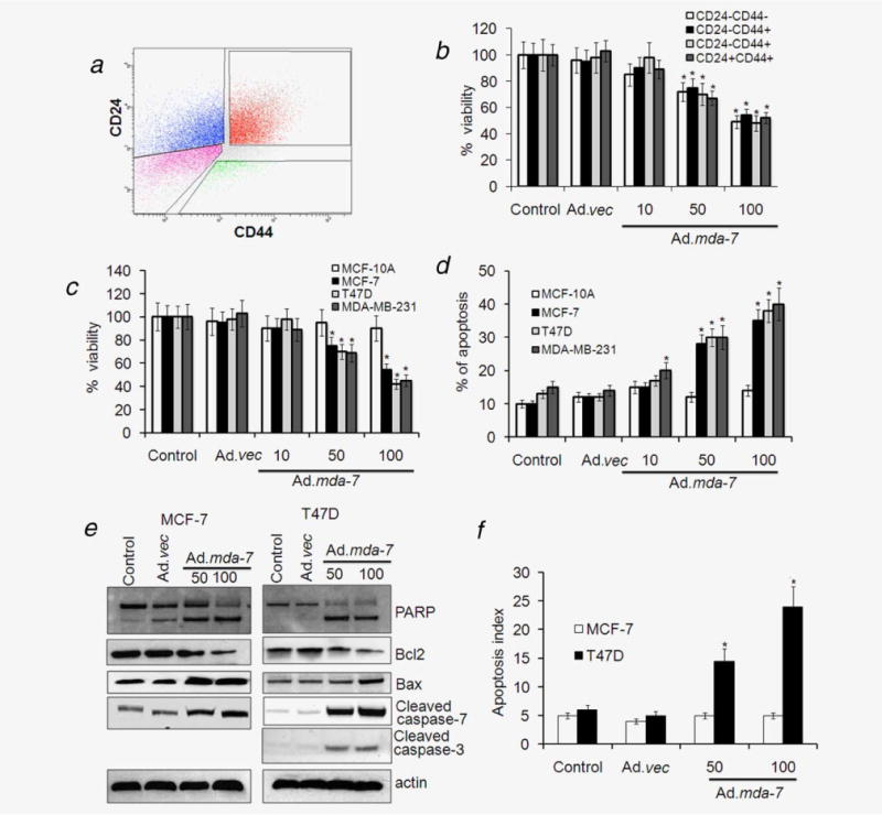
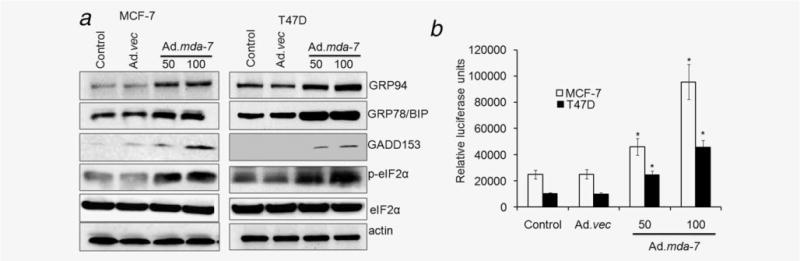
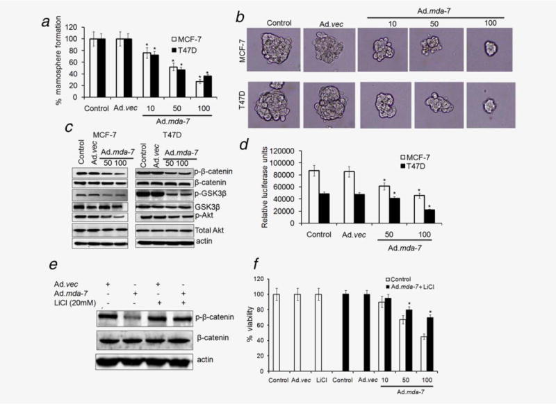
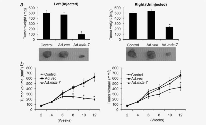
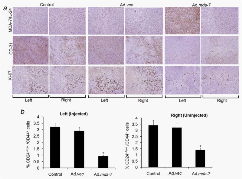
Melanoma differentiation-associated gene-7/interleukin-24 (mda-7/IL-24) displays a broad range of antitumor properties including cancer-specific induction of apoptosis, inhibition of tumor angiogenesis and modulation of antitumor immune responses. In our study, we elucidated the role of MDA-7/IL-24 in inhibiting growth of breast cancer-initiating/stem cells. Ad.mda-7 infection decreased proliferation of breast cancer-initiating/stem cells without affecting normal breast stem cells. Ad.mda-7 induced apoptosis and endoplasmic reticulum stress in breast cancer-initiating/stem cells similar to unsorted breast cancer cells and inhibited the self-renewal property of breast cancer-initiating/stem cells by suppressing Wnt/β-catenin signaling. Prevention of inhibition of Wnt signaling by LiCl increased cell survival upon Ad.mda-7 treatment, suggesting that Wnt signaling inhibition might play a key role in MDA-7/IL-24-mediated death of breast cancer-initiating/stem cells. In a nude mouse subcutaneous xenograft model, Ad.mda-7 injection profoundly inhibited growth of tumors generated from breast cancer-initiating/stem cells and also exerted a potent "bystander" activity inhibiting growth of distant uninjected tumors. Further studies revealed that tumor growth inhibition by Ad.mda-7 was associated with a decrease in proliferation and angiogenesis, two intrinsic features of MDA-7/IL-24, and a reduction in vivo in the percentage of breast cancer-initiating/stem cells. Our findings demonstrate that MDA-7/IL-24 is not only nontoxic to normal cells and normal stem cells but also can kill both unsorted cancer cells and enriched populations of cancer-initiating/stem cells, providing further documentation that MDA-7/IL-24 might be a safe and effective way to eradicate cancers and also potentially establish disease-free survival.

Keywords: MDA-7/IL-24; Wnt signaling; apoptosis; breast cancer; cancer-initiating/stem cells.


Conflict of interest statement

Conflicts of interest: Nothing to report

Figures

Figure 1
Figure 1
Ad.mda-7 infection results in expression of MDA-7/IL-24 in breast cancer-initiating/stem cells. MCF-7-, T47D-, MDA-MB-231- and MCF-10A-derived initiating/stem cells were sorted from their respective cell lines by flow cytometry and infected with either Ad.mda-7 (50 or 100 pfu) or Ad.vec (100 pfu) for 48 hr and total proteins were isolated. The expressions of MDA-7/IL-24 and actin (as a loading control) proteins were analyzed by Western blotting.
Figure 2
Figure 2
Ad.mda-7 infection induces apoptosis in breast cancer-initiating/stem cells. (a) MCF-7 cells were analyzed by flow cytometry for CD44 and CD24 expression. The gates shown were used for sorting of cells with the indicated phenotypes. (b) The sorted populations, based on surface markers, were infected with Ad.mda-7 (10, 50 and 100 pfu/cell) or Ad.vec (100 pfu/cell) and after 48 hr cell proliferation was measured by MTT assay. (c and d) Cancer-initiating/stem cells from MCF-7, T47D, MDA-MB-231 and MCF-10A were infected with Ad.mda-7 (50 or 100 pfu/cell) or Ad.vec (100 pfu/cell) and after 48 hr cell proliferation was measured by MTT assay (c) and Annexin V staining assay using flow cytometry (d) measured apoptosis. MCF-7- and T47D-initiating cells were infected with Ad.mda-7 (50 or 100 pfu/cell) or Ad.vec (100 pfu/cell) and cell lysates were prepared after 48 hr. (e) MCF-7- and T47D-initiating cells were infected with Ad.mda-7 (50 or 100 pfu/cell) or Ad.vec (100 pfu/cell) and cell lysates were prepared after 48 hr. Activation of poly(ADP-ribose) polymerase-1 (PARP), caspases 3 and 7 and expression of Bcl-2 and Bax were detected by Western blotting analysis. (f) After 48-hr infection with Ad.mda-7 or Ad.vec the apoptotic index as indicative of caspase 3/7 activity in MCF-7- and T47D-initiating cells was determined using a fluorescence microscope. The apoptotic index was calculated by dividing the number of caspase 3/7 fluorescent cells by the total number of DNA-containing cells after staining with Vybrant DyeCycle Green. Data represent mean ± SD (n = 3). Asterisk (*) represents significant difference (p < 0.5) with corresponding control.
Figure 3
Figure 3
Ad.mda-7 induces ER stress in breast cancer-initiating/stem cells. MCF-7- and T47D-initiating/stem cells were infected with Ad.mda-7 (50 or 100 pfu/cell) or Ad.vec (100 pfu/cell) and cell lysates were prepared after 48 hr. (a) Changes in BiP/GRP78, GRP94 GADD153 and activation of phospho-eukaryotic initiation factor 2 (p-eIF2α) proteins were evaluated by Western blotting. (b) MCF-7- and T47D-initiating/stem cells were transfected with ERSE reporter followed by infection with Ad.mda-7 or Ad.vec for 24 hr and dual Luciferase assays were performed and promoter activity values are expressed as arbitrary units using a Renilla reporter for internal normalization. Data represent mean ± SD (n = 3). Asterisk (*) represents significant difference (p < 0.5) with corresponding control.
Figure 4
Figure 4
Ad.mda-7 inhibits mammosphere formation and Wnt signaling in breast cancer-initiating/stem cells. (a) Cancer-initiating/stem cells from MCF-7 and T47D were infected with Ad.mda-7 (50 or 100 pfu/cell) or Ad.vec (100 pfu/cell) for 48 hr and the infected cells were seeded for formation of secondary mammospheres as described in “Material and Methods” section and quantified by counting the mammospheres by microscope. Data represent mean ± SD (n = 3). (b) Photomicrograph of primary mammospheres following the indicated treatments (magnification, ×100). (c) MCF-7- and T47D-initiating/stem cells were infected with Ad.mda-7 (50 or 100 pfu/cell) or Ad.vec (100 pfu/cell) and cell lysates were prepared after 48 hr and changes in phospho- and total β-catenin, GSK3β and Akt expression were evaluated by Western blotting. (d) MCF-7- and T47D-initiating/stem cells were infected with a TCF/LEF TOP-luc lentiviral reporter system for 48 hr followed by infection with Ad.mda-7 (50 or 100 pfu/cell) or Ad.vec (100 pfu) for 12 hr and relative luciferase activity was measured as described in the “Material and Methods” section. (e and f) T47D-initiating/stem cells were infected with 100 pfu/cell of Ad.mda-7 or Ad.vec in the presence of LiCl (20 mM) for 48 hr and expression of phospho- and total β-catenin was analyzed (e) and cell proliferation was measured by MTT assay (f). Asterisk (*) represents significant difference (p < 0.5) when compared to Ad.mda-7-infected cells without LiCl.
Figure 5
Figure 5
Ad.mda-7 eradicates primary tumors and inhibits distant tumors generated by breast cancer-initiating/stem cells in nude mice. Tumor xenografts from T47D-initiating/stem cells were established in athymic nude mice in the left and right flanks and only tumors on the left side were injected with PBS (control) or with the indicated Ad for 3 weeks (total of seven injections). (a) Measurement of tumor weight at the end of the study. Lower panel, picture of isolated tumors from the left-treated and right-untreated regions of animals. (b) Measurement of tumor volume at the end of the study. The data represent the mean ± SD with a minimum of five mice per group. Asterisk (*) represents significant difference (p < 0.5) with corresponding control.
Figure 6
Figure 6
Immunohistochemical analysis of tumor xenografts induced by T47D-initiating/stem cells infected with Ad.vec or Ad.mda-7 or controls injected with PBS. Tumor tissues were harvested and formalin-fixed and paraffin-embedded sections were immunostained for MDA-7/IL-24, CD-31 and Ki-67. The left and right tumors were from animals injected three times (left tumor) with PBS, Ad.vec or Ad.mda-7. Experimental details are given in “Material and Methods” section. (b) Single-cell suspensions were generated from the tumors of all groups and were fixed and stained with CD24 and CD44 antibodies to quantify the percentage of CD24−/low/CD44+ cells by fluorescence-activated cell sorting. The data represent the mean ± SD with a minimum of five mice per group. Asterisk (*) represents significant difference (p < 0.5) vs. corresponding control.

References

    1. Goldhirsch A, Colleoni M, Domenighetti G, et al. Systemic treatments for women with breast cancer: outcome with relation to screening for the disease. Ann Oncol. 2003;14:1212–14. - PubMed
    1. Charafe-Jauffret E, Monville F, Ginestier C, et al. Cancer stem cells in breast: current opinion and future challenges. Pathobiology. 2008;75:75–84. - PMC - PubMed
    1. Creighton CJ, Li X, Landis M, et al. Residual breast cancers after conventional therapy display mesenchymal as well as tumor-initiating features. Proc Natl Acad Sci USA. 2009;106:13820–5. - PMC - PubMed
    1. Al-Hajj M, Wicha MS, Benito-Hernandez A, et al. Prospective identification of tumorigenic breast cancer cells. Proc Natl Acad Sci USA. 2003;100:3983–8. - PMC - PubMed
    1. Han JS, Crowe DL. Tumor initiating cancer stem cells from human breast cancer cell lines. Int J Oncol. 2009;34:1449–53. - PubMed

Publication types