Review
. 2012 Oct;2(4):2443-69.
doi: 10.1002/cphy.c100015.

Pontine mechanisms of respiratory control

Affiliations
Review

Pontine mechanisms of respiratory control

Mathias Dutschmann et al. Compr Physiol. 2012 Oct.

Abstract

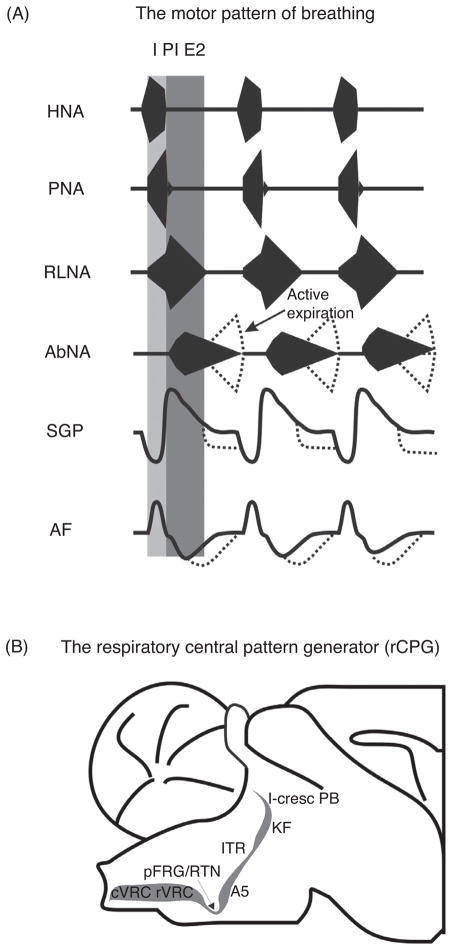
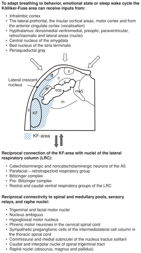
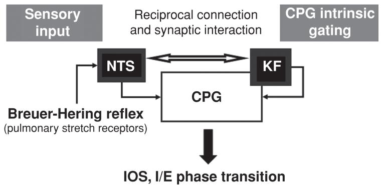
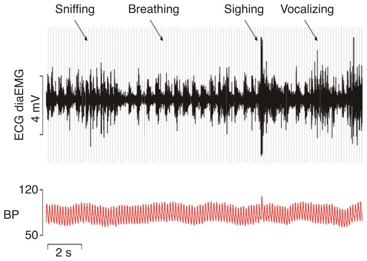
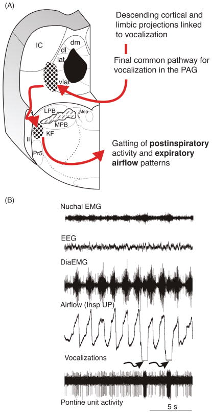
Pontine respiratory nuclei provide synaptic input to medullary rhythmogenic circuits to shape and adapt the breathing pattern. An understanding of this statement depends on appreciating breathing as a behavior, rather than a stereotypic rhythm. In this review, we focus on the pontine-mediated inspiratory off-switch (IOS) associated with postinspiratory glottal constriction. Further, IOS is examined in the context of pontine regulation of glottal resistance in response to multimodal sensory inputs and higher commands, which in turn rules timing, duration, and patterning of respiratory airflow. In addition, network plasticity in respiratory control emerges during the development of the pons. Synaptic plasticity is required for dynamic and efficient modulation of the expiratory breathing pattern to cope with rapid changes from eupneic to adaptive breathing linked to exploratory (foraging and sniffing) and expulsive (vocalizing, coughing, sneezing, and retching) behaviors, as well as conveyance of basic emotions. The speed and complexity of changes in the breathing pattern of behaving animals implies that "learning to breathe" is necessary to adjust to changing internal and external states to maintain homeostasis and survival.


Figures

Figure 1
Figure 1
(A) Respiratory motor outputs in relation to the three major phases of the respiratory cycle (I, inspiration, post-I, postinspiration, E2, late expiration). HNA, hypoglossal nerve activity; PNA, phrenic nerve activity; RLNA, recurrent laryngeal nerve activity; AbNA, abdominal nerve activity (lumbar segment 1). The lower two traces illustrate the dynamic changes in subglottal pressure (SGP) related to changes in upper airway resistance and airflow (AF). (B) Sagittal section of the anatomical organization of the lateral respiratory column (LRC) which includes the respiratory central pattern generator (rCPG) in the pontomedullary brainstem. Abbreviations: cVRC, caudal ventral respiratory column; rVRC, rostral ventral respiratory column; RTN/pFRG, retro-trapezoidal nucleus/parafacial respiratory group; A5, noradrenaline-containing neurons in the ventrolateral pons; ITR, intertrigeminal region; KF, Kölliker Fuse nucleus; l-cresc PB, lateral crescent nucleus of the parabrachial complex.
Figure 2
Figure 2
The efferent and afferent connections of the primary respiratory nuclei (shaded): Kölliker-Fuse nucleus and adjacent subnuclei of the parabrachial complex of the dorsolateral pons (KF-area). The data supporting the KF-area projecting to the parafacial nucleus are unpublished and supplied by Bellintani, Herbert, and Dutschmann. Abbreviations: c, central subnucleus of the PB; d, dorsal subnucleus of the PB; el, external lateral subnucleus of the PB; il, internal lateral subnucleus of the PB; ll, lateral lemniscus; m, medial subnucleus of the PB; me5, mesencephalic trigeminal nucleus; scp, superior cerebellar peduncle; v, ventral subnucleus of the PB.
Figure 3
Figure 3
Camera lucida drawings of coronal sections through the caudal pons and medulla oblongata from rostral to caudal (a–i) illustrating the pattern and distribution of anterogradely labeled descending fibers following PHA-L injection into the KF (see inset j, filled circles reflect neurons which have been filled with PHA-L, and therefore represent the probable neurons with projections). Figure published with permission of Horst Herbert, Tübingen, Germany. Orientation: Panels a–i are from rostral to caudal transverse sections through the pontomedullary brainstem. Abbreviations: 4V, 4th ventricle; 7, facial nucleus; 7L, lateral facial motor nucleus; 7n, facial nerve; 8n, vestibulocochlear nerve; 10, dorsal motor nucleus of vagus; 12, hypoglossal nucleus; A1, A1 noradrenergic cell group; A5, A5 noradrenergic cell group; Amb, nucleus ambiguus; AP, area postrema; Bo, Bötzinger complex; C1, C1 adrenergic cell group; CnF, cuneiform nucleus; Cu, cuneate nucleus; CVL, caudal ventrolateral reticular nucleus; DC, dorsal cochlear nucleus; DLL, dorsal nucleus of the lateral lemniscus; Gi, gigantocellular reticular nucleus; GiA, gigantocellular reticular nucleus, part alpha; Gr, gracile nucleus; icp, inferior cerebellar peduncle; IRt, intermediate reticular nucleus; KF, Kölliker-Fuse nucleus; LC, locus coeruleus; LPGi, lateral paragigantocellular nucleus; LRt, lateral reticular nucleus; LSO, lateral superior olive; LVe, lateral vestibular nucleus; me5, mesencephalic trigeminal tract; Mo5, trigeminal motor nucleus; NTS, nuclei of solitary tract; nts, solitary tract; PB, parabrachial nucleus; PnC, pontine reticular nucleus, caudal part; Pr5, principal sensory trigeminal nucleus; PrB, pre-Bötzinger complex; py, pyramidal tract; pyx, pyramidal decussation; RMg, raphé magnus nucleus; Rob, raphé obscurus nucleus; RPa, raphé pallidus nucleus; RVL, rostroventrolateral reticular nucleus; scp, superior cerebellar peduncle; sp5, spinal trigeminal tract; Sp5C, spinal trigeminal nucleus, caudal part; Sp5I, spinal trigeminal nucleus, interpolar part; Sp5O, spinal trigeminal nucleus, oral part; tz, trapezoid body; VCA, ventral cochlear nucleus, anterior part; VCP, ventral cochlear nucleus, posterior part subnuclei of the parabrachial complex (PB) (same as Fig. 3): c, central lateral; d, dorsal lateral; el, external lateral; eli, inner part of extern lateral; elo, outer part of extern lateral; exl, external lateral; exm, external medial; il, internal lateral; m, medial; s, superior; v, ventral; w, waist. Subnuclei of the nucleus of solitary tract (NTS): c, central; com, commissural; dm, dorsomedial; m, medial; vl, ventrolateral.
Figure 4
Figure 4
Schematic drawing illustrating the two convergent pathways involved in the mediation of inspiratory off-switch (IOS) and inspiratory/expiratory (I/E) phase transition. (A) An afferent pathway mediating Breuer-Hering reflex originates from pulmonary stretch receptor input via the vagus and terminates in the nuclei of the solitary tract (NTS). (B) A central pathway that requires the Kölliker-Fuse nucleus (KF) of the pons involves reciprocal interaction between the KF and central pattern generator (CPG). Plasticity in the expression of the breathing pattern depends on intrinsic gating mechanisms (for details see text) of the CPG attributed to the KF and results from interaction between reciprocal connections of the afferent and the network pathways.
Figure 5
Figure 5
Schematic illustration of the sequential phase of the pontine mediated inspiratory off-switch (IOS). The figure is adapted from Mörschel and Dutschmann 2009. Excitatory neurons and excitatory drive are highlighted in green; inhibitory neurons and interactions, red; and inactive cell populations and connections, gray. The model is based on the concept that ascending drive from the medullary respiratory neurons is required for the pontine mediated IOS. Panels on right show three time points in the cycle. (A) During the inspiratory phase, a pontine early-I population receives excitatory synaptic input (efference copy 1) from inspiratory driver neurons (I-driver) located in the pre-Bötzinger complex. The pontine early-I populations are inhibitory interneurons of the pons. The pontine early I neurons inhibit the pontine inspiratory/expiratory phase spanning neuron (I/E) and pontine postinspiratory premotoneurons (post-I) to prevent initiation of phase transition during early and mid-inspiration. With ongoing inspiration the pontine I/E neurons receive increasing excitatory drive (efference copy 2) from medullary augmenting-inspiratory premotor neurons that override the inhibition of early-I causing firing onset around late inspiration. (B) The pontine I/E neurons are excitatory pontobulbar neurons that activate the medullary late-I neurons to initiate the inspiratory/expiratory phase transition. (C) Finally, the inhibitory late-I neurons of the medulla terminate the activity of the medullary early-I neurons and release the medullary post-I population (inhibitory interneurons) from synaptic inhibition. These inhibitory post-I neurons inhibit the inspiratory populations (medullary and pontine). Consequently, the pontine and medullary postinspiratory premotor population innervating thyroarytenoid motoneurons starts firing.
Figure 6
Figure 6
Illustration of the effect of postsynaptic blockade of glutamatergic neurotransmission (e.g., NMDA-receptor antagonism) within the dorsolateral pons. Local blockade of excitatory synaptic interaction (black circles with white “x”) in the pons suppresses the efference copies 1–2 (see text and Fig. 5) causing blockade of the descending excitatory synaptic input from the pontine I/E neurons to the medullary late-I population. This abolishes the pontine mediated timing of the inspiratory/expiratory phase transition causing arrest in the inspiratory phase (apneusis). Figure adapted, with permission, from Mörschel and Dutschmann, 2009.
Figure 7
Figure 7
Conceptual model of a dynamic switch between either Breuer-Hering reflex (BHR, pulmonary stretch receptor) or Kölliker-Fuse complex (KF) controlled inspiratory off-switch (IOS). This switch occurs naturally in the process of postnatal maturation. The concept is based on the dual processing theory and involves habituation of the PSR input at level of the nuclei of the solitary tract (NTS) and sensitization of synaptic activity of the KF-area. (A) During PSR dominance the second-order neurons that receive PSR input (pump cells) of the NTS inhibit the efference copy (see also Fig. 7) to the KF and the sensory feedback mediates the IOS. (B) During KF dominance the PSR input habituates (H) at level of the NTS and in turn sensitizes (S) the descending KF input to CPG to mediate IOS.
Figure 8
Figure 8
(A) Illustration of the rhythmic vagal stimulation protocol. (B) Traces illustrating entrainment of phrenic nerve activity (PNA) to rhythmic vagal stimulation during the tenth stimulation trial in a neonatal perfused brainstem preparation. In the neonate, PNA entrains to stimulation trains (gray) occurring during the late inspiratory phase. Thus, the vagal stimuli provide the sensory trigger for IOS (details see reference 99). (C) Traces illustrating entrainment of phrenic nerve activity (PNA) to rhythmic vagal stimulation during the tenth stimulation trial in a juvenile rat. In juvenile rats, PNA is also entrained to the rhythmic vagal stimulation, but IOS occurs anticipatory before the onset of the stimulus train. This indicates that the KF-area has learned to pattern of vagal input and generates IOS prior to the sensory input (further details see text).
Figure 9
Figure 9
Radiotelemetry recordings of electrocardiogram (ECG, gray lines), diaphragm electromyogram (DiaEMG, black) and arterial blood pressure (BP, red) in a conscious rat illustrating the continuous modulation of breathing in the context of behaviors such as sniffing, vocalizing, and sighing. Figure is published with permission of Julian Paton, Bristol.
Figure 10
Figure 10
(A) Schematic illustration of the proposed pathway for adaptation of the breathing pattern during vocalization. (B) Original recording of an inspiratory modulated respiratory pontine unit showing pronounced activation during vocalization. Recording was obtained from the KF-area of cat. The data are unpublished and supplied by Dick TE, Anderson C, and Orem, JM.

References

    1. Abbott SB, Stornetta RL, Fortuna MG, Depuy SD, West GH, Harris TE, Guyenet PG. Photostimulation of retrotrapezoid nucleus phox2b-expressing neurons in vivo produces long-lasting activation of breathing in rats. J Neurosci. 2009;29:5806–5819. - PMC - PubMed
    1. Abdala AP, Dutschmann M, Bissonnette JM, Paton JF. Correction of respiratory disorders in a mouse model of Rett syndrome. Proc Natl Acad Sci U S A. 107:18208–18213. - PMC - PubMed
    1. Abdala AP, Rybak IA, Smith JC, Paton JF. Abdominal expiratory activity in the rat brainstem-spinal cord in situ: Patterns, origins and implications for respiratory rhythm generation. J Physiol. 2009;587:3539–3559. - PMC - PubMed
    1. Abdala AP, Rybak IA, Smith JC, Zoccal DB, Machado BH, St-John WM, Paton JF. Multiple pontomedullary mechanisms of respiratory rhythmogenesis. Respir Physiol Neurobiol. 2009;168:19–25. - PMC - PubMed
    1. Adrian ED, Bronk DW. Discharges in mammalian sympathetic nerves. J Physiol. 1932;74:115–133. - PMC - PubMed

Publication types

MeSH terms

LinkOut - more resources