Review
. 2012 Oct;2(4):2683-731.
doi: 10.1002/cphy.c110033.

The cellular building blocks of breathing

Affiliations
Review

The cellular building blocks of breathing

J M Ramirez et al. Compr Physiol. 2012 Oct.

Abstract

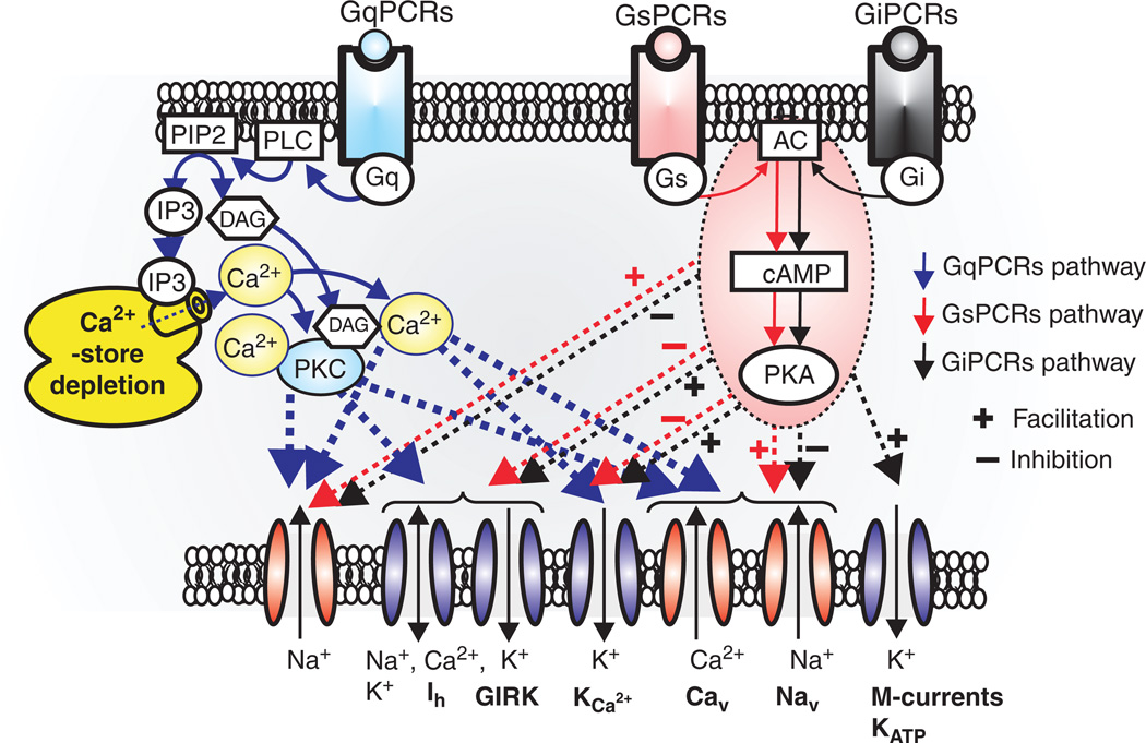
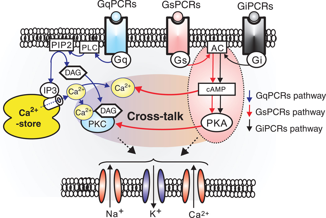
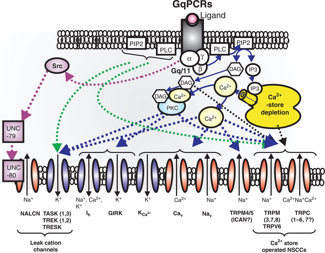
Respiratory brainstem neurons fulfill critical roles in controlling breathing: they generate the activity patterns for breathing and contribute to various sensory responses including changes in O2 and CO2. These complex sensorimotor tasks depend on the dynamic interplay between numerous cellular building blocks that consist of voltage-, calcium-, and ATP-dependent ionic conductances, various ionotropic and metabotropic synaptic mechanisms, as well as neuromodulators acting on G-protein coupled receptors and second messenger systems. As described in this review, the sensorimotor responses of the respiratory network emerge through the state-dependent integration of all these building blocks. There is no known respiratory function that involves only a small number of intrinsic, synaptic, or modulatory properties. Because of the complex integration of numerous intrinsic, synaptic, and modulatory mechanisms, the respiratory network is capable of continuously adapting to changes in the external and internal environment, which makes breathing one of the most integrated behaviors. Not surprisingly, inspiration is critical not only in the control of ventilation, but also in the context of "inspiring behaviors" such as arousal of the mind and even creativity. Far-reaching implications apply also to the underlying network mechanisms, as lessons learned from the respiratory network apply to network functions in general.


Figures

Figure 1
Figure 1
Anatomical and physiological characterization of the pre-Bötzinger complex (preBötC) in the ventrolateral medulla. (A) Anatomical maps of brainstem regions from rodent [left (436)] and human (right, modified, with permission, from reference (489) containing the preBötC. The location of the preBötC is anatomically characterized by the same transverse section as the nucleus ambiguus (Amb), inferior olive (IO), nucleus tractus solitarius (NTS), and hypoglossal nucleus (XII). (B) Isolating the preBötC in a single medullary brainstem slice from rodents preserves rhythmic neuronal activity implicated in the generation of inspiratory activity. Heat maps of activity show both an anatomical and physiological overlap of neuronal activities representing fictive eupnea (left), sighs (center), and gasps (right). Modified, with permission, from reference .
Figure 2
Figure 2
Simultaneous intracellular whole cell recordings and integrated extracellular population recordings from pre-Bötzinger (preBötC) respiratory neurons (A) in vitro from a mouse brain slice exhibiting “fictive” eupneic activity and (B) in vivo from an anesthetized rat, during “eupneic activity” (modified, wih permission, from reference 490). (C) Integrated population recordings in vitro of a fictive sigh recorded with a surface electrode from the preBötC in a mouse brain slice and (E) from a working heart-brainstem preparation (WHBP). (D) A sigh recorded in vivo from the phrenic nerve (PN) from an anesthetized cat (modified, with permission, from reference 76).
Figure 3
Figure 3
Putative inspiratory neurons of the preBötC integrate modulatory, synaptic, intrinsic, and intracellular mechanisms that give rise to bursting. Modulation commonly involves a cascade of mechanisms mediated through metabotropic glutamate receptors (mGLUR-I, II, III), noradrenergic receptors (α1, α2, and β-NE), serotonergic receptors (5HT-1A, 2, 4, and 7), peptidergic (NK1-R), and purinergic receptors (P2Y1) that represent G-protein-coupled protein receptors (Gs, Gi, and Gq) and act on various intracellular signal transductions. Synaptic mechanisms involve ionotropic glutamatergic (AMPA-R, NMDA-R), GABAergic (GABAA-R), and glycinergic receptors (GLY-R) that rapidly change membrane potential and hence, neuronal excitability when activated. Intrinsic mechanisms refer to other membrane conductances that are not strictly synaptic, but also regulate neuronal excitability. These include, but are not limited to: (1) leak conductances, (TREK, TWIK, TASK, and NALCN); (2) calcium conductances (T, P, and L-Ca2+); (3) K+ conductances (KATP, BK, SK); (4) sodium conductances (Nav, INaP); and nonspecific cation conductances, ICAN (TRPC-3, 7, and TRPM-4,5). Intracellular mechanisms refer to the molecules and ions regulating intracellular signaling cascades and ultimately lead to changes in excitability. For example, Ca2+ and ATP influence neuronal excitability through indirect mechanisms affecting the conductances generated through various channels such as the KATP, and ICAN. Blue outlines represent hyperpolarizing conductances, while red outlines represent depolarizing conductances.
Figure 4
Figure 4
Autonomously active neurons are part of the spectrum of intrinsic building blocks responsible for rhythmic population bursts in the preBötC. At the cellular level, a diversity of mechanisms exist to produce several forms of bursting that have different pharmacological properties. Some neurons express cadmium-insensitive bursting (CI) that is riluzole sensitive, and therefore, appears to be mediated by INaP (blue). Other neurons express cadmium-sensitive bursting (CS), predominantly mediated by both voltage-dependent Ca2+ currents (Cav) and a FFA-sensitive ICAN. Moreover, pharmacological blockade of 5HT2A receptors can turn CI-bursting into tonic spiking while activation of a1-noradrenergic receptors in tonic spiking neurons into CS-bursting. This type of conditional bursting demonstrates that the role of autonomously active neurons in the preBötC may not be fixed. Hence, while both forms of bursting, CI and CS, depend on dominant current(s) to drive spontaneous bursting, all putative inspiratory neurons of the preBötC appear to possess tonic boosting currents (grey), the INaP (blue), ICAN (yellow), and Cav (purple).
Figure 5
Figure 5
Synaptic modulation of bursting properties. (A) Pharmacological removal of synaptic inhibition can alter the propensity of a respiratory neuron to switch from a tonic spiking into a bursting mode. (B) In a simultaneous intracellular and population recording, a burst of action potentials can be triggered in the cell by a brief positive current injection (rd) or by synaptic input occurring during the population burst.
Figure 6
Figure 6
Generation of fictive sighs depends on the activation of P/Q-type calcium channels. Pharmacological blockade of the P/Q type channels with ω-agatoxin TK specifically abolishes sighs. (A) Sighs recorded under control conditions in population recordings from the preBötC (sighs indicated by red arrows) and (B) after bath application of ω-agatoxin TK, at low concentrations (modified, with permission, from reference 297). (C) Reduction of the amplitude of intracellularly recorded evoked EPSPs (by electrical stimulation of the contralateral preBötC) in respiratory neurons by pharmacological blockade of P/Q-type calcium channels. Individual responses of five neurons to ω-agatoxin TK [120 nmol/L] show a variable response, with a minimum of 8% reduction and a maximum of 90% reduction. (D) Individual responses to the N-type specific calcium channel blocker GVIA [0.5 µmol/L], showing a homogeneous 40% reduction of evoked EPSPs (modified, with permission, from reference 297). (E) Activation of the metabotropic glutamate receptors (mGluR8) leads to specific inhibition of “fictive sighs” recorded from the preBötC (modified, with permission, from reference 296). (F) Hypothesized mechanism of action for the inhibition of P/Q-type calcium channels by activated mGluR8 receptors, through a direct inhibitory interaction with the β-subunit of the heterotrimeric G-protein.
Figure 7
Figure 7
Pacemaker neurons are able to burst throughout a range of extracellular potassium concentrations with the contribution of the persistent sodium current. (A) An intracellular recording from an individual pacemaker neuron illustrating autonomous bursting throughout a range of extracellular potassium concentrations (3–8 mmol/L) without significant changes to membrane potential. (B) Traces expanded from A of the autonomously bursting pacemaker at 3 mmol/L (left) and 8 mmol/L (right) extracellular K+. (C) Autonomous bursting involves INaP as revealed by long-lasting hyperpolarizing current injections that cause the neuron to cease bursting, but as it intrinsically depolarizes bursting is resumed. Hence, pacemaker neurons do not require artificial elevation of extracellular potassium to autonomously burst (77, 564, 566).
Figure 8
Figure 8
Synaptic inhibition and its potential role in the generation of rhythmic network activity. (A) Schematic cartoon of a half center model with neurons connected by reciprocal inhibition. (B) Neurons connected to a dynamic clamp establish an artificial half center and can produce a variety of patterned outputs, as shown. Examples of a half-center model exhibiting (B1) alternating slow oscillations, (B2) antiphasic spiking, or (B3) high-frequency half-center oscillation. Output of the half-center model is highly dependent upon the synaptic and ionic conductance parameters defined by the dynamic clamp (modified, with permission, from reference 503).
Figure 9
Figure 9
Network and cellular responses of respiratory neurons to muscarinic receptor activation. (A) Bath application of the muscarinic agonist oxotremorine stimulates fictive sigh activity (red arrows), while inhibiting eupneic activity (blue arrows). (B) Bath application of oxotremorine induces the generation of two distinct burst patterns recorded from a CI pacemaker neuron, isolated from fast synaptic transmission (modified, with permission, from reference 563).
Figure 10
Figure 10
Network reconfiguration of the preBötC during hypoxia, at the cellular and network level. (A) Typical biphasic network activity response of the preBötC during hypoxia, assessed by integrated population recording from the preBötC. After an early augmentation phase, activity enters a late depression phase. During this response, activity changes from a “fictive eupneic” to a “fictive gasping” mode of activity. Note the generation of multiple “fictive sighs” during the augmentation phase. (B1) Plot of spontaneous excitatory and inhibitory postsynaptic currents (ESPCs and IPSCs) recorded from a preBötC respiratory neuron in response to hypoxia. Note the dramatic reduction of IPSCs during hypoxia. (B2) IPSCs recorded in vivo from a respiratory neuron of an anesthetized cat, before and during hypoxia (modified, with permission, from reference 486). (C) Average activity of three subsets of autonomous active neurons, in response to hypoxia. Tonically firing autonomous spiking and cadmium-sensitive (CS) pacemaker neurons hyperpolarize and cease firing during hypoxia (State I and State II). (D) By contrast, cadmium-insensitive (CI) pacemaker neurons continue to fire under hypoxia, even with persistent exposure to hypoxic conditions (State III) (modified, with permission, from references and 553).
Figure 11
Figure 11
Examples of activity pattern changes in respiratory neurons during hypoxia. (A1) Current-clamp recording from an inspiratory neuron showing an augmenting burst pattern under control normoxic conditions, switching to a sharply rising and decrementing burst under hypoxia. (A2) This change in network-initiated burst patterns coincides with a decrease in spontaneous inhibitory postsynaptic currents (IPSCs), measured in voltage clamp. (A3) Example of an expiratory neuron that receives inhibitory synaptic input under normoxic conditions, switching to excitatory synaptic input under hypoxia. (B) Examples of autonomous activity in three subsets of respiratory neurons isolated from fast synaptic transmission, before and during hypoxia. Tonic firing neurons and CS-pacemaker neurons become silent, while CI-pacemaker neurons continue to generate bursting activity under hypoxia.
Figure 12
Figure 12
The diverse interaction and relative contribution of multiple mechanisms (see Figure 3) in a single neuron gives rise to a neuronal population that possesses heterogeneous properties used to generate bursting and ultimately contribute to the eupneic rhythm. This integrated respiration during eupnea provides both stability and dynamic responsiveness. Hypoxia reconfigures the network to a state dominated by the INaP, KATP, and the AMPA receptor (AMPA-R) and various forms of neuromodulation. At the cellular level, INaP appears to be the basis for bursting involved with the gasping rhythm (i.e., INaP-driven gasping).
Figure 13
Figure 13
Downstream pathways and effector channels for Gq/11, Gs, and Gi protein coupled receptors. Gs protein coupled receptors (GsPCRs) and Gq/11 protein coupled receptors (GqPCRs) are in general excitatory systems, and Gi protein coupled receptors (GiPCRs) are inhibitory systems. Both GsPCRs and GiPCRs regulate AC, cAMP, and PKA, and GqPCRs facilitate Ca2+ release from Ca2+ store and protein kinase C (PKC). These second messenger proteins up- and downregulate open probability of several voltage-dependent cation channels. PIP2 (phosphatidylinositol 4,5-biphosphate), PLC (phospholipase C), IP3 (inositol-1,4,5-trisphosphate), DAG (diacylglycerol), PKC, AC (adenylyl cyclase), cAMP (cyclic AMP), PKA (protein kinase A), ICAN (calcium-activated nonspecific cation current), Ih (hyperpolarization activated non-selective cation channels), GIRK (G protein coupled inward rectifier K+ channels), KCa2+ (Ca2+-activated K+ channels), Cav (voltage-dependent Ca2+ channels), Nav (voltage-dependent Na+ channels), M-current (muscarinic receptor activated K+ channels), and KATP (ATP-sensitive K+ channels).
Figure 14
Figure 14
Cross-talk between Gq/11 and Gs system. It has been thought that each of the GqPCRs and Gs(i)PCRs systems are separated. However, recent studies suggest GsPCRs-related cAMP production or activation of PKA stimulates elevation of intracellular Ca2+ and activation of PKC (17, 151, 272, 457, 608).
Figure 15
Figure 15
GqPCRs regulates not only voltage-dependent cation channels, but also transient receptor potential (TRP) and leak cation channels. GqPCRs-induced depletion of Ca2+-store facilitates activation of Ca2+-store operated NSCCs, such as TRPC (–6, 7), TRPM (3, 7, 8) and TRPV6. On the other hand, TRPM4/5 may be directly activated by elevation of internal Ca2+ concentration (300, 576). GqPCRs seem to modulate activity of K2P (TASK, TREK, and TRESK) and sodium-leak-channel-nonspecific (NALCN) channels. In particular, GqPCRs act through Src protein controls NALCN channels through both UNC-79 and UNC-80 (316, 317, 491, 540). Abbreviations; P2K (“two-pore” potassium channels, TASK, TREK, and TRESK channels), TRPM (transient receptor potential melastatin), TRPV(transient receptor potential vanilloid), TRPC (transient receptor potential canonical), Ca2+ store operated NSCCs (nonselective cation currents), and Src (sarcoma) is a proto-oncogenic and nonreceptor tyrosine kinase.

References

    1. Aarts M, Iihara K, Wei WL, Xiong ZG, Arundine M, Cerwinski W, MacDonald JF, Tymianski M. Akey role for TRPM7 channels in anoxic neuronal death. Cell. 2003;115:863–877. - PubMed
    1. Abbott SB, Stornetta RL, Fortuna MG, Depuy SD, West GH, Harris TE, Guyenet PG. Photostimulation of retrotrapezoid nucleus phox2b-expressing neurons in vivo produces long-lasting activation of breathing in rats. J Neurosci. 2009;29:5806–5819. - PMC - PubMed
    1. Abdala AP, Rybak IA, Smith JC, Zoccal DB, Machado BH, St-John WM, Paton JF. Multiple pontomedullary mechanisms of respiratory rhythmogenesis. Respir Physiol Neurobiol. 2009;168:19–25. - PMC - PubMed
    1. Aguilar-Bryan L, Nichols CG, Wechsler SW, Clement JP, th, Boyd AE, 3rd, Gonzalez G, Herrera-Sosa H, Nguy K, Bryan J, Nelson DA. Cloning of the beta cell high-affinity sulfonylurea receptor: A regulator of insulin secretion. Science. 1995;268:423–426. - PubMed
    1. Ahmmed GU, Mehta D, Vogel S, Holinstat M, Paria BC, Tiruppathi C, Malik AB. Protein kinase Calpha phosphorylates the TRPC1 channel and regulates store-operated Ca2+ entry in endothelial cells. J Biol Chem. 2004;279:20941–20949. - PubMed

Publication types