Review
. 2012 Oct;2(4):2733-52.
doi: 10.1002/cphy.c120002.

Nonclassical renin-angiotensin system and renal function

Affiliations
Review

Nonclassical renin-angiotensin system and renal function

Mark C Chappell. Compr Physiol. 2012 Oct.

Abstract

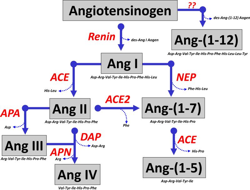
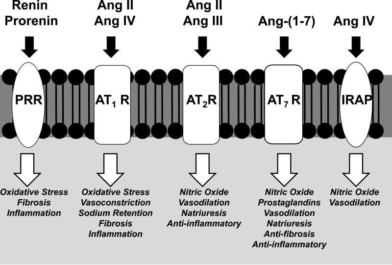
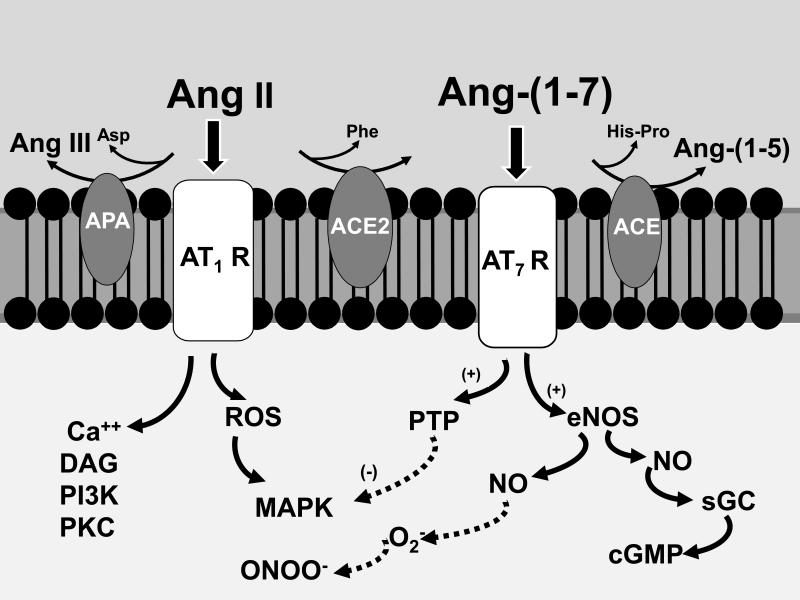
The renin-angiotensin system (RAS) constitutes one of the most important hormonal systems in the physiological regulation of blood pressure through renal and nonrenal mechanisms. Indeed, dysregulation of the RAS is considered a major factor in the development of cardiovascular pathologies, including kidney injury, and blockade of this system by the inhibition of angiotensin converting enzyme (ACE) or blockade of the angiotensin type 1 receptor (AT1R) by selective antagonists constitutes an effective therapeutic regimen. It is now apparent with the identification of multiple components of the RAS within the kidney and other tissues that the system is actually composed of different angiotensin peptides with diverse biological actions mediated by distinct receptor subtypes. The classic RAS can be defined as the ACE-Ang II-AT1R axis that promotes vasoconstriction, water intake, sodium retention, and other mechanisms to maintain blood pressure, as well as increase oxidative stress, fibrosis, cellular growth, and inflammation in pathological conditions. In contrast, the nonclassical RAS composed primarily of the AngII/Ang III-AT2R pathway and the ACE2-Ang-(1-7)-AT7R axis generally opposes the actions of a stimulated Ang II-AT1R axis through an increase in nitric oxide and prostaglandins and mediates vasodilation, natriuresis, diuresis, and reduced oxidative stress. Moreover, increasing evidence suggests that these non-classical RAS components contribute to the therapeutic blockade of the classical system to reduce blood pressure and attenuate various indices of renal injury, as well as contribute to normal renal function.


Figures

Figure 1
Figure 1. Enzymatic cascade of angiotensin peptide formation and metabolism
Renin cleaves the precursor protein angiotensinogen (Aogen) at the Leu10-Leu11 bond to angiotensin-(1-10) (Ang I) which is further processed to the biologically active peptides Ang-(1-8) (Ang II) by angiotensin converting enzyme (ACE) and Ang-(1-7) by endopeptidases such as neprilysin (NEP). Ang II undergoes further processing at the carboxy terminus by the carboxypeptidase ACE2 to yield Ang-(1-7) and at the amino terminus by aminopeptidase A (APA) to form Ang-(2-8) or Ang III. Ang-(1-7) is metabolized by ACE to form Ang-(1-5) and Ang III is further hydrolyzed by aminopeptidase N (APN) to yield Ang-(3-8) or Ang IV. Ang II can be directly cleaved by dipeptidyl aminopeptidase IV (DAP) to Ang IV. The novel peptide Ang-(1-12) is derived from the hydrolysis of the Tyr12-Tyr12 bond of Aogen although the identity of the enzyme that forms the peptides is not known to date. Adapted from Chappell (27).
Figure 2
Figure 2. Renal actions of the classical and non-classical components of the reninangiotensin system
Ang II interacts with the AT1 receptor (AT1R) to increase renal vasoconstriction, sodium reabsorption and promote inflammation and fibrosis. Ang IV may stimulate vasoconstriction through an interaction with the AT1R. Renin or prorenin binds to the prorenin receptor (PRR) to directly promote oxidative stress, fibrosis and inflammation. Ang II or Ang III stimulates vasodilation, reduced vascular resistance and natriuresis through activation of the AT2R and the generation of nitric oxide. Ang-(1-7) recognizes the AT7R to stimulate vasodilation, diuresis and natriuresis, but reduce inflammation and fibrosis through increased generation of nitric oxide and prostaglandins. Ang IV may interact with the insulin regulated aminopeptidase (IRAP) to reduce vascular resistance through an increase in nitric oxide.
Figure 3
Figure 3. Enzymatic metabolism of 125I-Ang II in isolated sheep proximal tubules
125I-Ang II (AII) was incubated with 50 Qg of proximal tubules membranes for 30 minutes at 37°C and the metabolites separated by HPLC. Panel A: Quantification of the peptidase activities for 125I-AII metabolism from the sheep proximal tubule membranes expressed as the rate of metabolism products formed (fmol/mg/min). Conditions: Control (no inhibitors); +AP,CYS,CHM-I (inhibitors for aminopeptidase, chymase, cysteine proteases); +NEP-I (addition of neprilysin inhibitor); +ACE-I (addition of ACE inhibitor); +ACE2-I (addition of ACE2 inhibitor). Data are means; n=4. Panel B: Influence of ACE2 inhibition on half-life (t1/2) of 125I-Ang II (AII) in proximal tubules. Conditions: Control (no inhibitors); +MLN (only the ACE2 inhibitor). Data are means; n=5; *P < 0.05 vs Control. Panel C: Metabolism pathway for Ang II and Ang-(1-7) in sheep proximal tubules. Adapted from Shaltout et al (129).
Figure 4
Figure 4. Deletion of tissue ACE significantly reduces levels of Ang II but not Ang-(1-7) in mouse kidney
HPLC/RIA analysis of pooled mouse kidney samples from Wildtype (upper panel) and tissue ACE knockout (tisACE−/−) mice (lower panel). The HPLC fractions were measured with Ang-(1-7) (fractions 1-20) and Ang II (fractions 21-40) RIAs, respectively. The arrows indicate the elution peak times for Ang-(1-7), Ang-(4-8) and Ang II. Inset: intrarenal concentration of Ang II and Ang-(1-7) expressed as fmol/mg protein in Wildtype and tisACE−/− mice, n = 8 per group; *P < 0.001 vs Wildtype. Adapted from Modrall et al. (88).
Figure 5
Figure 5. Autoradiography of Ang II binding sites in the fetal and adult sheep kidney
Frozen-thawed kidney sections were incubated with receptor antagonists to the AT2 receptor (PD123319) or the AT1 receptor (losartan) in the presence of the non-selective antagonist 125I Sarthran (0.2 nM). Nonspecific labeling was obtained by pre-incubation with the unlabeled Sarthran antagonist (5 VM). Adapted from Gwathmey et al (57).
Figure 6
Figure 6. Sex differences in systolic blood pressure, proteinuria and components of the renin-angiotensin system in the renal cortex of mRen2.Lewis congenic rats
Systolic blood pressure is expressed in mm Hg and proteinuria as mg per kilogram body weight per day (mg/kg/day). Intrarenal concentrations of Ang II and Ang-(1-7) are expressed as fmol peptide per mg protein (fmol/mg) and enzyme activities as fmol product per mg protein per min (fmol/mg/min) in 15 week old hemizygous mRen2.Lewis, n = 5-8 per group; **P < 0.01 or *P < 0.01. Adapted from Pendergrass et al. (105).
Figure 7
Figure 7. Immunocytochemical distribution of the Mas receptor in the adult sheep kidney and natriuretic influence of Ang-(1-7).
Upper panel: Signal for Mas receptor in proximal tubules (PT) and distal tubules but not glomerulus in renal cortex (A); positive staining of collecting ducts in cortex (B); Mas staining of thick ascending limb of Henle (TAL) and vasa recta (VR) in renal medulla; antigenic peptide for primary antibody abolishes Mas staining in adjacent tissue sections (D-F). Binding of the primary antibody against the Mas receptor protein was followed by the secondary antibody conjugated to Alexa Fluor 488 (green fluorescence) and the nuclear marker stain DAPI (blue). Adapted from Gwathmey et al. (59). Lower panel: Ang-(1-7) infusion increases sodium excretion (% of an acute sodium load) in control sheep as compared to saline infusion (Vehicle). The natriuretic response to Ang-(1-7) was absent in sheep prenatally exposed to the glucocorticoid betamethasone (Beta). Data are means, n=11-12 sheep. Adapted from Tang et al (142).
Figure 8
Figure 8. Ang-(1-7) increases nitric oxide and attenuates Ang II-dependent increase in reactive oxygen species in isolated nuclei from renal cortex.
Panel A: Ang-(1-7) exhibits greater potency than Ang II at the AT2R to stimulate nitric oxide (NO) as detected by diaminofluorescein [DAF; *P<0.05 vs. Ang-(1-7)]. Panel B: The AT1R antagonist losartan (LOS) blocks the Ang II stimulation of ROS; the AT7R antagonist D-Ala7-Ang-(1-7) (DALA) and ACE2 inhibitor MLN4760 (MLN) exacerbate the Ang II response (δ P<0.05 vs. Ang II); the AT2R antagonist PD had no effect. Panel C: HPLC chromatograph of conversion of Ang II to Ang-(1-7) [Ang7] in isolated nuclei from sheep proximal tubules and inhibition by the ACE2 inhibitor MLN. Adapted from Gwathmey et al (56).
Figure 9
Figure 9. Scheme for the attenuation of the Ang II-AT1 receptor signaling by Ang-(1-7)
Ang II stimulates various signaling pathways including reactive oxygen species (ROS) that culminates in the activation of intracellular kinases (MAPK). Attenuation of Ang II signaling within the kidney occurs through amino and carboxy terminal metabolism to Ang III and Ang-(1-7) by aminopeptidase A (APN) and ACE2, respectively. Formation of Ang-(1-7) will stimulate the generation of nitric oxide (NO) and cGMP that may antagonize the actions of Ang II, as well as complex superoxide (O2) to form peroxynitrite (ONOO). In addition, Ang-(1-7) may activate intracellular phosphatases (PTP) to attenuate the Ang II-induced phosphorylation of kinases. ACE may abrogate Ang-(1-7) signaling by enzymatic conversion to Ang-(1-5) which likely does not interact with the AT7/Mas receptor. Although not depicted, generation of Ang III from Ang II may contribute to increased formation of NO by stimulation of the AT2 receptor pathway. Additional abbreviations: calcium (Ca++); diacylglycerol (DAG); phosphoinositol 3 kinase (PI3 kinase); protein kinase C (PKC); endothelial NO synthase (eNOS); soluble guanylate cyclase (sGC). Adapted from Chappell (27).

Similar articles

Cited by

References

    1. Abadir PM, Carey RM, Siragy HM. Angiotensin AT2 receptors directly stimulate renal nitric oxide in bradykinin B2-receptor null mice. Hypertension. 2003;42:600–604. - PubMed
    1. Abadir PM, Foster DB, Crow M, Cooke CA, Rucker JJ, Jain A, Smith BJ, Burks TN, Cohn RD, Fedarko NS, Carey RM, O'Rourke B, Walston JD. Identification and characterization of a functional mitochondrial angiotensin system. Proc Natl Acad Sci U S A. 2011 - PMC - PubMed
    1. Ahmad A, Ward PE. Role of aminopeptidase activity in the regulation of the pressor activity of circulating angiotensins. J Pharmacol Exp Ther. 1990;252:643–650. - PubMed
    1. Ahmad S, Simmons T, Varagic J, Moniwa N, Chappell MC, Ferrario CM. Chymase-dependent generation of angiotensin II from angiotensin-(1-12) in human atrial tissue. PLoS One. 2011;6:e28501. - PMC - PubMed
    1. Akhtar S, Yousif MH, Dhaunsi GS, Chandrasekhar B, Al-Farsi O, Benter IF. Angiotensin-(1-7) inhibits epidermal growth factor receptor transactivation via a Mas receptor-dependent pathway. Br J Pharmacol 2011. Epub Aug. 11 2011. - PMC - PubMed

Publication types

MeSH terms

LinkOut - more resources