Review
. 2013 Jan;3(1):243-87.
doi: 10.1002/cphy.c120009.

Hepatocyte polarity

Affiliations
Review

Hepatocyte polarity

Aleksandr Treyer et al. Compr Physiol. 2013 Jan.

Abstract

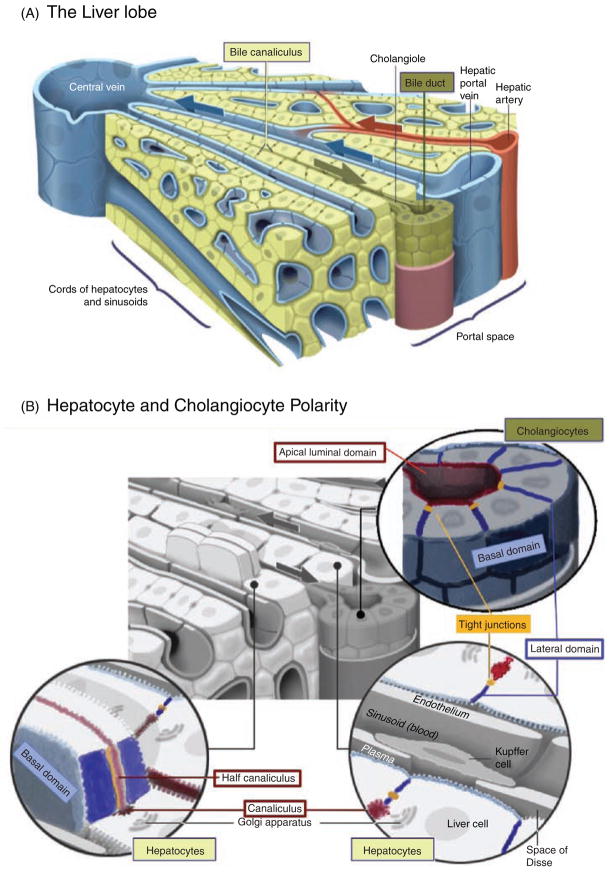
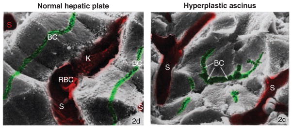
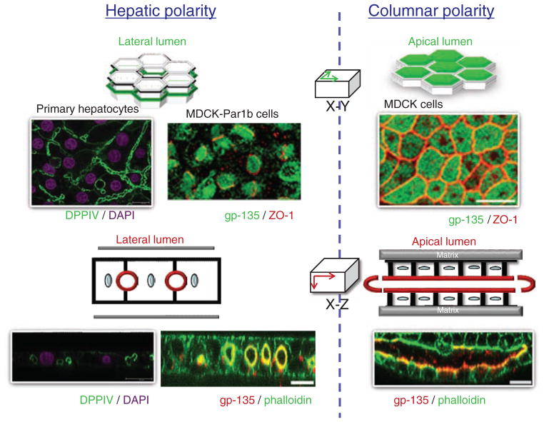
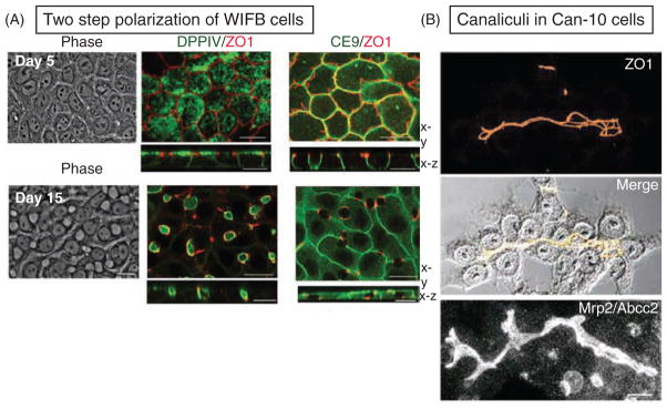
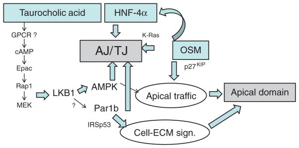
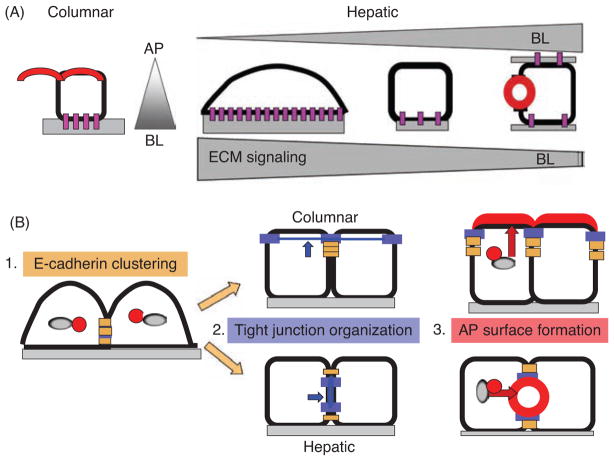
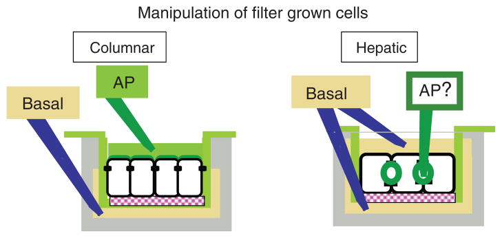
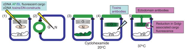
Hepatocytes, like other epithelia, are situated at the interface between the organism's exterior and the underlying internal milieu and organize the vectorial exchange of macromolecules between these two spaces. To mediate this function, epithelial cells, including hepatocytes, are polarized with distinct luminal domains that are separated by tight junctions from lateral domains engaged in cell-cell adhesion and from basal domains that interact with the underlying extracellular matrix. Despite these universal principles, hepatocytes distinguish themselves from other nonstriated epithelia by their multipolar organization. Each hepatocyte participates in multiple, narrow lumina, the bile canaliculi, and has multiple basal surfaces that face the endothelial lining. Hepatocytes also differ in the mechanism of luminal protein trafficking from other epithelia studied. They lack polarized protein secretion to the luminal domain and target single-spanning and glycosylphosphatidylinositol-anchored bile canalicular membrane proteins via transcytosis from the basolateral domain. We compare this unique hepatic polarity phenotype with that of the more common columnar epithelial organization and review our current knowledge of the signaling mechanisms and the organization of polarized protein trafficking that govern the establishment and maintenance of hepatic polarity. The serine/threonine kinase LKB1, which is activated by the bile acid taurocholate and, in turn, activates adenosine monophosphate kinase-related kinases including AMPK1/2 and Par1 paralogues has emerged as a key determinant of hepatic polarity. We propose that the absence of a hepatocyte basal lamina and differences in cell-cell adhesion signaling that determine the positioning of tight junctions are two crucial determinants for the distinct hepatic and columnar polarity phenotypes.


Figures

Figure 1
Figure 1. The two epithelial cell types in the liver
(A) The mammalian biliary tree is characterized by a network of bile canaliculi, the luminal domains of adjacent hepatocytes, which are organized in one or two-cell-thick cords. The bile canaliculi connect to the bile ducts located in the portal triad that also encompasses the hepatic artery and portal vein. Ducts are composed of biliary epithelial cells that exhibit columnar polarity. (B) The domain organization of hepatocytes and cholangiocytes. Red: luminal domains, dark blue: lateral domains engaged in cell-cell adhesion, gray: basal domain in contact with a basal lamina (cholagiocytes) or facing the space of Disse (hepatocytes). Adapted, with permission, from Color Textbook of Histology by Leslie Gartner and James Hiatt, 2nd Edition, Chapter 18: Digestive System III. Glands (140), Copyright Elsevier (2001).
Figure 2
Figure 2. The organization of hepatocytes in the injured liver
Scanning electron micrographs of left: normal hepatic plate. Note the continuous, linear and lateral canaliculi (BC); Sinusoids (S) are separated by single-cell thick plates of hexagonal hepatocytes. Right: hyperplastic acinus. Notice a prominent central lumen (BC with arrows) shared by several neighboring hepatocytes; other canaliculi are short and discontinuous; sinusoids (S) are separated by multiple cells. K, Kupffer cell; RBC, red blood cell; x1530. Adapted by permission from Macmillan Publishers Ltd. on behalf of Cancer Research UK: [Br J Cancer] (Ogawa et al., Vol. 40: 782–90) (343), copyright (1979).
Figure 3
Figure 3. The hepatic and columnar polarity phenotypes
columnar epithelia, for example, kidney-derived Mardin Darby Canine kidney (MDCK) cells establish their luminal domains at the apex. When cultured in three-dimensional (3D) collagen matrices, they organize a luminal domain (labeled by the apical marker gp135) between two layers of cells. Phalloidin, an actin filament label, outlines all cell surfaces. Hepatocyte luminal domains are grooves interrupting the lateral surfaces of neighboring cells. When cultured in collagen sandwiches, primary rat hepatocytes remain monolayered and form an elaborate network of bile canaliculi labeled by the luminal protein DPPIV. MDCK cells overexpressing the kinase Par1b organize with hepatic polarity, they form their gp135-positive luminal surface between neighboring cells and remain monolayered in 3D collagen matrices. Unlike in hepatocytes, however, MDCK-Par1b lumina do not align to form interconnected bile canaliculi but remain cyst-like extracellular spheres between two cells. MDCK, MDCK-Par1b images are, with permission, from Cohen D et al., originally published in J Cell Biol. 164(5):717–27 (81).
Figure 4
Figure 4. Examples of hepatic cell culture models
(A) In two-dimensional (2D) cultures, WIFB cells initially acquire columnar polarity (day 5 after plating) with apical DPPIV and with a “chickenwire” tight junction belt (ZO1). The lateral protein CE9 lines the sites of cell-cell contacts. Cells subsequently repolarize with hepatic lumen organization over a time course of 10 days. The spherical luminal domains between neighboring cells appear as translucent holes in phase images and are surrounded by the tight junction marker ZO1. Note also that CE9 remains present at the cell-contacting surfaces but is absent where they are interrupted by the lumina. Adapted, with permission, from Cohen D et al. 2004; originally published in J Cell Biol. 164(5): 717–27 (81). (B) In 2D cultures of Can10, the luminal domains of adjacent cells align to form elaborate canalicular networks that label for the bile acid transporter MRP2 and the tight junction marker ZO1. Adapted, with permission, from Peng X et al. Cell Tissue Res 2006; 323:233–243 (357); reproduced, with permission, from Springer Verlag.
Figure 5
Figure 5. Signaling Pathways that determine hepatocyte polarization
Bile acids, the transcription factor HNF-4α and the cytokine Oncostatin M (OSM) have all been linked to two main aspects of hepatocyte polarization, the formation of tight (TJ) and adherens (AJ) junctions and the generation of an apical domain. The kinase LKB1 and its downstream target adenosine monophosphate kinase (AMPK) have evolved as key effecters of taurocholic acid signaling to promote both polarity features. LKB1 can also act as activating kinase for Par1 paralogues but has not yet been shown to do so in hepatocytes. Par1b regulates cell-cell adhesion and promotes a hepatic polarity phenotype in MDCK cells, in part by inhibiting the rho-GTPase adaptor protein IRSp53 in its role in cell-matrix signaling. For details, see text.
Figure 6
Figure 6. Models for the role of cell adhesion and E-cadherin-mediated junction formation in epithelial polarization
(A) In cultured columnar epithelial cells, the basal lamina provides a polarity cue to establish the apical (AP) domain on the free surface. In cultured hepatic cells, strong asymmetric integrin signaling on collagen or matrigel causes a nonpolarized, spread phenotype; on adhesive nonextracellular matrix (ECM) substrates and even more so in spheroid culture without any substrate contact, hepatic cells acquire a cuboidal cell shape and might develop luminal domains; best lumen polarity is achieved in ECM sandwich cultures. (B) In the absence of E-cadherin-mediated adhesion, cultured nonpolarized columnar and hepatic cells maintain luminal proteins in an intracellular organelle. In polarizing cells, adhesion (brown) and tight junction (blue) proteins are not yet separated (1). Strong E-cadherin clustering, induced by myosin II, promotes tight junction maturation with a chickenwire phenotype, parallel to the basal domain (columnar). Weak E-cadherin clustering results in a tight junction belt parallel to the cell-cell contacting surface (hepatic) (2). Apical surface formation follows the established tight junction pattern (3). AP, apical; BL, basolateral.
Figure 7
Figure 7. Experimental access to the apical and basolateral domain and secretory compartments in columnar and hepatic cells
Columnar: when grown on polycarbonate filter inserts that are suspended in clusterwell culture dishes, the tight junctions of Mardin Darby Canine kidney cells provide a diffusion barrier for small molecules that generate separate apical and basal chambers. These culture conditions allow for the quantitative study of apical (AP) and basolateral membrane and secretory proteins. Hepatic: filter cultures allow experimental access to the entire basolateral but not apical domain, but the luminal/apical domain is sequestered from experimental access.
Figure 8
Figure 8. Studying protein targeting by live cell imaging
Microinjection of cDNAs encoding interfering cDNAs and proteins into individual cells to study their effect on cargo protein transport has proven an extremely effective strategy for the discovery of the apical and basolateral protein targeting machinery in Mardin Darby Canine kidney cells. To set a pulse of newly synthesized apical and/or basolateral proteins, cDNAs encoding fluorescent cargo proteins are comicroinjected with the interfering cDNAs into the nucleus [(1) for columnar or (2) for hepatic cells]. Upon protein expression from the cDNAs 1 to 3 h postmicroinjection (3), the cargo protein (an apical marker is shown here in green) is chased to the trans-Golgi network (TGN) in the presence of cyclohexamide to prevent further protein synthesis (4). At this point, toxins or function-blocking antibodies can be injected into the cytosol of cells. Protein exit from the TGN and arrival at the cell surface are determined after release of the TGN block (4). Cargo exit from the TGN is measured as decrease of Golgi-associated cargo (5). A fluorescent Golgi marker serves to define the Golgi area. Arrival at the cell surface is assessed with antibodies to the extracellular domain of the apical protein, added to unpermeabilized cells (5).
Figure 9
Figure 9. Example of a domain-selective targeting experiment in filter-grown columnar Mardin Darby Canine kidney cells
(A) Monovalent antibodies (Fab) to the extracellular domain of a membrane protein are bound to its basolateral population at 4°C (1). The fate of the labeled membrane protein after a chase period at 37°C (2) is determined by the addition of anti-Fab antibodies to either the apical (3) or basolateral (4) domain. (B) In hepatic cells, a similar quantitative assessment cannot be performed. Basolateral-to-apical transcytosis has only been documented in a qualitative manner by the appearance of basolaterally added antibodies in the luminal domain.
Figure 10
Figure 10. Targeting pathways from the trans-Golgi network (TGN) to the apical/canalicular and basolateral surfaces in Mardin Darby Canine kidney (MDCK) and hepatic cells
(A) Known pathways in MDCK cells. Apical (AP) pathways (green arrows): Depending on their presence in detergent-insoluble microdomains, raft-dependent and independent apical routes have been distinguished. Membrane and secretory proteins have been shown to traverse the apical recycling endosome (ARE) (in a MyoVb and Rab11-dependent manner) or the apical early endosome (AEE) before reaching the apical domain. Basolateral (BL) pathways (blue arrows): basolateral protein exit from the TGN is clathrin and AP-1A mediated. Multiple pathways have been distinguished: some basolateral proteins reach the basolateral domain without intermediate, others traversing either the basolateral early endosome (BEE) or the common recycling endosome (CRE) (in an AP-1B-dependent manner) prior to arriving at the basolateral domain. (B) Known and hypothetical pathways in hepatic cells. Two classes of apical proteins have been distinguished: those traveling from the TGN to the apical domain directly and those that first reach the basolateral domain. It is likely that, as in MDCK cells additional distinctions apply. Thus, some proteins might pass the ARE en route to the apical domain and there is evidence for raft and nonraft association in the TGN of apical proteins that take the transcytotic route. It is not resolved whether (A) apical and basolateral proteins are sorted into distinct transport carriers in the TGN and if so, whether basolateral proteins follow the same pathways as in MDCK cells, or (B) whether apical and basolateral proteins travel in common carriers to the basolateral domain.
Figure 11
Figure 11. Proposed targeting pathways from the apical proteins (AP) and basolateral (BL) plasma membrane in hepatocytes
Model 1: upon endocytosis from the canalicular domain APs are sorted in the apical early endosome (AEE) into either the late endosomal/lysosomal pathway or for recycling to the apical recycling endosome (ARE). Apical proteins that have reached the basolateral surface from the trans-Golgi network are internalized via clathrin-coated vesicles (nonraft) or in a clathrin-independent, flotillin-dependent manner (raft), and reach the (subapical endosome) SAC via basolateral early endosomes (BEE). From the SAC they are targeted to the apical surface via the ARE (green arrows). BL resident proteins (blue arrows) undergo fast recycling from the BEE directly or via the SAC where they are sorted from apical proteins that reach the SAC in the transcytotic pathway. As the AEE, the BEE also sorts proteins for degradation to the lysosome (LYS). Pathways similar to model 1 have been described in MDCK cells. Model 2: differs from model 1 in the definition of the SAC and in the itinerary of apical proteins undergoing basolateral-to-apical transcytosis. Apical proteins reach the bile canalicular membrane via the SAC after being sorted from basolateral proteins in the CRE. The SAC is also involved in the targeting of some lysosomal proteins such as endolyn-78. It is distinct from the ARE, which handles apical protein recycling.

Similar articles

Cited by

References

    1. Agnello V, Abel G, Elfahal M, Knight GB, Zhang QX. Hepatitis C virus and other flaviviridae viruses enter cells via low density lipoprotein receptor. Proc Natl Acad Sci U S A. 1999;96:12766–12771. - PMC - PubMed
    1. Ait-Slimane T, Galmes R, Trugnan G, Maurice M. Basolateral internalization of GPI-anchored proteins occurs via a clathrin-independent flotillin-dependent pathway in polarized hepatic cells. Mol Biol Cell. 2009;20:3792–3800. - PMC - PubMed
    1. Ala A, Walker AP, Ashkan K, Dooley JS, Schilsky ML. Wilson’s disease. Lancet. 2007;369:397–408. - PubMed
    1. Alessi DR, Sakamoto K, Bayascas JR. LKB1-dependent signaling pathways. Annu Rev Biochem. 2006;75:137–163. - PubMed
    1. Almeida CG, Yamada A, Tenza D, Louvard D, Raposo G, Coudrier E. Myosin 1b promotes the formation of post-Golgi carriers by regulating actin assembly and membrane remodelling at the trans-Golgi network. Nat Cell Biol. 2011;13:779–789. - PubMed

Publication types

LinkOut - more resources