Review
. 2013 Jan;3(1):403-28.
doi: 10.1002/cphy.c110063.

The influence of exercise on cognitive abilities

Affiliations
Review

The influence of exercise on cognitive abilities

Fernando Gomez-Pinilla et al. Compr Physiol. 2013 Jan.

Abstract

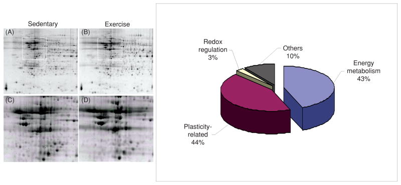
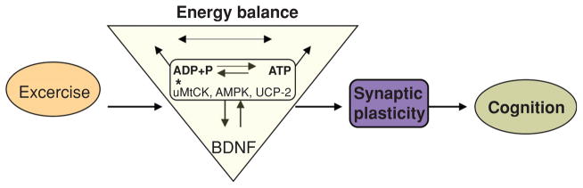
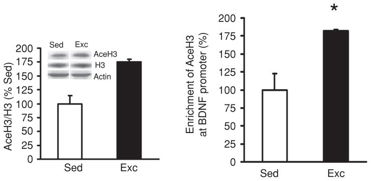
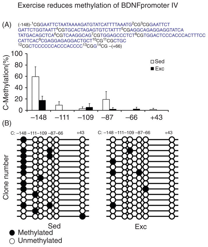
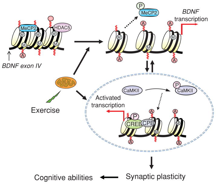
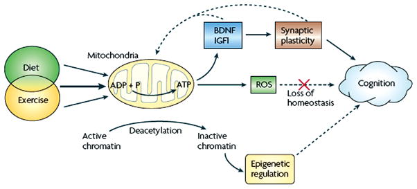
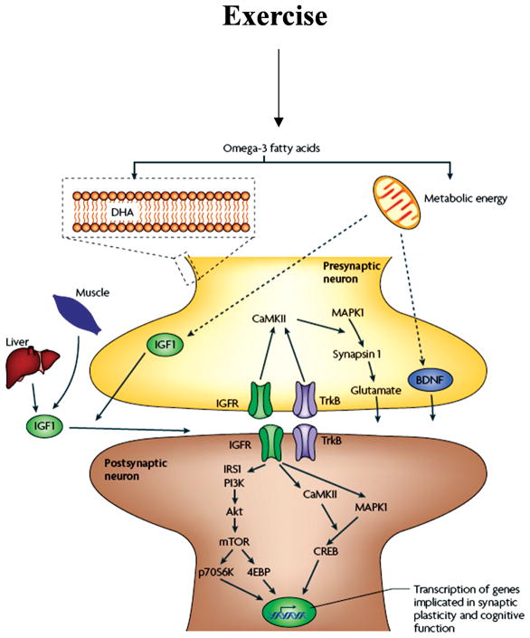
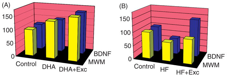
Scientific evidence based on neuroimaging approaches over the last decade has demonstrated the efficacy of physical activity improving cognitive health across the human lifespan. Aerobic fitness spares age-related loss of brain tissue during aging, and enhances functional aspects of higher order regions involved in the control of cognition. More active or higher fit individuals are capable of allocating greater attentional resources toward the environment and are able to process information more quickly. These data are suggestive that aerobic fitness enhances cognitive strategies enabling to respond effectively to an imposed challenge with a better yield in task performance. In turn, animal studies have shown that exercise has a benevolent action on health and plasticity of the nervous system. New evidence indicates that exercise exerts its effects on cognition by affecting molecular events related to the management of energy metabolism and synaptic plasticity. An important instigator in the molecular machinery stimulated by exercise is brain-derived neurotrophic factor, which acts at the interface of metabolism and plasticity. Recent studies show that exercise collaborates with other aspects of lifestyle to influence the molecular substrates of cognition. In particular, select dietary factors share similar mechanisms with exercise, and in some cases they can complement the action of exercise. Therefore, exercise and dietary management appear as a noninvasive and effective strategy to counteract neurological and cognitive disorders.


Figures

Figure 1
Figure 1
Grand averaged response-locked waveforms for subjects exposed to cardiorespiratory fitness (left side) and acute exercise (right side) showing error and correct trials at the FCz and Pz electrode sites. Reprinted, with permission, from reference (172), p. 763.
Figure 2
Figure 2
Characterization of a stimulus-locked event-related potential denoting the N1, P2, N2, and P3 components.
Figure 3
Figure 3
P3 latencies for young and older men undergoing low and high aerobic fitness. While P3 latency occurred significantly later for the older as compared to young men, the age effect was mostly due to very long P3 latencies of the older low fit men. Each mean was based on data for 15 subjects. The error bars are standard errors of the mean. Reprinted, with permission, from reference (55), p. 198.
Figure 4
Figure 4
P3 latency by group across both conditions of the Eriksen flankers task. Reprinted, with permission, from reference (86), p. 180.
Figure 5
Figure 5
Topographical amplitude maps for the P3a and P3b components for each age and fitness from each stimulus. Note different voltage scales for each task. Reprinted, with permission, from reference (143), p. 384.
Figure 6
Figure 6
Distribution of P3 amplitude across both conditions of the Eriksen flankers at midline sites by group. Reprinted, with permission, from reference (86), p. 181.
Figure 7
Figure 7
Role of brain-derived neurotrophic factor (BDNF) on the action of exercise on learning and memory assessed in the Morris Water Maze task. (A) Exercise improved learning ability as depicted by the enhanced aptitude of exercised animals to locate the platform in a significantly shorter time (shorter escape latencies in the exc/cytC group). Blocking BDNF action during exercise resulted in escape latency comparable to sedentary control animal (exc/TrkB-IgG vs. sed/cytC). The BDNF receptor blocker TrkB-IgG was injected into the hippocampus and cytochrome C (cytC) was used as a vehicle control. Data are expressed as mean ± SEM (ANOVA; Fischer test; Scheffe Fischer test; *, P < 0.05; **,‡‡, P < 0.01; * represents comparison between groups, ‡‡ represents comparison within groups). (B) Exercise increased the memory retention as indicated by significantly more time in quadrant P than sedentary controls (exc/cytC vs. sed/cytC). Blocking BDNF action during exercise abolished this exercise-induced preference for the P quadrant (exc/TrkB-IgG vs. exc/cytC), to sedentary control levels (exc/TrkB-IgG vs. sed/cytC). Representative samples of trials traveled during the probe test (B, begin, E, end, P, quadrant which previously housed the platform). Each value represents the mean ± SEM (ANOVA; Fischer test; *, P < 0.05). Reprinted, with permission, from reference (190), pp. 2582, 2584.
Figure 8
Figure 8
Potential mechanism through which insulin-like growth factor 1 (IGF-1) may interface with brain-derived neurotrophic factor (BDNF)-mediated synaptic plasticity in the hippocampus during exercise. Exercise can induce IGF-1 production in the hippocampus. IGF-1 and BDNF are shown to have similar downstream signaling mechanisms, incorporating both p-CAMKII and p-MAPKII signaling cascades. In turn, these affect the state of vesicular release and gene expression by modulating synapsin I and CREB, respectively. IGF-1 may modulate BDNF possibly at the pro-BDNF level. The regulation of IGF-1 and BDNF mRNA expression, BDNF, and pro-BDNF protein is illustrated on the postsynaptic membrane for concise purposes, although this type of regulation likely occurs on the presynaptic neuron as well. Reprinted, with permission, from reference (47), p. 831.
Figure 9
Figure 9
Proteomic analysis showing preponderant action of exercise on proteins associated with energy metabolism and synaptic plasticity. Representative two-dimensional gels of the hippocampus from sedentary (panel A) and exercise (panel B) rats. The boxes in A and B represent the areas enlarged in C and D showing the position of protein spots. The diagram on the right illustrates the relative proportion of protein types stimulated by voluntary exercise. Modified, with permission, from reference (48), p. 1270
Figure 10
Figure 10
Proposed mechanism by which exercise enhances cognitive function by engaging aspects of cellular energy metabolism. There is a crucial association between metabolic energy and synaptic plasticity, in which brain-derived neurotrophic factor (BDNF) plays a crucial role. The effects of exercise on hippocampal BDNF would activate several molecular systems involved in the metabolism of energy, thereby modulating the capacity of the synapse to process information relevant to cognitive function. In particular, molecular systems such as uMtCK, AMPK, and UCP-2 may work at the interface between energy and synaptic plasticity. Energy related-molecules can interact with BDNF to modulate synaptic plasticity and cognitive function. Therefore, BDNF appears to be a central integrator for the effects of exercise on synaptic markers and energy metabolic processes to affect cognitive function. Reprinted, with permission, from reference (74), p. 2284.
Figure 11
Figure 11
Exercise regulates brain-derived neurotrophic factor (BDNF) using epigenetic mechanisms. (A) Voluntary exercise increased histone H3 acetylation in hippocampi of rats assessed using chromatin immunoprecipitation (ChIP) assay. Primers specific to BDNF promoter IV were used to amplify the DNA from the AceH3 immunoprecipitates, and the relative enrichments of the BDNF promoter IV in the AceH3 immunoprecipitates were measured using real-time PCR. Equal amounts of DNA from sedentary (Sed) or exercised (Exc) rat hippocampi were used for immunoprecipitation. Data are presented as means ± SEM; *, P < 0.05. (B) Levels of AceH3 and H3 were assessed by Western blot analysis in the same hippocampal tissue used for the ChIP assay, and found a significant (**, P < 0.01) increase in the ratio AceH3/H3 in the exercise group compared to sedentary rats. Reprinted, with permission, from reference (75), p. 387.
Figure 12
Figure 12
Exercise regulates brain-derived neurotrophic factor (BDNF) using epigenetic mechanisms. Exercise reduced DNA methylation of BDNF exon IV promoter in rat hippocampus. (A) The bar graph shows the DNA methylation levels of exercise and sedentary control animals on six CpG sites. Bisulfite sequencing analysis showed that the DNA methylation level was less in animals exposed to exercise, −148 CpG site showing the most dramatic DNA demethylation. (B) The number on top of the diagram labels the position of CpG sites relative to the transcription starting site (+1), and each horizontal line represents result for one clone (opened circles: unmethylated CpGs, filled circles: methylated CpGs). The DNA methylation level was calculated by the number of methylated CpG divided by the total number of CpGs analyzed, values represent the mean ± SEM; *, P < 0.05. (Sed: Sedentary; Exc: exercise). Reprinted, with permission, from reference (75), p. 385.
Figure 13
Figure 13
Proposed mechanism by which exercise impacts synaptic plasticity and cognitive abilities by engaging aspects of epigenetic regulation. As discussed in the text, changes in energy metabolism may be an important mediator for the effects of exercise on synaptic plasticity, in a process engaging mechanisms of epigenetic regulation. Exercise promotes DNA demethylation in BDNF promoter IV, involving phosphorylation of methyl CpG binding protein 2 (MeCP2), and acetylation of histone H3. These events may result in dissociation of MeCP2 and chromatin remodeling events leading to BDNF gene transcription. The effects of exercise on brain-derived neurotrophic factor (BDNF) regulation may also involve the action of histone deacetylases (HDACs) such as HDAC5 implicated in the regulation of BDNF gene (Tsankova et al., 2006). Exercise elevates the activated stages of calcium/calmodulin-dependent protein kinase II (p-CaMKII) and cAMP response element binding protein (p-CREB), which in turn can contribute to regulate BDNF transcription, as well as participate in the signaling events by which BDNF influences synaptic plasticity and cognitive abilities. The impact of exercise on the remodeling of chromatin containing the BDNF gene emphasizes the importance of exercise on the control of gene transcription in the context of brain function and plasticity. Reprinted, with permission, from reference (75), p. 388.
Figure 14
Figure 14
Hypothetical mechanism by which the interaction of exercise with other aspects of lifestyle such as feeding would affect cognitive abilities. Exercise activates molecular systems involved in energy metabolism and synaptic plasticity, and the interaction between these systems influences cognitive function. The same type of interaction may involve epigenetic mechanisms with long-lasting effects on cognition. Diet and exercise can affect mitochondrial energy production, which is important for maintaining neuronal excitability and synaptic function. The combined applications of select diets and exercise can have synergistic effects on synaptic plasticity and cognitive function. Specific energy events may regulate the activation of molecules such as BDNF and IGF-1 that support synaptic plasticity and cognitive function. The mitochondrion manages the balance of energy so that excess energy production caused by high caloric intake or strenuous exercise results in formation of reactive oxygen species (ROS). When ROS levels exceed the buffering capacity of the cell, synaptic plasticity and cognitive function are compromised. Failure to maintain energy homeostasis can gradually affect the cellular machinery associated with cognitive function, and increase the risk for mental disorders. Healthy diets and physiological levels of exercise, which have the capacity to reestablish cellular homeostasis, that is, energy metabolism and buffer ROS, can help to maintain cognitive function under challenging situations. Reprinted, with permission, from reference (73), p. 571.
Figure 15
Figure 15
Brain-derived neurotrophic factor (BDNF) works at the interface of energy and cognition. Dietary omega-3 fatty acids can affect synaptic plasticity and cognition. The omega-3 fatty acid DHA that is mainly found in fish, can affect synaptic function and cognitive abilities by providing plasma membrane fluidity at synaptic regions. The fact that docosahexaenoic (DHA) constitutes more than the 30% of the total phospholipids composition in brain plasma membranes, makes DHA crucial for maintaining neuronal excitability and synaptic function that rely on membrane integrity. Dietary DHA is indispensable for maintaining membrane ionic permeability and function of transmembrane receptors that support synaptic transmission and cognitive abilities. Omega-3 fatty acids also activate energy-generating metabolic pathways that subsequently affect molecules such as BDNF and insulin-like growth factor 1 (IGF-1). IGF-1 can also be produced in the gastrointestinal system (liver) and skeletal muscle such that IGF-1 can convey peripheral messages to the brain in the context of diet and exercise. BDNF and IGF-1 signaling can activate pathways associated with learning and memory such as the mitogen-activated protein (MAP) kinase, and CaMKII signaling systems and modulations of synapsin I and cAMP response element binding protein (CREB). BDNF has also been involved in modulating synaptic plasticity and neuronal function through the PI3K/Akt and the mTOR-PI3K signaling systems. The activity of the mTOR and Akt signaling pathways are also modulated by metabolic signals such as insulin and leptin. Reprinted, with permission, from reference (73), p. 572.
Figure 16
Figure 16
Cartoonish representation that illustrates the interaction between exercise and diet on the regulation of brain plasticity and cognitive function. (A) Based on experimental evidence (73), exercise or a diet rich in the omega-3 fatty acid docosahexaenoic can increase the expression of genes involved in synaptic plasticity and function while a high-saturated fat and sucrose (HF) diet has the opposite effects. Dietary supplementation with omega-3 fatty acids elevates levels of brain-derived neurotrophic factor (BDNF)-mediated synaptic plasticity in the hippocampus, a brain region important for learning and memory. Molecular changes are associated with an enhancement in hippocampal-dependent spatial learning performance in the Morris water maze (MWM). (B) In turn, animals exposed for three weeks to a HF diet showed opposite effects to the omega-3 fatty acid diet on BDNF levels and cognitive capacity. Concomitant exposure of the animals to voluntary running wheel exercise enhanced the effects of the omega-3 fatty acid diet, while counteracted the effects of the HF diet on synaptic markers and cognitive ability. Values are expressed as a percentage of control (regular diet, no exercise). Modified, with permission, from reference (73), p. 575
Figure 17
Figure 17
Exercise contributes to the action of an omega-3 diet by supporting plasma membrane homeostasis. Exercise enhanced the effects of docosahexaenoic (DHA) dietary supplementation on syntaxin 3 (STX-3), a protein associated with synaptic membrane (A). The values were converted to percent of RD-Sed controls (mean ± SEM; ANOVA; **, P < 0.01). (B) The levels of STX-3 changed proportionally to the amount of exercise in animals fed the DHA diet (DHA-Exc). (C–F) Immunofluorescence for STX-3 in coronal sections of the hippocampus after DHA diet combined with exercise. Representative sections show STX-3 red fluorescence label (Cy3 secondary antibody) in RD-Sed (C and E) controls and DHA-Exc (D and F) rats. High magnification photomicrographs of CA3 hippocampal areas highlighted in E and F show a marked increase in STX-3 immunofluorescence (white arrows) in a (F) DHA-Exc rat compared to a (E) RD-Sed control. Immunofluorescence for myelin-associated glycoprotein was performed in the same brain sections to label myelinated axons in green (FITC secondary antibody). Reprinted, with permission, from reference (27), p. 34.

References

    1. Ang ET, Tai YK, Lo SQ, Seet R, Soong TW. Neurodegenerative diseases: Exercising toward neurogenesis and neuroregeneration. Front Aging Neurosci. 2010;2 pii. 25. - PMC - PubMed
    1. Anlar B, Sullivan KA, Feldman EL. Insulin-like growth factor-I and central nervous system development. Horm Metab Res. 1999;31:120–125. - PubMed
    1. Baker LD, Frank LL, Foster-Schubert K, Green PS, Wilkinson CW, McTiernan A, Plymate SR, Fishel MA, Watson GS, Cholerton BA, Duncan GE, Mehta PD, Craft S. Effects of aerobic exercise on mild cognitive impairment: A controlled trial. Arch Neurol. 2010;67:71–79. - PMC - PubMed
    1. Barrientos RM, Frank MG, Crysdale NY, Chapman TR, Ahrendsen JT, Day HE, Campeau S, Watkins LR, Patterson SL, Maier SF. Little exercise, big effects: Reversing aging and infection-induced memory deficits, and underlying processes. J Neurosci. 2011;31:11578–11586. - PMC - PubMed
    1. Baylor AM, Spirduso WW. Systematic aerobic exercise and components of reaction time in older women. J Gerontol. 1988;43:121–126. - PubMed

Publication types

LinkOut - more resources