Review
. 2013 Apr;3(2):567-98.
doi: 10.1002/cphy.c120011.

Liver immunology

Affiliations
Review

Liver immunology

Dimitrios P Bogdanos et al. Compr Physiol. 2013 Apr.

Abstract

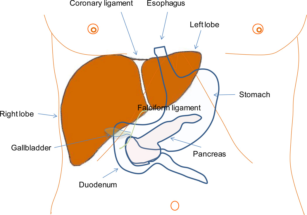
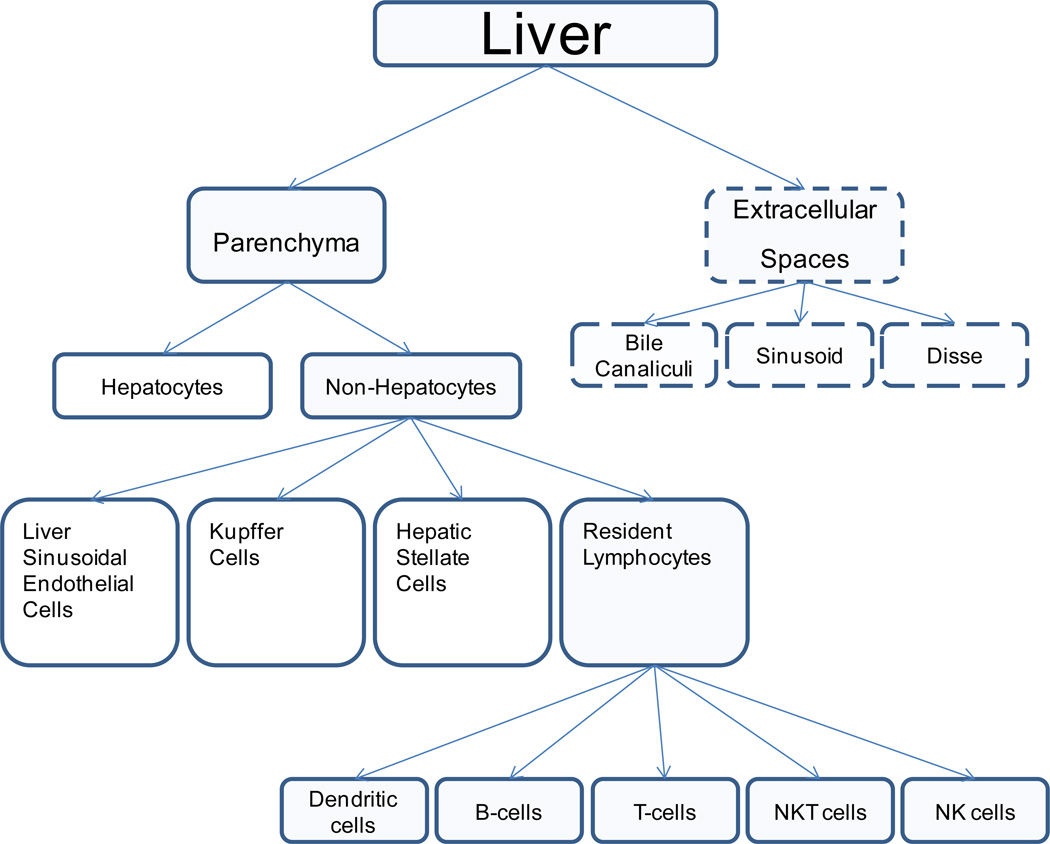
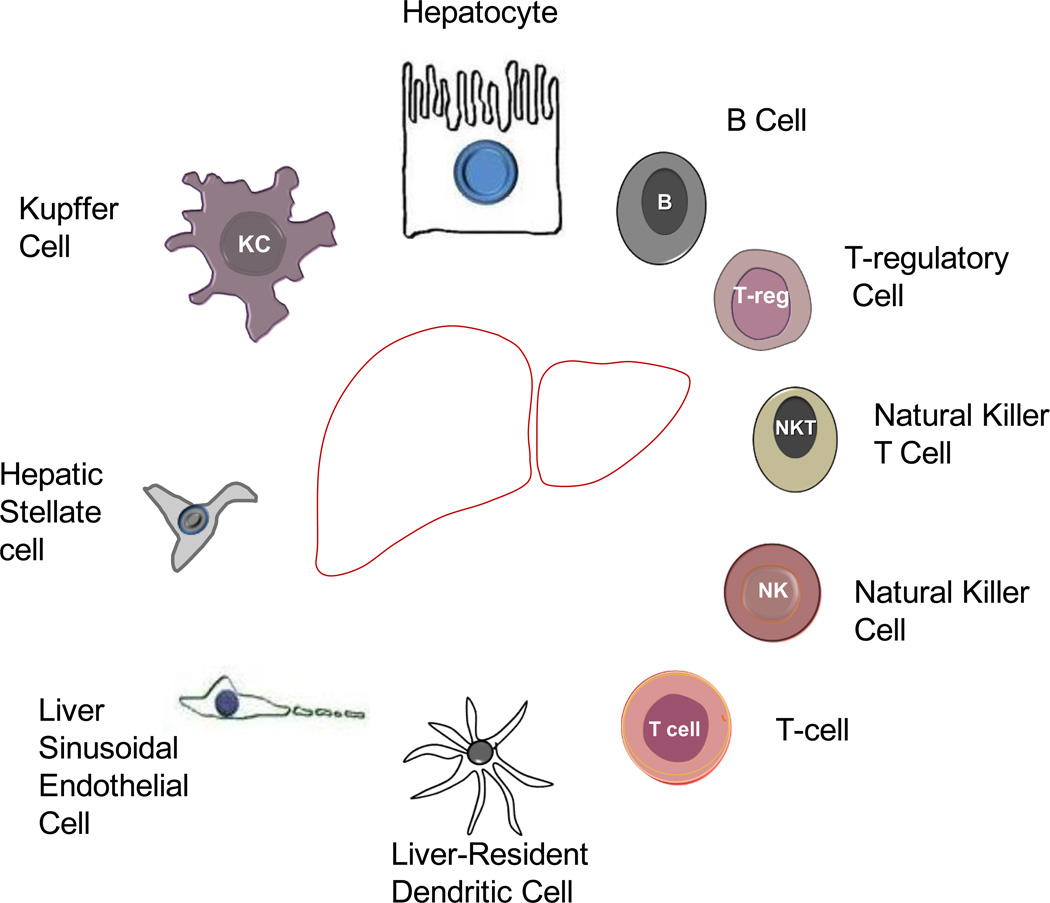
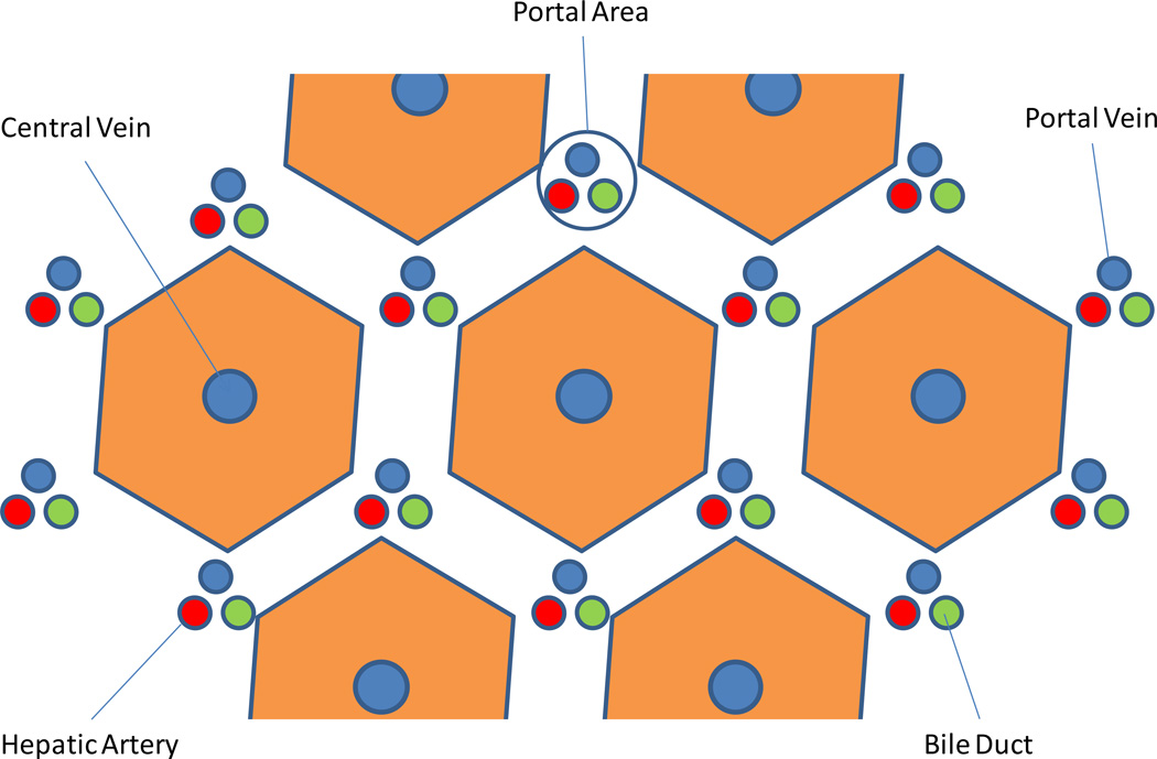
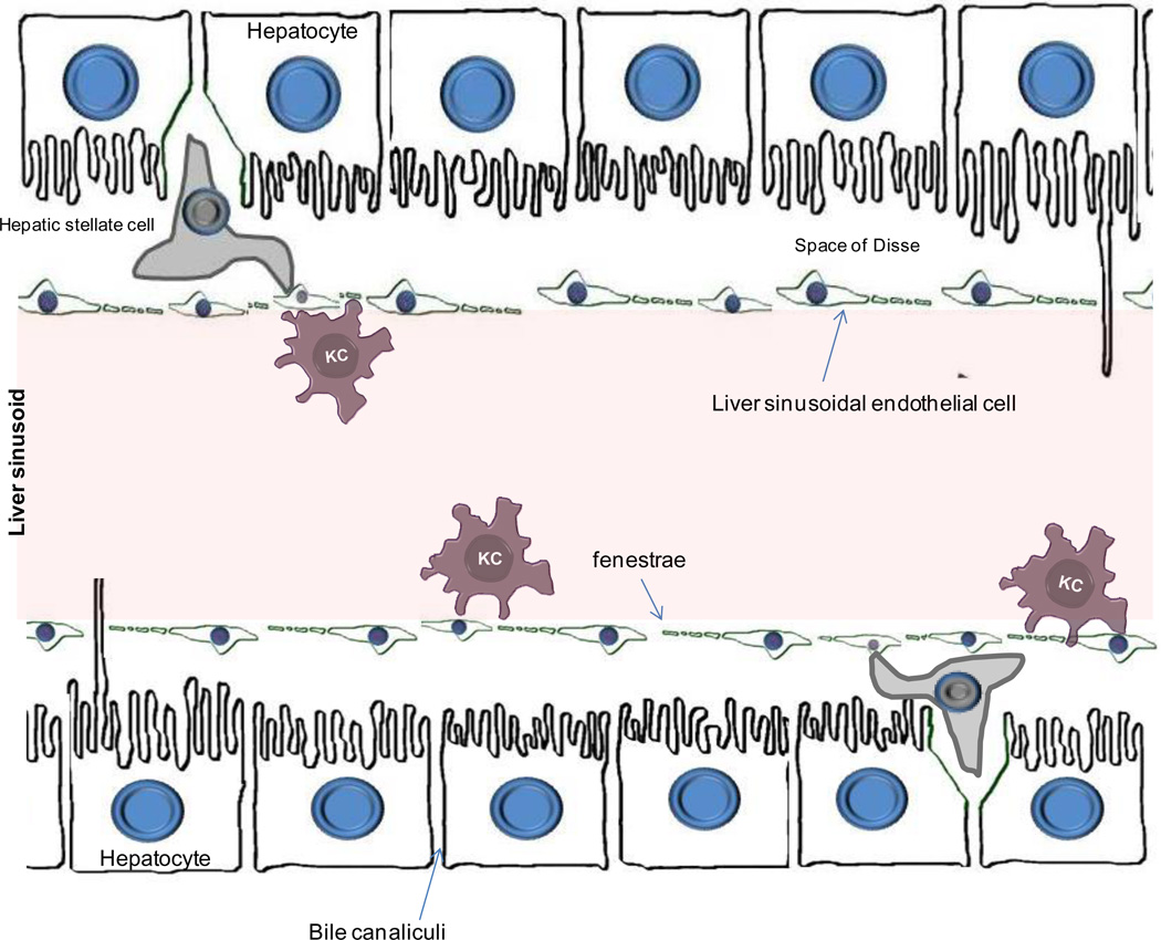
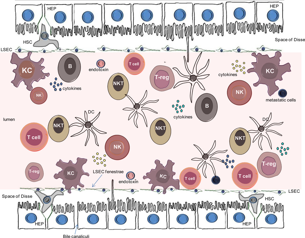
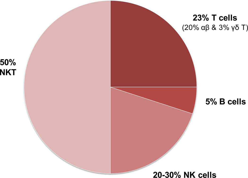
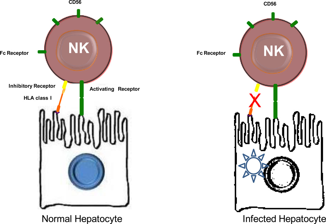
The liver is the largest organ in the body and is generally regarded by nonimmunologists as having little or no lymphoid function. However, such is far from accurate. This review highlights the importance of the liver as a lymphoid organ. Firstly, we discuss experimental data surrounding the role of liver as a lymphoid organ. The liver facilitates tolerance rather than immunoreactivity, which protects the host from antigenic overload of dietary components and drugs derived from the gut and it is instrumental to fetal immune tolerance. Loss of liver tolerance leads to autoaggressive phenomena, which if not controlled by regulatory lymphoid populations, may lead to the induction of autoimmune liver diseases. Liver-related lymphoid subpopulations also act as critical antigen-presenting cells. The study of the immunological properties of liver and delineation of the microenvironment of the intrahepatic milieu in normal and diseased livers provides a platform to understand the hierarchy of a series of detrimental events that lead to immune-mediated destruction of the liver and the rejection of liver allografts. The majority of emphasis within this review will be on the normal mononuclear cell composition of the liver. However, within this context, we will discuss selected, but not all, immune-mediated liver disease and attempt to place these data in the context of human autoimmunity.


Conflict of interest statement

Conflict of Interest: None of the authors has a conflict of interest to declare.

Figures

Figure 1
Figure 1
Anatomical location and external appearance of the liver. The falciform ligament, on the surface of the diaphragm, splits the liver into right and left lobe. The anatomical relationship of the liver with organs such as the gallbladder, stomach, duodenum, and pancreas is illustrated.
Figure 2
Figure 2
Cellular and extracellular composition of the liver
Figure 3
Figure 3
The morphological appearance of cells within the liver.
Figure 4
Figure 4
The hepatic lobule is the structural unit of the liver. It consists of an hexagonal arrangement of hepatocyte plates with the central vein located in the center of the structure and the portal triads distributed at the vertices of the lobule. The portal triad consists of terminal branches of the portal vein and the hepatic artery and a bile duct.
Figure 5
Figure 5
Illustration of the microanatomical localization of hepatocytes, liver sinusoidal endothelial cells, Kupffer cells, and hepatic stellate cells. The space of Disse separates hepatocytes from the liver sinusoids. The endothelium of liver sinusoids is discontinued (fenestrated) and is formed by a layer of liver sinusoidal endothelial cells. These cells act as scavenger cells and form a physical filtering barrier between the sinusoidal blood and plasma, . Kupffer cells are resident macrophages, that are attached to the layer of liver sinusoidal endothelial cells. The hepatic stellate cells are located in the sub-endothelial space of Disse and play vital role in fibrogenesis.
Figure 6
Figure 6
Cells comprising the liver including hepatocytes (HEP), liver sinusoidal ensothelial cells (LSEC), Kupffer cells (KC), hepatic stellate cells (HSC) and lymphoid cell sub-populations. NK, natural killer; NKT, natural killer T-cells; DC, dendritic cell; Treg, T-regulatory cell
Figure 7
Figure 7
Distribution of cell sub-populations within intrahepatic lymphocytes
Figure 8
Figure 8
Schematic illustration of NK cell receptors and killing of viral hepatitis infected cells. Under normal conditions, non-infected cells are not killed because inhibitory signals from HLA class I molecules prevail over activating signals. Virus-infected cells are characterized by altered expression of HLA class I molecules. This disrupts the inhibitory signals and allows activation of NK cells and subsequent lysis of the infected hepatocytes. NK-mediated killing of infected hepatocytes is not operated in viral hepatitides.
Figure 9
Figure 9
Cell-cell interaction which can lead to activation of naïve T lymphocytes within the liver include contact with Kupffer cells (KC), liver sinusoidal endothelial cells (LSEC) or hepatocytes (indicated by the respective arrows).
Figure 10
Figure 10
Flow cytometric analysis of peripheral blood reflects the relative proportion of bright and low NK (CD36+) cells, NKT (CD3+CD56+) and T-cells (CD3+CD56) in a representative donor. Bright and low NK cells can be seen.
Figure 11
Figure 11
Hepatic dendritic subsets in mouse
Figure 12
Figure 12
Cell-Cell interaction of biliary epithelial cells (BECs) as antigen presenting cells, effector and regulatory T-cells
Figure 13
Figure 13
CYP2D6, CYP1A2, and CYP2D6 amino acid homology. The three cytochromes appear highly conserved but autoantibody responses against the one does not invoke cross-reactive immunity targeting the other. Amino acid analysis has been performed using the T-coffee software. Highlights of red, yellow and green correspond to areas of good, average and bad degree of homology; cons, conservation of amino acids (* indicate identical amino acids, : indicate conserved and . semi-conservative substitutions).
Figure 14
Figure 14
3D-prediction model of the three major linear epitopic regions of human cytochrome P450IID6 (CYP2D6). The B-cell epitopes of anti-CYP2D6 antibodies (also known as anti-liver kidney microsomal type 1 antibodies- anti-LKM1) has been studied and the three main epitopic regions recognized span CYP2D6254–271, CYP2D6193–212 and CYP2D6321–351 sequences, being targeted by more than 55% of the patients with CYP2D6 autoantibodies. The antigenicity of this area may in part been explained by the exposure of these sequences to the surface of the molecule as it is illustrated in Figure 7. Aminoacids of the autoepitopic regions are presented in the form of space fill in different colours and the remaining in a wire worm backbone (grey); in red and yellow are the dominant CYP2D6254–271 and CYP2D6193–212 epitopes. Prediction analysis anticipates that the epitopes are exposed on the surface of the molecule. The structure was analyzed with the Cn3D visualization tool.
Figure 15
Figure 15
The tetraspin CD81 is used by hepatitis C virus to entry the hepatocyte. Expression of CD81 by NK suppresses the induction of pro-inflammatory cytokines such as interferon-γ and inhibits the cytotoxic capability of these cells allowing for the persistence of the virus. Other HCV co-receptors (for further details, see main text), expressed by cells of the innate immune system resident within the sinusoids, may participate in a similar fashion facilitating the inability of the host to clear the virus.

References

    1. Kita H, Mackay IR, Van De Water J, Gershwin ME. The lymphoid liver: considerations on pathways to autoimmune injury. Gastroenterology. 2001;120:1485–1501. - PubMed
    1. Starzl TE, Marchioro TL, Vonkaulla KN, Hermann G, Brittain RS, Waddell WR. Homotransplantation of the Liver in Humans. Surg Gynecol Obstet. 1963;117:659–676. - PMC - PubMed
    1. Calne RY, Rolles K, White DJ, Thiru S, Evans DB, McMaster P, Dunn DC, Craddock GN, Henderson RG, Aziz S, Lewis P. Cyclosporin A initially as the only immunosuppressant in 34 recipients of cadaveric organs: 32 kidneys, 2 pancreases, and 2 livers. Lancet. 1979;2:1033–1036. - PubMed
    1. Zimmermann FA, White DJ, Gokel JM, Calne RY. Orthotopic liver transplantation in rats. Prolonging of survival time of allotransplants using cyclosporin A in an acute rejection model. Chir Forum Exp Klin Forsch. 1979:339–344. - PubMed
    1. Calne RY, Sells RA, Pena JR, Davis DR, Millard PR, Herbertson BM, Binns RM, Davies DA. Induction of immunological tolerance by porcine liver allografts. Nature. 1969;223:472–476. - PubMed

Publication types

LinkOut - more resources