Review
. 2013 Apr;3(2):977-1010.
doi: 10.1002/cphy.c120020.

Apoptosis and necrosis in the liver

Affiliations
Review

Apoptosis and necrosis in the liver

Maria Eugenia Guicciardi et al. Compr Physiol. 2013 Apr.

Abstract

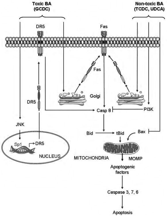
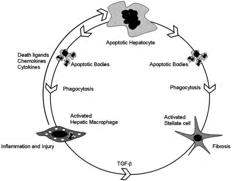
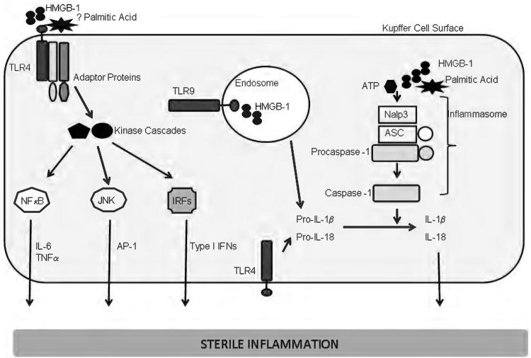
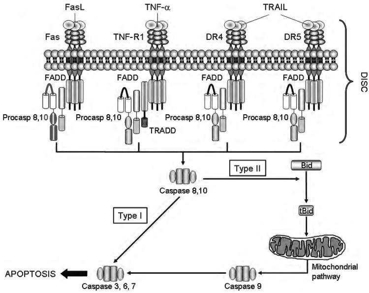
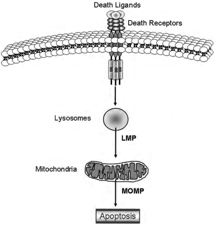
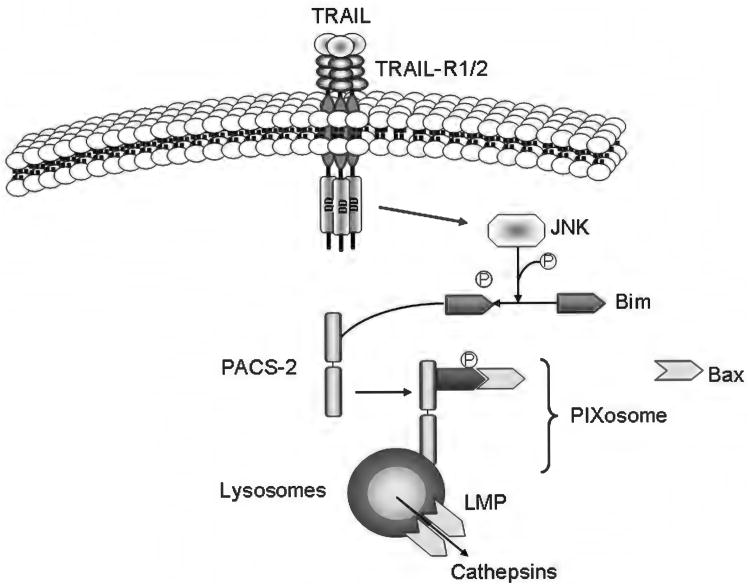
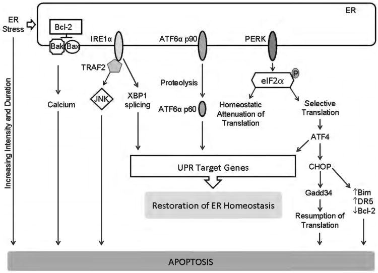
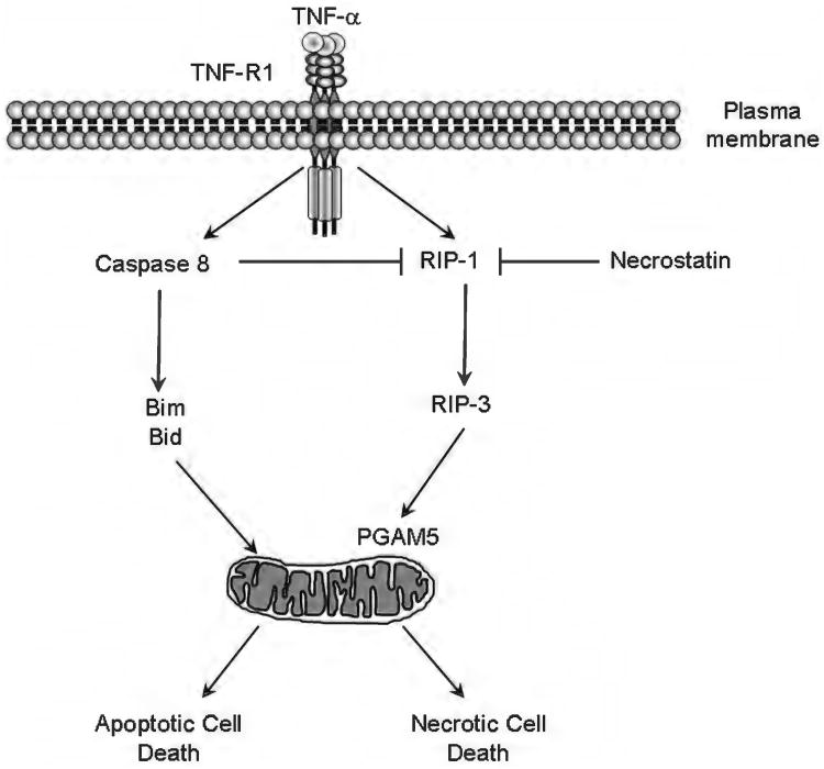
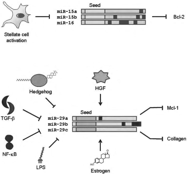
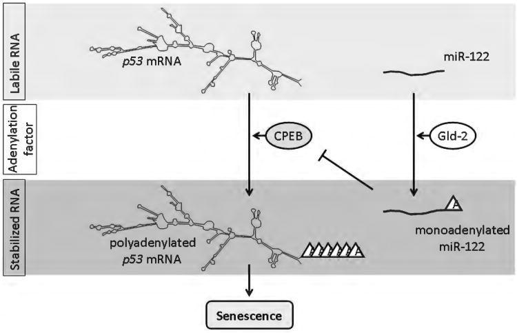
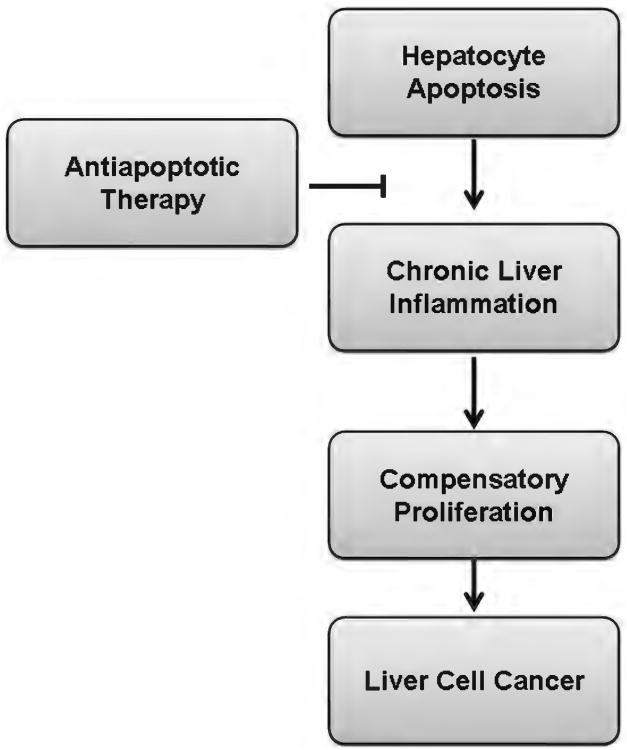
Because of its unique function and anatomical location, the liver is exposed to a multitude of toxins and xenobiotics, including medications and alcohol, as well as to infection by hepatotropic viruses, and therefore, is highly susceptible to tissue injury. Cell death in the liver occurs mainly by apoptosis or necrosis, with apoptosis also being the physiologic route to eliminate damaged or infected cells and to maintain tissue homeostasis. Liver cells, especially hepatocytes and cholangiocytes, are particularly susceptible to death receptor-mediated apoptosis, given the ubiquitous expression of the death receptors in the organ. In a quite unique way, death receptor-induced apoptosis in these cells is mediated by both mitochondrial and lysosomal permeabilization. Signaling between the endoplasmic reticulum and the mitochondria promotes hepatocyte apoptosis in response to excessive free fatty acid generation during the metabolic syndrome. These cell death pathways are partially regulated by microRNAs. Necrosis in the liver is generally associated with acute injury (i.e., ischemia/reperfusion injury) and has been long considered an unregulated process. Recently, a new form of "programmed" necrosis (named necroptosis) has been described: the role of necroptosis in the liver has yet to be explored. However, the minimal expression of a key player in this process in the liver suggests this form of cell death may be uncommon in liver diseases. Because apoptosis is a key feature of so many diseases of the liver, therapeutic modulation of liver cell death holds promise. An updated overview of these concepts is given in this article.


Figures

Figure 1
Figure 1
Bile acid (BA)-induced apoptotic and prosurvival signaling. Internalized toxic (more hydrophobic) bile acids, such as glycine-conjugated form of chenodeoxycholic acid (GCDC), trigger death-receptor-mediated apoptosis. BA stimulate microtubule-dependent trafficking of Fas from the Golgi compartment to the plasma membrane, increasing Fas density on the cell surface and promoting spontaneous Fas oligomerization independent of FasL. This results in activation of caspase 8, which, in turn, cleaves Bid, whose truncated form translocates to mitochondria and cooperates with Bax to induce mitochondrial outer membrane permeabilization (MOMP). Following MOMP, several apoptogenic factors, such as cytochrome c, apoptosis-inducing factor and second mitochondrial activator of caspases, are released into the cytosol, which ultimately promote the activation of effector caspases-3, 6, and 7, and cell death. Moreover, BA activate c-jun N-terminal kinase, resulting in activation of the transcription factor Sp1 (specificity protein 1), which, in turn, upregulates death receptor 5 sensitizing the hepatocytes also to TRAIL-induced apoptosis. Nontoxic BA, such as tauro-conjugate of chenodeoxycholic acid and ursodeoxycholic acid, can also trigger plasma membrane trafficking and activation of Fas. However, the simultaneous activation of cytoprotective pathways which prevent activation of key players such as caspase 8, Bid, or Bax, blocks the apoptotic cascade and inhibits cell death.
Figure 2
Figure 2
Apoptosis-inflammation-fibrosis in the liver. This cartoon depicts the circular relationship between apoptosis, inflammation, and fibrosis in the liver. Hepatocyte apoptosis is the central event in the model shown. In the setting of an apoptotic stimulus, for example, toxic bile salts or palmitic acid, a vulnerable hepatocyte undergoes cell death. Apoptotic bodies are formed. These can be engulfed both by hepatic macrophages, also known as Kupffer cells and hepatic stellate cells (54, 57). Macrophages, upon activation, in turn release death ligands, such as Fas ligand and TRAIL, both of which can induce hepatocyte apoptosis. Inflammatory cytokines such as TNF-α, IL-1β, and IL-6 are also released by activated macrophages (54). These result in liver inflammation and injury. Engulfment of apoptotic bodies in a permissive milieu (inflamed liver with increased fibrogenic signals, such as Transforming growth factor (TGF)-β) results in activation of hepatic stellate cells to myofibroblasts (57). These cells remodel the extracellular matrix resulting in fibrosis and cirrhosis.
Figure 3
Figure 3
Sterile inflammation in liver diseases. A model is presented for sterile inflammation in liver diseases. Palmitic acid, a toxic free fatty acid, which can activate the NLRP3 inflammasome and high mobility group box 1 (HMGB-1), a nuclear protein released from dead cells, are shown as activating damage associated molecular patterns (DAMPs). Activation of cel surface toll like receptors (TLRs)-1, 2, 4, 6, 5, or endosomal TLRs (7, 9) leads to recruitment of adaptor proteins, activation of kinase cascades that result in activation of nuclear factor κ B, c-jun N-terminal kinase, and interferon (IFN) regulatory factors. These result in transcriptional activation of several proinflammatory mediators including interleukin (IL)-6, TNF-α and Type IFNs. The inflammasome can also be activated by many endogenous DAMPs. The mechanism for this activation is not fully elucidated. Shown here is the NLRP3 (nucleotide oligomerization domain [NOD]-like receptor, pyrin domain containing 3) inflammasome. The NLRP3 gene product is the intracellular protein, Nalp3 (NACHT, LRR, and pyrin domain-containing 3). Nalp3, upon activation, recruits ASC (apoptosis-associated speck-like protein containing a CARD, also known as PYCARD), and procaspase-1, leading to the activation of caspase-1. Caspase-1 cleaves and activates the precursor forms on IL-1 β and L-18; both are subsequently secreted, and activate their receptors on target cells, resulting in the activation of proinflammatory pathways.
Figure 4
Figure 4
The death receptors and the extrinsic pathway of apoptosis. Binding of a death ligand to its cognate receptor results in the recruitment of adaptor proteins, such as Fas-associated protein with death domain and Tumor necrosis factor receptor 1-associated death domain protein (TRADD), and procaspases 8 and/or 10, to form a multiprotein receptor complex named death inducing signaling complex (DISC). This complex provides a platform for caspase 8 and 10 to undergo autoactivation. In type I cells, active caspase 8/10 directly activate caspase 3, an effector caspase, whereas in type II cells, caspase 8 (and, perhaps, caspase 10) cleaves the BH3-only protein Bid to generate truncated Bid (tBid). tBid, in turn, cooperates with Bax or Bak to induce mitochondrial outer membrane permeabilization and to initiate the mitochondrial pathway of apoptosis (see Figure 5 for details).
Figure 5
Figure 5
The intrinsic pathway of apoptosis. Various stimuli, including UV and gamma-irradiation, endoplasmic reticulum (ER) stress, growth factor deprivation, and oxidative stress trigger the intrinsic pathway of apoptosis. This pathway requires the oligomerization of the proapoptotic members of the Bcl-2 family of protein Bax and/or Bak on the outer mitochondrial membrane, resulting in mitochondrial outer membrane permeabilization (MOMP) and release of apoptogenic factors. The oligomerization of Bax and Bak can be directly stimulated by the BH3-only proteins Bid, Bim, or PUMA (activators). Bax and Bak, as well as Bid, Bim and PUMA, are bound to and inhibited by the prosurvival Bcl-2 proteins, Bcl-2, Bcl-xL, and Mcl-1. The prosurvival function of these proteins can be repressed by the BH3-only proteins Bad, Bik, Hrk, Bmf, and NOXA (sensitizers), which displace Bid, Bim, and PUMA by binding to the prosurvival proteins. Release of Bid, Bim, and PUMA then allows activation of Bax and/or Bak. MOMP can also be achieved by the so-called mitochondrial permeability transition (MPT) initiated at the inner mitochondrial membrane through the opening of a multiprotein complex known as permeability transition pore (PTP). Several apoptogenic factors, including cytochrome c and SMAC/DIABLO, are released from the mitochondrial intermembrane space into the cytosol as a consequence of MOMP. Cytochrome c binds to the adaptor Apaf-1, and recruits procaspase-9 in a complex named apoptosome, which promotes the activation of caspase-9. Caspase-9, in turn, activates the effector caspases (caspase-3, 6, and 7). SMAC/DIABLO contributes to caspase activation by binding and inactivating the endogenous inhibitor of caspases X-chromosome linked inhibitor of apoptosis protein.
Figure 6
Figure 6
The lysosomal pathway of apoptosis. In some cells, including hepatocytes and cholangiocytes, the binding of a death ligand to its cognate receptor results in early lysosomal membrane permeabilization associated with release of lysosomal enzymes into the cytosol. These lysosomal enzymes, and in particular the highly abundant lysosomal cathepsins, then trigger mitochondrial outer membrane permeabilization and mitochondrial dysfunction likely by cleaving and/or activating members of the Bcl-2 family. This apoptotic cascade as been referred to as the lysosomal pathway.
Figure 7
Figure 7
Model of tumor necrosis factor-related apoptosis-inducing ligand (TRAIL)-induced lysosomal membrane permeabilization mediated by members of the Bcl-2 family of proteins. Binding of TRAIL to death receptor 5 results in c-jun N-terminal kinase-mediated phosphorylation of Bim and its release from the cytoskeleton. At the same time, phosphofurin acidic cluster sorting protein-2 (PACS-2) associates with the internalized receptor complex in the endosomal/lysosomal compartment and then binds the cytosolic Bim, promoting Bax association to the Bim:PACS-2 complex and subsequent Bax activation. This complex has been named the PIXosome (PACS-2:BIM:BAX). After translocating to the lysosomes, Bax inserts into the membrane, homo-oligomerizes and induces lysosomal membrane permeabilization. Cathepsins are released into the cytosol where they contribute to cell death.
Figure 8
Figure 8
Endoplasmic reticulum (ER) stress and apoptosis. ER stress activates, in parallel, three distinct ER-to-nucleus signaling pathways that are aimed at attenuating ER stress via activation of unfolded protein response (UPR) target genes with restoration of ER homeostasis. However, ER stress of increasing duration and intensity results in failure of restoration of homeostasis and apoptosis. The three canonical UPR mediators are ER transmembrane proteins; they are inositol-requiring protein 1α (IRE1α), activating transcription factor (ATF) 6α, and protein kinase RNA-like ER kinase (PERK). Active PERK phosphorylates eukaryotic translation initiation factor 2α (eIF2α) leading to an attenuation of translation. This reduces the load of nascent proteins entering the ER. Selective translation of activating ATF4 also occurs. ATF4 promotes adaptation, but also transcriptionally activates C/EBP homologous protein (CHOP). CHOP promotes ER stress-induced apoptosis via several pathways, including increasing the proapoptotic proteins Bim and death receptor 5 (DR5) and decreasing the antiapoptotic protein Bcl-2. IRE1α splices X box-binding protein 1 (XBP-1) mRNA to generate a transcription factor which leads to ER adaptation by activating a large number of UPR genes. The stress kinase, c-jun N-terminal kinase (JNK) is activated by IRE1α via the adaptor protein TNF receptor-associated factor 2 (TRAF2). Activating transcription factor 6α (ATF6α) is proteolytically cleaved in the Golgi to generate a transcription factor which also drives expression of UPR target genes. Failure of restoration of ER homeostasis due to increasing intensity and duration of ER stress results in apoptosis. Some of the recognized mediators of ER stress-induced apoptosis are the transcription factor CHOP which can repress Bcl-2 expression, and increase expression of the proapoptotic proteins Bim and DR5. ER stress-induced apoptosis can also me mediated by ER calcium release, which can be regulated by Bax, Bak, and Bcl-2. The stress kinase JNK can also activate the intrinsic apoptosis machinery.
Figure 9
Figure 9
TNF-α triggers both apoptotic and necrotic pathways. In conditions where activation of the transcription factor NF-κB is impaired, binding of TNF-α to TNF-R1 results in activation of caspase 8, which triggers an apoptotic signaling cascade mediated by the BH3-only proteins Bid and Bim. Active caspase 8 also cleaves and inactivates the kinase RIP-1. When caspase 8 is inhibited and RIP-1 is not ubiquitinated, TNF-α stimulation induces RIP-1 association with RIP-3, promoting RIP-3 phosphorylation and activation. RIP-3, in turn, phosphorylates and activates the mitochondrial phosphatase phosphoglycerate mutase 5 (PGAM5), which promotes mitochondrial permeability transition and necroptosis. Necrostatin, a pharmacologic RIP-1 inhibitor, effectively prevents activation of the necroptotic signaling cascade.
Figure 10
Figure 10
Stabilization of microRNAs, like mRNAs, by adenylation. It is well known that mRNAs are stabilized by polyadenylation, a posttranscriptional modification that is dependent on multiple protein factors. For p53, cytoplasmic polyadenylation element binding protein (CPEB) is necessary for polyadenylation. Similarly, microRNAs can be stabilized by modification at the 3′ terminus by monoadenylation; shown is the Gld-2-dependent adenylation of miR-122. Gld-2 mediated miR-122 stabilization results in increased miR-122, decreased CPEB, and less stabilization of p53. Conversely, decreased Gld-2 (or decreased miR-122) results in increased CPEB protein levels and activity, thus increasing p53 mRNA stability and promoting senescence.
Figure 11
Figure 11
Repression of microRNAs influences survival and function of liver cells, especially hepatic stellate cells. Among microRNAs altered in HSC activation or liver fibrogenesis are members of the miR-15 family and the miR-29 family. Upper: the miR-15 family contains three microRNAs which have identical seed sequences and scattered base differences in the 3′ end of the microRNA (indicated by colored blocks on the diagram). These microRNAs are decreased in culture-activated stellate cells, which permits derepression of Bcl-2, promoting cell survival. Lower: the miR-29 family also contains three family members with seed identity and a few base differences in the 3′end. Several studies have demonstrated molecular signals that either inhibit [Hedgehog, transforming growth factor (TGF)-β, nuclear factor kappa B (NF-κB), and lipopolysaccharide (LPS)] or activate [hepatocyte growth factor (HGF) and estrogen] miR-29 expression. Decreased miR-29 allows excess collagen expression, promoting fibrosis, and excess Mcl-1, promoting cell survival.
Figure 12
Figure 12
Model of liver disease progression. In pathologic conditions, persistent hepatocyte apoptosis promotes chronic liver inflammation and associated compensatory cellular proliferation, increasing the risk of carcinogenesis in the liver. Inhibition of apoptosis in these conditions should protect against the development of liver cancer.

References

    1. Abu-Amara M, Yang SY, Tapuria N, Fuller B, Davidson B, Seifalian A. Liver ischemia/reperfusion injury: Processes in inflammatory networks—a review. Liver Transplant. 2010;16:1016–1032. - PubMed
    1. Adachi M, Suematsu S, Kondo T, Ogasawara J, Tanaka T, Yoshida N, Nagata S. Targeted mutation in the Fas gene causes hyperplasia in peripheral lymphoid organs and liver. Nat Genet. 1995;11:294–300. - PubMed
    1. Afford SC, Ahmed-Choudhury J, Randhawa S, Russell C, Youster J, Crosby HA, Eliopoulos A, Hubscher SG, Young LS, Adams DH. CD40 activation-induced, Fas-dependent apoptosis and NF-kappaB/AP-1 signaling in human intrahepatic biliary epithelial cells. FASEB J. 2001;15:2345–2354. - PubMed
    1. Agarwal R, Hennings L, Rafferty TM, Letzig LG, McCullough S, James LP, MacMillan-Crow LA, Hinson JA. Acetaminophen-induced hepatotoxicity and protein nitration in neuronal nitric-oxide synthase knockout mice. J Pharmacol Exp Ther. 2012;340:134–142. - PMC - PubMed
    1. Akazawa Y, Gores GJ. Death receptor-mediated liver injury. Semin Liver Dis. 2007;27:327–338. - PubMed

Publication types