. 2013 Aug 1;305(3):F255-64.
doi: 10.1152/ajprenal.00160.2013. Epub 2013 May 29.

Mitochondria-targeted heme oxygenase-1 decreases oxidative stress in renal epithelial cells

Affiliations

Mitochondria-targeted heme oxygenase-1 decreases oxidative stress in renal epithelial cells

Subhashini Bolisetty et al. Am J Physiol Renal Physiol. .

Abstract

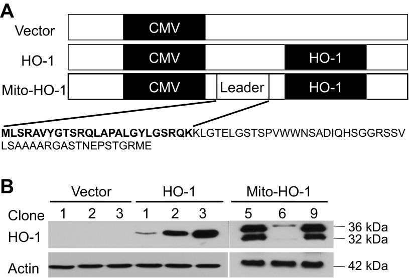
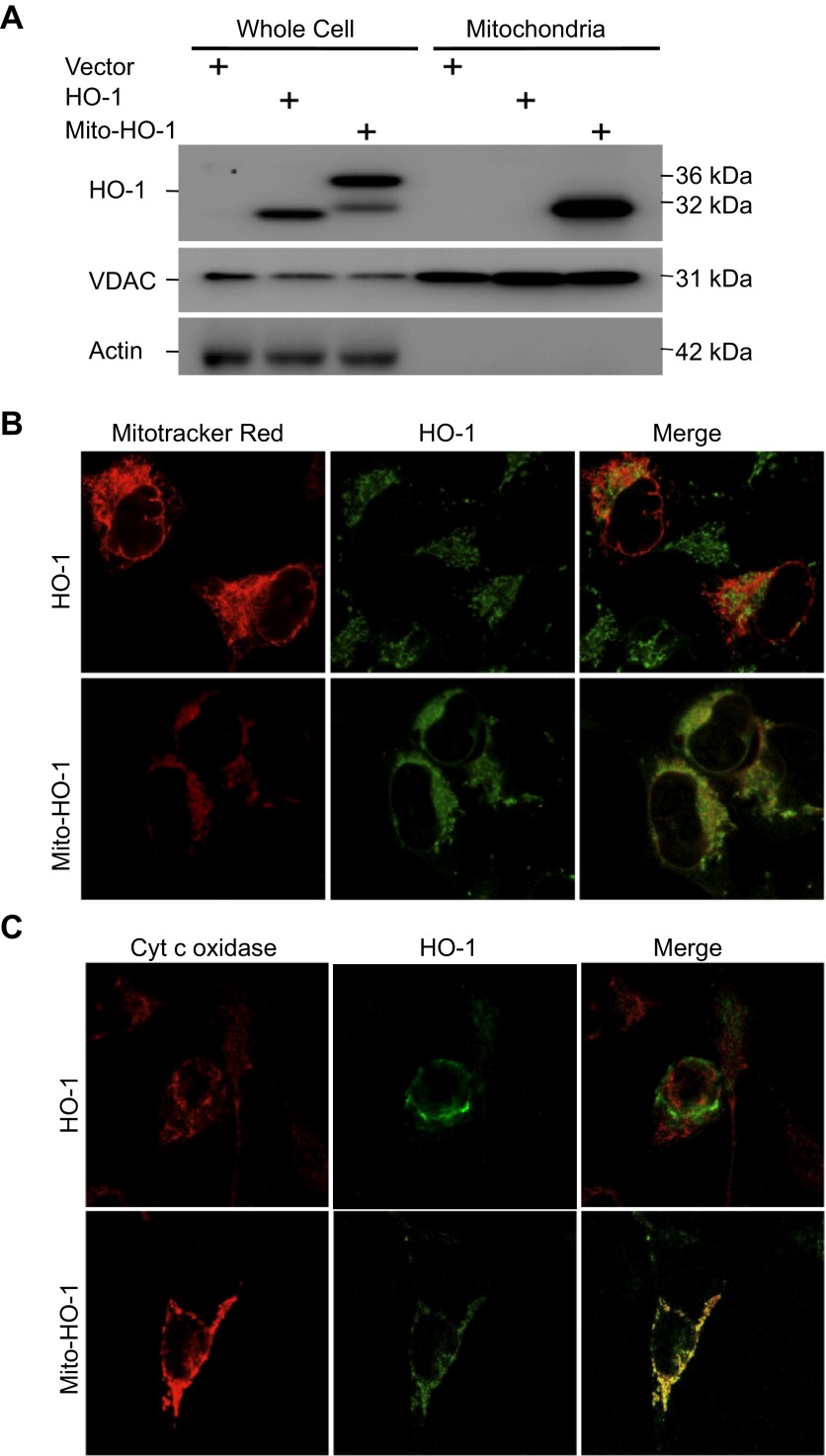
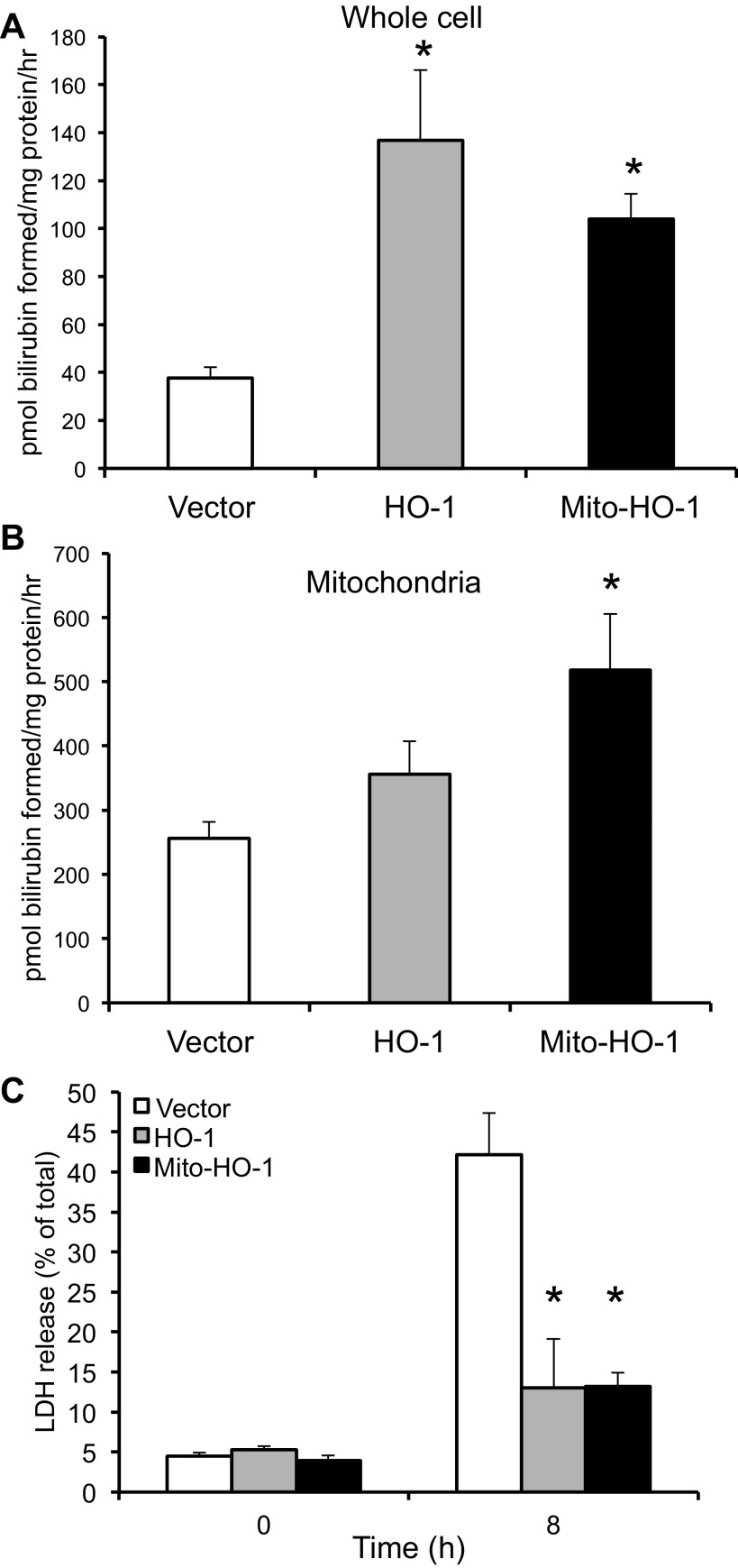
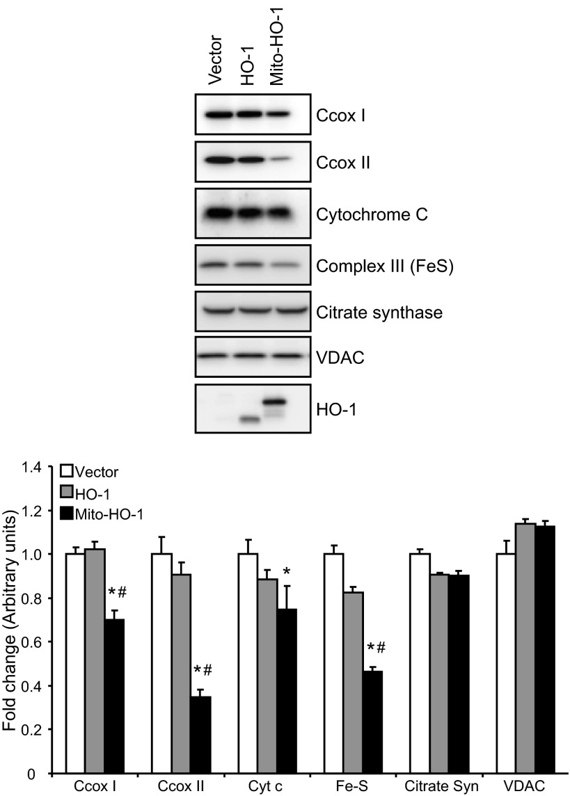
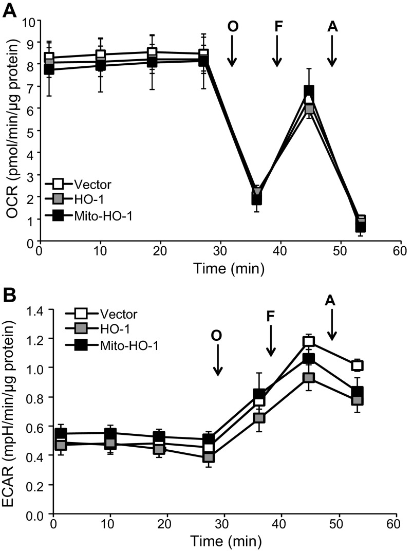
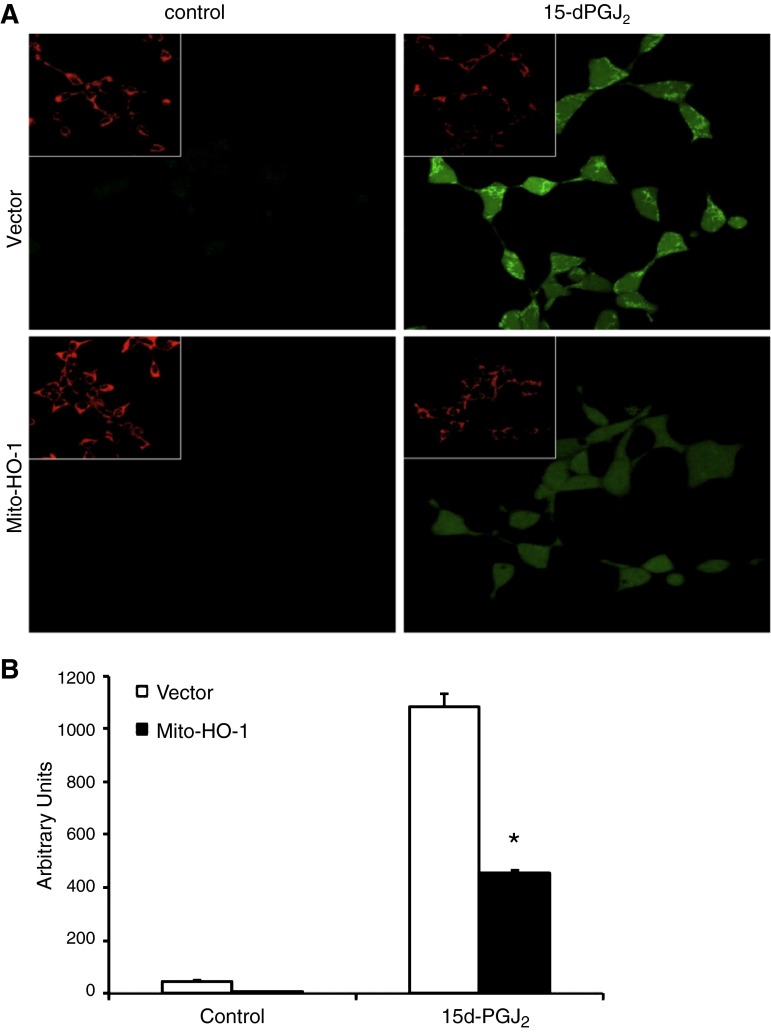
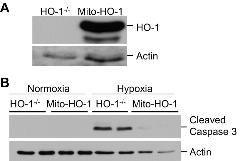
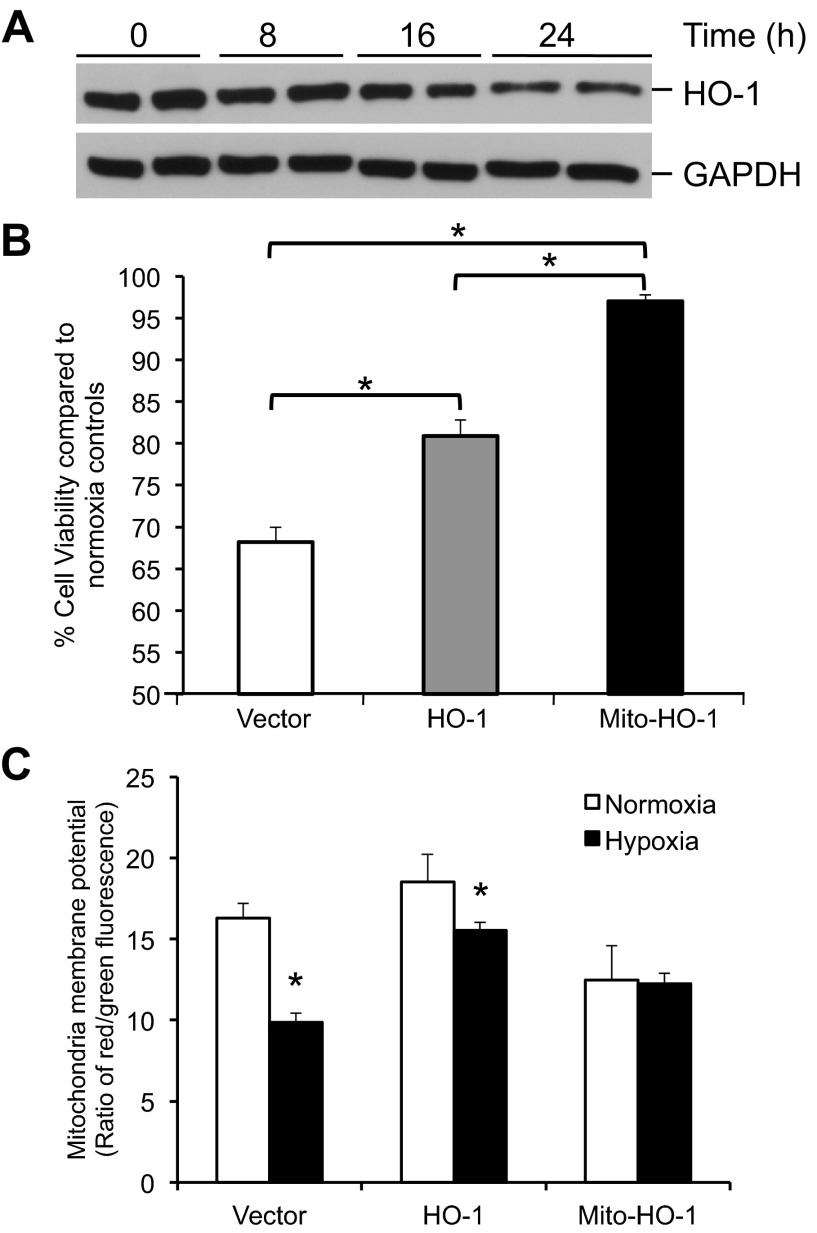
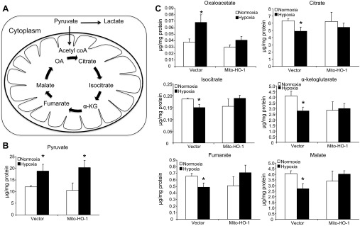
Mitochondria are both a source and target of the actions of reactive oxygen species and possess a complex system of inter-related antioxidants that control redox signaling and protect against oxidative stress. Interestingly, the antioxidant enzyme heme oxygenase-1 (HO-1) is not present in the mitochondria despite the fact that the organelle is the site of heme synthesis and contains multiple heme proteins. Detoxification of heme is an important protective mechanism since the reaction of heme with hydrogen peroxide generates pro-oxidant ferryl species capable of propagating oxidative stress and ultimately cell death. We therefore hypothesized that a mitochondrially localized HO-1 would be cytoprotective. To test this, we generated a mitochondria-targeted HO-1 cell line by transfecting HEK293 cells with a plasmid construct containing the manganese superoxide dismutase mitochondria leader sequence fused to HO-1 cDNA (Mito-HO-1). Nontargeted HO-1-overexpressing cells were generated by transfecting HO-1 cDNA (HO-1) or empty vector (Vector). Mitochondrial localization of HO-1 with increased HO activity in the mitochondrial fraction of Mito-HO-1 cells was observed, but a significant decrease in the expression of heme-containing proteins occurred in these cells. Both cytosolic HO-1- and Mito-HO-1-expressing cells were protected against hypoxia-dependent cell death and loss of mitochondrial membrane potential, but these effects were more pronounced with Mito-HO-1. Furthermore, decrement in production of tricarboxylic acid cycle intermediates following hypoxia was significantly mitigated in Mito-HO-1 cells. These data suggest that specific mitochondrially targeted HO-1 under acute pathological conditions may have beneficial effects, but the selective advantage of long-term expression is constrained by a negative impact on the synthesis of heme-containing mitochondrial proteins.

Keywords: heme; hypoxia.


Figures

Fig. 1.
Fig. 1.
Plasmid construction for targeted heme oxygenase-1 (HO-1) overexpression. A: HO-1 cDNA was cloned into XbaI of pcDNA3.1 plasmid (vector) to generate HO-1 vector. The mitochondria leader sequence of the manganese superoxide dismutase gene was cloned 5′ to the HO-1 cDNA (HindIII and XbaI) to generate a plasmid construct containing the manganese superoxide dismutase mitochondria leader sequence fused to HO-1 cDNA (Mito-HO-1) vector. The leader sequence is indicated in bold. B: HEK293 cells were transfected with the indicated vectors, and stable transfectants were selected using specific antibiotics. Whole cell lysates from three different clones per vector were analyzed for HO-1 expression by Western blot analysis. Unprocessed HO-1 protein (leader sequence attached to the HO-1 protein) is represented as the 36 kDa.
Fig. 2.
Fig. 2.
Cellular localization of HO-1 expression. A: whole cell lysates and mitochondria were analyzed for HO-1 expression by Western blot analysis. HO-1 is overexpressed in the HO-1 and Mito-HO-1 cell lysates. A higher-molecular-weight protein in the Mito-HO-1 cell lysates represents the unprocessed HO-1. HO-1 is expressed in the mitochondria of only Mito-HO-1 cells. Voltage-dependent anion channel (VDAC, mitochondrial marker) and actin (cytosol marker) serve as loading controls. The absence of actin in the mitochondrial fraction indicates the purity of the mitochondria prep. B and C: mitochondria were labeled with Mitotracker Red (B) or cytochrome c oxidase (C), and cells were immunostained with anti-HO-1 antibody. HO-1 colocalized with mitochondria only in Mito-HO-1 cells.
Fig. 3.
Fig. 3.
Functional assessment of targeted HO-1 expression. A and B: HO activity was determined by measuring the amount of bilirubin formed per mg protein in whole cell lysates and mitochondria and expressed as pmol bilirubin formed per mg protein. *P < 0.05 compared with vector cells. C: cells were treated with 100 μM heme for 8 h, and %cytotoxicity was evaluated by measuring lactate dehydrogenase (LDH) release. *P < 0.01 compared with vector cells treated with hemin.
Fig. 4.
Fig. 4.
Expression of mitochondrial heme-containing proteins in the stable cells. Western blot analysis of Vector, HO-1, and Mito-HO-1 cell lysates for the expression of cytochrome c, cytochrome c oxidase subunit I and II (C cox), complex III, citrate synthase, and voltage-dependent anion channel. Densitometry was performed on two independent cell lysates and expressed as fold difference compared with vector cells. *P ≤ 0.05 vs. vector control; #P ≤ 0.05 vs. HO-1.
Fig. 5.
Fig. 5.
Bioenergetic function is not changed by targeting HO-1 to the mitochondrion. HEK293 from vector, HO-1, and Mito-HO-1 were seeded at 20,000 cells/well into specialized seahorse plates to determine oxygen consumption rate (OCR) and extracellular acidification rate (ECAR) simultaneously under normoxic conditions. Basal OCR was established, and then subsequent injections of oligomycin (O; 1 μg/ml), carbonyl cyanide 4-(trifluoromethoxy)phenylhydrazone (F; 1 μM), and antimycin-A (A; 10 μM) were made to determine the mitochondrial function (A) and ECAR profile (B) under the same conditions. Data were normalized to protein and represented as means ± SE, n = 6–7 experiments/group.
Fig. 6.
Fig. 6.
Mitochondria-targeted HO-1 overexpression inhibits ROS generation. Vector and Mito-HO-1 cells were treated with 15d-PGJ2 (10 μM) for 30 min, and mitochondria were labeled with Mitotracker Red. The fluorescent probe 2′,7′-dichlorodihydrofluorescein diacetate (H2DCF-DA) was added 1 h before imaging. Images were taken (A), blinded and quantitated for ROS generation (B). Mito-HO-1 cells had a significant decrease in ROS generation compared with vector cells. *P < 0.05 vs. 15d-PGJ2-treated vector cells.
Fig. 7.
Fig. 7.
Mitochondria-targeted HO-1 prevents apoptosis in HO-1-deficient cells. HO-1-deficient proximal tubular epithelial cells were transiently transfected with Mito-HO-1 plasmid. A: Western blot analysis to confirm the expression of targeted HO-1. B: Western blot analysis of cleaved caspase 3 expression in control and transfected HO-1-deficient cells following hypoxia.
Fig. 8.
Fig. 8.
Mitochondria-targeted HO-1 expression protects against hypoxic injury and cell death. Stable cells were incubated in normoxia or 1% oxygen (hypoxia). A: Western blot analysis of HO-1 expression in Vector cells exposed to hypoxia for indicated times. B: cell viability was measured using 3-[4,5-dimethylthiazol-2-yl]-2,5-diphenyltetrazolium bromide (MTT) assay and expressed as viability under hypoxic conditions compared with normoxia controls. *P < 0.05. C: mitochondrial membrane potential was measured following incubation in normoxia or hypoxia using 5,5′,6,6′-tetrachloro-1,1′,3,3′-tetraethylbenzimidazolylcarbocyanine iodide and is expressed as the ratio of red to green fluorescence. *P < 0.05 vs. normoxia controls.
Fig. 9.
Fig. 9.
Analysis of aerobic and anaerobic metabolic pathways. A: schematic representation of glycolysis and tricarboxylic acid (TCA) cycle. Vector and Mito-HO-1 cells were incubated in normoxic or hypoxic conditions, and metabolites were analyzed. B and C: pyruvate (B) and TCA cycle metabolites (C). The results were normalized to protein content and expressed as μg/mg protein. OA, oxaloacetate; α-KG, α-ketoglutarate. *P ≤ 0.05 vs. normoxia.

References

    1. Baliga R, Ueda N, Walker PD, Shah SV. Oxidant mechanisms in toxic acute renal failure. Drug Metab Rev 31: 971–997, 1999 - PubMed
    1. Bolisetty S, Traylor AM, Kim J, Joseph R, Ricart K, Landar A, Agarwal A. Heme oxygenase-1 inhibits renal tubular macroautophagy in acute kidney injury. J Am Soc Nephrol 21: 1702–1712, 2010 - PMC - PubMed
    1. Brookes P, Darley-Usmar VM. Hypothesis: the mitochondrial NO(*) signaling pathway, and the transduction of nitrosative to oxidative cell signals: an alternative function for cytochrome C oxidase. Free Radic Biol Med 32: 370–374, 2002 - PubMed
    1. Brookes PS, Levonen AL, Shiva S, Sarti P, Darley-Usmar VM. Mitochondria: regulators of signal transduction by reactive oxygen and nitrogen species. Free Radic Biol Med 33: 755–764, 2002 - PubMed
    1. Brooks C, Wei Q, Cho SG, Dong Z. Regulation of mitochondrial dynamics in acute kidney injury in cell culture and rodent models. J Clin Invest 119: 1275–1285, 2009 - PMC - PubMed

Publication types

MeSH terms

LinkOut - more resources