. 2013 Nov;67(11):757-72.
doi: 10.1002/syn.21683. Epub 2013 Jul 17.

Stress differentially alters mu opioid receptor density and trafficking in parvalbumin-containing interneurons in the female and male rat hippocampus

Affiliations

Stress differentially alters mu opioid receptor density and trafficking in parvalbumin-containing interneurons in the female and male rat hippocampus

Teresa A Milner et al. Synapse. 2013 Nov.

Abstract

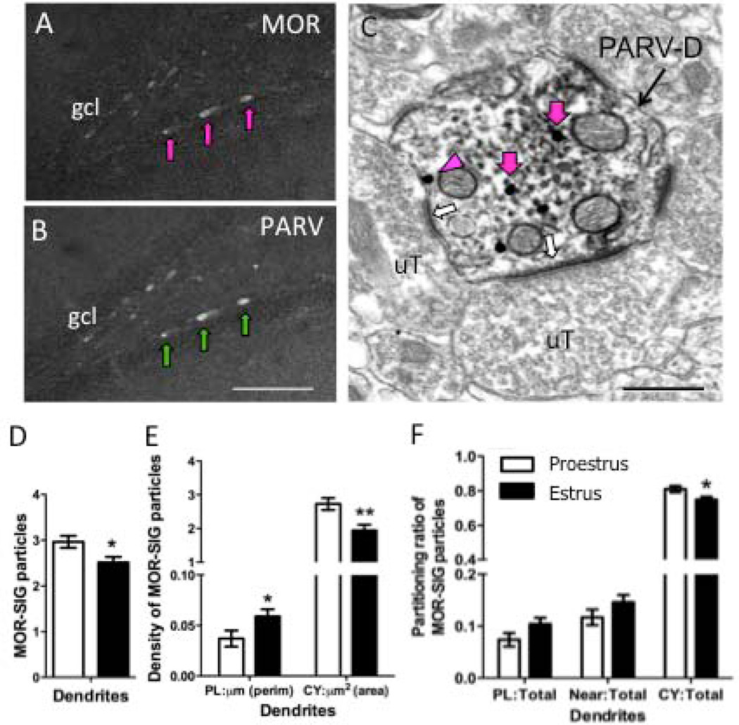
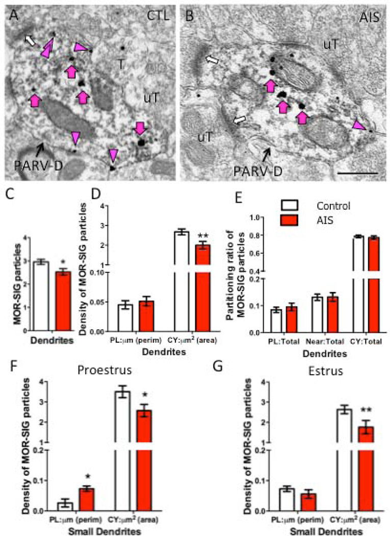
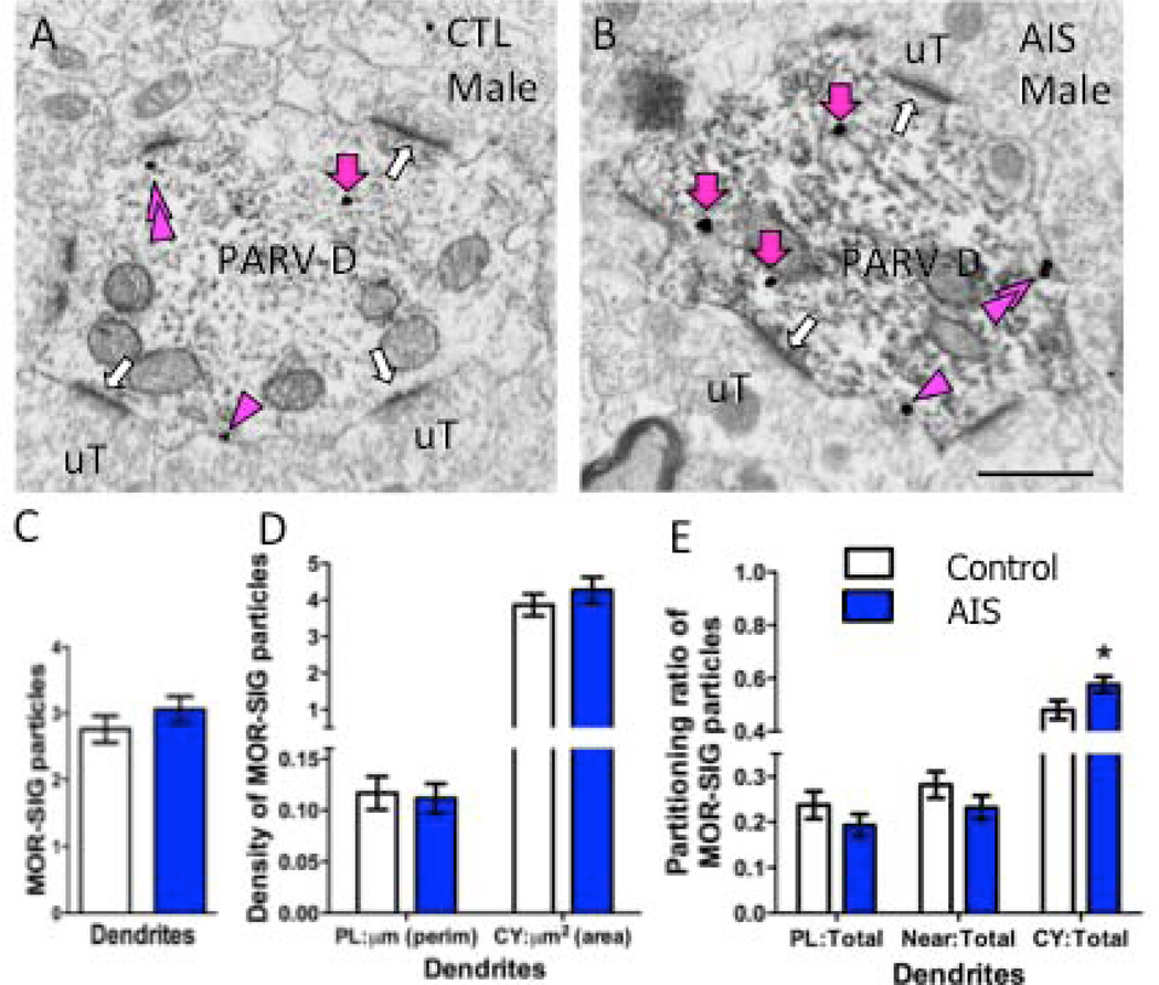
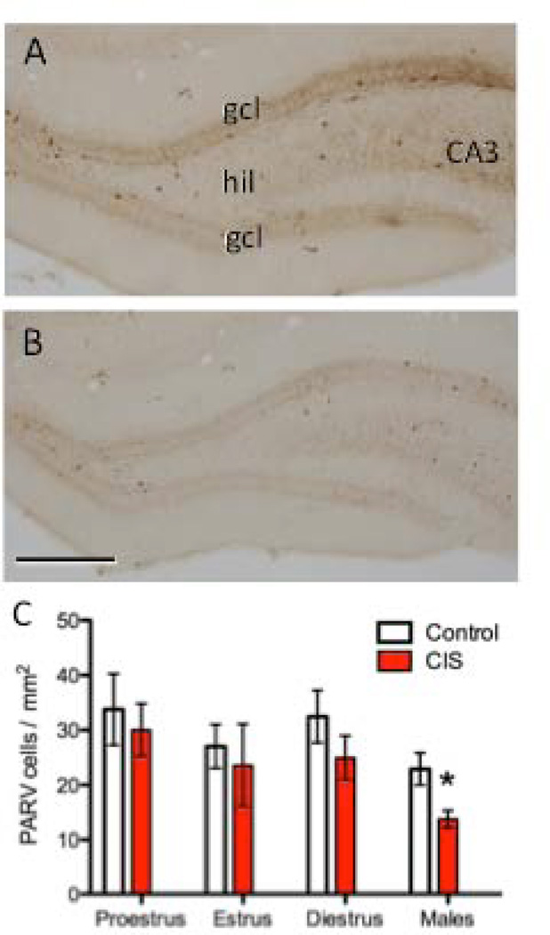
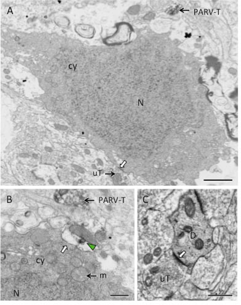
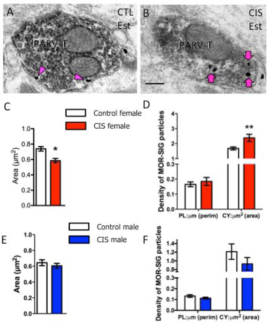
Stress differentially affects hippocampal-dependent learning relevant to addiction and morphology in male and female rats. Mu opioid receptors (MORs), which are located in parvalbumin (PARV)-containing GABAergic interneurons and are trafficked in response to changes in the hormonal environment, play a critical role in promoting principal cell excitability and long-term potentiation. Here, we compared the effects of acute and chronic immobilization stress (AIS and CIS) on MOR trafficking in PARV-containing neurons in the hilus of the dentate gyrus in female and male rats using dual label immunoelectron microscopy. Following AIS, the density of MOR silver-intensified gold particles (SIGs) in the cytoplasm of PARV-labeled dendrites was significantly reduced in females (estrus stage). Conversely, AIS significantly increased the proportion of cytoplasmic MOR SIGs in PARV-labeled dendrites in male rats. CIS significantly reduced the number of PARV-labeled neurons in the dentate hilus of males but not females. However, MOR/PARV-labeled dendrites and terminals were significantly smaller in CIS females, but not males, compared with controls. Following CIS, the density of cytoplasmic MOR SIGs increased in PARV-labeled dendrites and terminals in females. Moreover, the proportion of near-plasmalemmal MOR SIGs relative to total decreased in large PARV-labeled dendrites in females. After CIS, no changes in the density or trafficking of MOR SIGs were seen in PARV-labeled dendrites or terminals in males. These data show that AIS and CIS differentially affect available MOR pools in PARV-containing interneurons in female and male rats. Furthermore, they suggest that CIS could affect principal cell excitability in a manner that maintains learning processes in females but not males.

Keywords: acute stress; chronic stress; estrogens; opioids; sex differences.


Figures

Figure 1
Figure 1. Ovarian hormones influence MOR subcellular distribution in PARV-labeled dendrites in the hilus
Light micrographs show MOR-ir alone (A) and PARV-ir alone (B) from the hilus of the dentate gyrus of a proestrus female rat. Arrows indicate examples of cells that contain both MOR- and PARV-labels. C. Electron micrograph showing an example of a PARV-labeled dendrite (PARV-D) that contains MOR SIG particles in the cytoplasm (magenta arrows) or on the plasma membrane (magenta arrowhead) from a proestrus rat. Unlabeled terminals (uT) form asymmetric synapses (white arrows) on the dendrite. Scale bars A, B = 50 µm; C = 500 nm. D. Significantly fewer total MOR SIG particles are in PARV-labeled dendrites in estrus females compared to proestrus females. E. Both proestrus and estrus females have a greater density of MOR SIG particles in the cytoplasm compared to the plasma membrane. However, estrus females have significantly more MOR SIG particles on the plasma membrane (*p < 0.05) and fewer MOR-SIG particles (**p < 0.001) in the cytoplasm of PARV-labeled dendrites. F. Proestrus females showed an increased distribution of MOR SIG particles to the cytoplasm of PARV-labeled dendritic profiles in comparison to estrus females (*p<0.05).
Figure 2
Figure 2. AIS reduces the number and density of MOR SIG particles in PARV-labeled dendrites in female rats
Examples of PARV-labeled dendrites (PARV-D) containing MOR SIG particles in the cytoplasm (magenta arrows), on the plasma membrane (magenta arrowhead) and near the plasma membrane (double magenta arrowheads) from control (A) and AIS (B) proestrus females. Unlabeled terminals (uT) contact (white arrows) dual labeled dendrites. Scale bars = 500 nm. C. AIS significantly decreased the total number of MOR SIG particles in PARV-labeled dendrites of females, regardless of their stage in the estrous cycle (*p < 0.05). D. AIS significantly reduced the cytoplasmic density of MOR SIG particles in PARV-labeled dendrites of females, regardless of estrous cycle stage (**p < 0.01). E. AIS did not alter MOR SIG particle distribution on the plasma membrane, near the plasma membrane, or in the cytoplasm of PARV-labeled dendrites (p > 0.05), regardless of estrous cycle stage. F, G. After AIS, proestrus females displayed increased plasma membrane density (*p < 0.05; F) and decreased cytoplasmic density (*p < 0.05; G) of MOR SIG particles in small PARV-labeled dendrites whereas estrus females had a significant decrease in cytoplasmic density of MOR SIG particles in small PARV-labeled dendrites (**p < 0.01; G). N = 3 rats per group; n = 50 dendrites per rat
Figure 3
Figure 3. AIS increases the density of MOR SIG particles in PARV dendrites in male rats
Examples of PARV-labeled dendrites (PARV-D) containing MOR SIG particles in the cytoplasm (magenta arrows), on the plasma membrane (magenta arrowhead) and near the plasma membrane (double magenta arrowheads) from control (A) and AIS (B) male rats. Unlabeled terminals (uT) contact (white arrows) dual labeled dendrites. Scale bars = 500 nm. C, D. AIS did not alter the total number (C) or the plasma membrane and cytoplasmic density of MOR SIG particles (D) in PARV-labeled dendrites of males (p>0.05). E. AIS increased the MOR SIG particle distribution to the cytoplasm of PARV-labeled dendrites (*p < 0.05) in males. N = 3 rats per group; n = 50 dendrites per rat
Figure 4
Figure 4. CIS decreases the number of PARV-labeled cells in the hilus of male, but not female, rats
Representative photomicrographs show the distribution of PARV-labeled cells in the dentate gyrus from control (A) and CIS (B) male rats. CA3, cornu ammonis 3 region; gcl, granule cell layer; hil, hilus. Scale bar = 50 µm. C. The number of PARV-labeled cells was not significantly different in CIS and control female rats, regardless of estrous stage. Conversely, the number of PARV-labeled cells was significantly less (*p < 0.05) in CIS compared to control males. N = 6 rats per group.
Figure 5
Figure 5. Electron microscopy reveals necrotic cell profiles in the hilus of male CIS rats
A. A neuronal cell body, identified by its size and the presence of unlabeled terminals (uT) synapsing on the plasma membrane (white arrow), contains a dark nucleus (N) and dark cytoplasm (cy). A PARV-labeled terminal (PARV-T) is found nearby. B. At high magnification, a swollen profile with a patch of PARV-labeling (green arrowhead) abuts (white arrow) another neuron cell body with dark cytoplasm (cy) and nucleus (N). A PARV-labeled terminal (PARV-T) is found nearby. Numerous swollen mitochondria (m) are found in the cytoplasm of the profiles in A and B. C. A dendrite (D) with dark cytoplasm is contacted (white arrow) by an unlabeled terminal (uT). Scale bars = 500 nm.
Figure 6
Figure 6. CIS affects the number and density of MOR SIG particles in PARV-labeled dendrites in female rats
Examples of PARV-labeled dendrites (PARV-D) containing MOR SIG particles in the cytoplasm (magenta arrows), on the plasma membrane (magenta arrowhead) and near the plasma membrane (double magenta arrowheads) from control (A) and CIS (B) estrus females. Unlabeled terminals (uT) contact (white arrows) dual labeled dendrites. Scale bars = 500 nm. C. Following CIS, the area of total PARV-labeled dendrites was less (*p < 0.05) and tended to be less in large dendrites (p = 0.06) in CIS estrus females compared to controls. D. CIS significantly increased the total density of MOR SIG particles in large and total PARV-labeled dendrites of estrus females (*p < 0.05). E. CIS significantly increased (*p < 0.05) the cytoplasmic density of MOR SIG particles in small and total PARV-labeled dendrites and tended (p = 0.06) to increase the cytoplasmic density of MOR SIG particles in large PARV-labeled dendrites. F. CIS significantly decreased (**p < 0.01) the near plasma membrane MOR SIG particle distribution in of PARV-labeled dendrites in estrus females. N = 3 rats per group; n = 50 dendrites per rat.
Figure 7
Figure 7. CIS affects the number and density of MOR SIG particles in PARV-labeled terminals in female, but not male, rats
Examples of PARV-labeled terminals (PARV-T) containing MOR SIG particles in the cytoplasm (magenta arrows) and on the plasma membrane (magenta arrowhead) from control (A) and CIS (B) estrus females. Scale bars = 500 nm. C. Following CIS, the area of PARV-labeled terminals was significantly less (*p < 0.05) in CIS estrus females compared to controls. D. CIS significantly increased the cytoplasmic density of MOR SIG particles in PARV-labeled terminals of females (**p < 0.01). E. Following CIS, the area of PARV-labeled terminals was not significantly different (p > 0.05) in CIS males compared to controls. F. No significant differences were seen in either the plasma membrane or cytoplasmic density of MOR SIG particles in PARV-labeled terminals of CIS and control males (p > 0.05). N = 3 rats per group; n = 30–50 terminals per rat.
Figure 8
Figure 8. Schematic diagram summarizing the effects of acute and chronic stress on the density and trafficking of MORs in PARV dendrites in the hippocampus of female and male rats
MORs are located on the plasmalemma (circle), near-plasmalemma (triangle) and in the cytoplasm (square) of PARV-containing dendrites in female and male rats. For ease of comparison, only estrus females are shown. Dashed line indicated division between small and large PARV-labeled dendrites and gray shadings on membranes indicate synapses. After AIS, fewer MORS are detected in the cytoplasm of small PARV-containing dendrites in females (red) whereas the proportion of MORs in cytoplasm compared to the total is greater in small and large PARV-containing dendrites in males (blue). After CIS, the numbers of PARV-labeled neurons decrease by about 30% in males but do not change in females (not shown). However, the size of large PARV-labeled dendrites decreases about 20% in females but not males. Following CIS, the density of MORs increases in the cytoplasm of small and large PARV labeled dendrites in females. Moreover, the proportion of near-plasmalemmal MORs compared to the total in large PARV-labeled dendrites decreases in females. After CIS, no changes in the density or trafficking of MORs are seen in the PARV-labeled dendrites in males.

References

    1. Abbadie C, Pan YX, Drake CT, Pasternak GW. Comparative immunohistochemical distributions of carboxy terminus epitopes from the mu-opioid receptor splice variants MOR-1D, MOR-1 and MOR-1C in the mouse and rat CNS. Neurosci. 2000;100:141–153. - PubMed
    1. Akaishi T, Saito H, Ito Y, Ishige K, Ikegaya Y. Morphine augments excitatory synaptic transmission in the dentate gyrus through GABAergic disinhibition. Neurosci Res. 2000;38:357–363. - PubMed
    1. Alreja M, Shanabrough M, Liu WM, Leranth C. Opioids suppress IPSCs in neurons of the rat medial septum/diagonal band of Broca: Involvement of m-opioid receptors and septohippocampal GABAergic neurons. J Neurosci. 2000;20:1179–1189. - PMC - PubMed
    1. Arvidsson U, Riedl M, Chakrabarti S, Lee JH, Nakano AH, Dado RJ, Loh HH, Law PY, Wessendorf MW, Elde R. Distribution and targeting of a mu-opioid receptor (MOR1) in brain and spinal cord. J Neurosci. 1995;15:3328–3341. - PMC - PubMed
    1. Atkins AL, Mashhoon Y, Kantak KM. Hippocampal regulation of contextual cue-induced reinstatement of cocaine-seeking behavior. Pharmacol Biochem Behav. 2008;90:481–491. - PMC - PubMed

Publication types