Review
. 2013 Aug;163C(3):157-68.
doi: 10.1002/ajmg.c.31366. Epub 2013 May 29.

Embryonic cardiac chamber maturation: Trabeculation, conduction, and cardiomyocyte proliferation

Affiliations
Review

Embryonic cardiac chamber maturation: Trabeculation, conduction, and cardiomyocyte proliferation

Leigh Ann Samsa et al. Am J Med Genet C Semin Med Genet. 2013 Aug.

Abstract

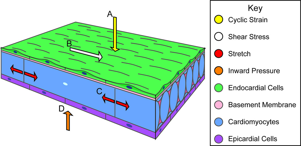
Congenital heart diseases are some of the most common human birth defects. Though some congenital heart defects can be surgically corrected, treatment options for other congenital heart diseases are very limited. In many congenital heart diseases, genetic defects lead to impaired embryonic heart development or growth. One of the key development processes in cardiac development is chamber maturation, and alterations in this maturation process can manifest as a variety of congenital defects including non-compaction, systolic dysfunction, diastolic dysfunction, and arrhythmia. During development, to meet the increasing metabolic demands of the developing embryo, the myocardial wall undergoes extensive remodeling characterized by the formation of muscular luminal protrusions called cardiac trabeculae, increased cardiomyocyte mass, and development of the ventricular conduction system. Though the basic morphological and cytological changes involved in early heart development are clear, much remains unknown about the complex biomolecular mechanisms governing chamber maturation. In this review, we highlight evidence suggesting that a wide variety of basic signaling pathways and biomechanical forces are involved in cardiac wall maturation.

Keywords: BMP; Ephrin; FGF; Left Ventricular Non-Compaction (LVNC); Neuregulin; Notch; Retinoic acid; Semaphorin; cardiac chamber maturation; cardiac trabeculation; cardiomyocyte proliferation; conduction; endothelin; extracellular matrix signaling.


Conflict of interest statement

CONFLICT OF INTEREST

None.

Figures

Figure 1
Figure 1. Cardiac chamber maturation
Figure 1A–D features a schematized cross-section of a theoretical ventricle wall with the developing atrio-ventricular canal represented as the open break in the ventricle wall, such that the outer curvature is on the left and the inner curvature is on the right. (A) Early in development, the cardiac chamber wall is smooth and consists of endocardial cells and myocardial cells. (B) Emergence: Myocardial protrusions called trabeculae begin to appear in the outer curvature of the ventricle, projecting into to the lumen. The trabeculae are lined by a continuous layer of endocardium. (C) Trabeculation: Trabeculae increase in length and the chamber wall becomes topologically more complex as additional trabeculae form throughout the outer curvature, creating a meshwork network of interconnected trabeculae. The compact myocardium does not thicken appreciably. A third layer of cells, the epicardium, surrounds the developing heart. (D) Compaction/Remodeling. Trabeculae cease luminal growth, thicken radially, and their base coalesces to form part of the solid myocardial wall. The compact myocardium increases in mass concomitant with the coronary vessel formation in the myocardial wall. The compact myocardium is shown in dark blue, trabecular cardiomyocytes in cerulean, endocardial cells in green, and epicardial cells in purple. The developing cardiac vasculature is represented by gray circles outlined in green.
Figure 2
Figure 2. Signaling pathways in cardiac chamber maturation
Several signaling pathways have been identified as key regulators of cardiac chamber morphogenesis. Please see below for abbreviations. (A) Canonical NOTCH ligands including Delta and Jagged family members bind to NOTCH family receptors. Upon binding, ADAM17 cleaves the extracellular domain of NOTCH and γ-secretase cleaves the intracellular domain of NOTCH, releasing the NICD into the cytoplasm. NICD translocates into the nucleus and modulates gene transcription. NOTCH activation leads to stimulation of EphrinB2 signaling through EPH4 and NRG1 signaling through ERBB2/4, both of which are essential for trabeculation. NOTCH activation also leads to activation of BMP signaling through BMP10/BMPR interactions and FGF signaling through FGFR. BMP and FGF signaling are essential for cardiomyocyte proliferation and expansion of the compact myocardium. (B) Other signaling pathways essential for cardiac chamber maturation. SEMA6D signaling though PLXNA1 activates the enabled homolog MENA, modulating both trabeculation and compact myocardium proliferation/expansion. Vitamin A is oxidized into retinoic acid. Retinoic acid family members, RXRs, bind retinoic acid and translocate into the nucleus where they influence gene transcription involved in compact cardiomyocyte proliferation. Pro-endothelin secreted into extracellular space is converted into ET-1 by ECE1. ET-1 binding activates the G-protein coupled receptor EDNRA, leading to downstream signaling and gene transcription essential for Purkinje Fiber formation. Diverse extracellular matrix molecules collectively referred to as ECM, either whole or after proteolysis by MMPs, interact with α/β integrin heterodimers. This induces conformation changes in the integrin heterodimer that activate downstream signal transduction that ultimately modulates all elements cardiac chamber maturation. Abbreviations: NRG1; Neuregulin-1: ERBB2/4; heterodimer with ErbB2 (v-erb-b2 erythroblastic leukemia viral oncogene homolog 2) and ErbB4 (v-erb-a erythroblastic leukemia viral oncogene homolog 4): EFNB2; Ephrin-B2: EPHB4; EPH receptor B4: ADAM17; ADAM metallopeptidase domain 17: NOTCH; NOTCH family receptors: γ-secretase; gamma-secretase: NICD; NOTCH intracellular domain: BMP10; Bone Morphogenic Protein 10: BMPR2; Bone morphogenic Protein receptor, type II: SMAD; SMAD family transcription factors: FGF; Fibroblast Growth Factors: FGFR; Fibroblast Growth Factor Receptors: SEMA6D; Semaphorin 6D: PLXNA1; Plexin A1: MENA; Enabled homolog (mammalian): RXR; Retinoic Acid Receptor family: Pro-ET; Pro-endothelin: ECE1; Endothelin-converting enzyme 1: ET-1; Endothelin 1: EDNRA; Endothelin receptor type A: ECM; Extra Cellular Matrix components: MMP; Matrix Metalloprotease.
Figure 3
Figure 3. Biomechanical forces in Cardiac Wall Maturation
Biomechanical forces are important for normal developmental patterning. Forces exerted on the wall from blood flow include (A) cyclic strain, a force perpendicular to the vessel wall, and (B) shear stress, the frictional force parallel to the vessel wall. (C) Force from cardiac contraction exerts strain on myocardial and endothelial cell-cell junctions. (D) The splachnopleural membrane interacts with the myocardial wall during development and may exert an inward pressure on the myocardial wall.

References

    1. Angelini A, Melacini P, Barbero F, Thiene G. Evolutionary persistence of spongy myocardium in humans. Circulation. 1999;99:2475–2475. - PubMed
    1. Beis D, Stainier DYR. In vivo cell biology: Following the zebrafish trend. Trends Cell Biol. 2006;16:105–112. - PubMed
    1. Berdougo E, Coleman H, Lee DH, Stainier DYR, Yelon D. Mutation of weak atrium/atrial myosin heavy chain disrupts atrial function and influences ventricular morphogenesis in zebrafish. Development. 2003;130:6121–6129. - PubMed
    1. Bhatia NL, Tajik AJ, Wilansky S, Steidley DE, Mookadam F. Isolated noncompaction of the left ventricular myocardium in adults: A systematic overview. J Card Fail. 2011;17:771–778. - PubMed
    1. Breckenridge RA, Anderson RH, Elliott PM. Isolated left ventricular non-compaction: The case for abnormal myocardial development. Cardiol Young. 2007;17:124–129. - PubMed

Publication types

MeSH terms