. 2013 Oct 1;22(19):3869-82.
doi: 10.1093/hmg/ddt242. Epub 2013 May 29.

A role of mitochondrial complex II defects in genetic models of Huntington's disease expressing N-terminal fragments of mutant huntingtin

Affiliations

A role of mitochondrial complex II defects in genetic models of Huntington's disease expressing N-terminal fragments of mutant huntingtin

Maria Damiano et al. Hum Mol Genet. .

Abstract

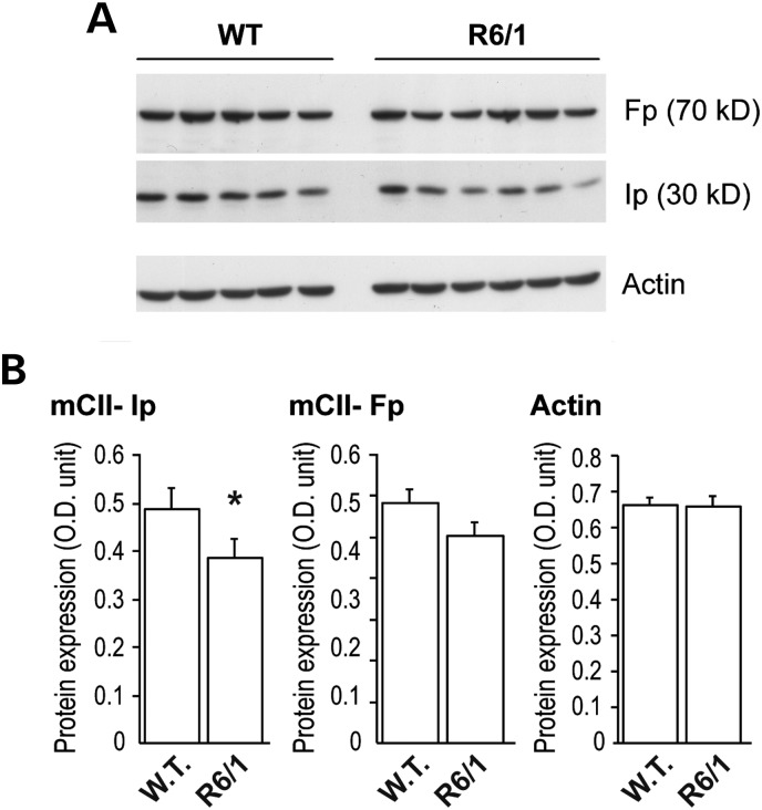
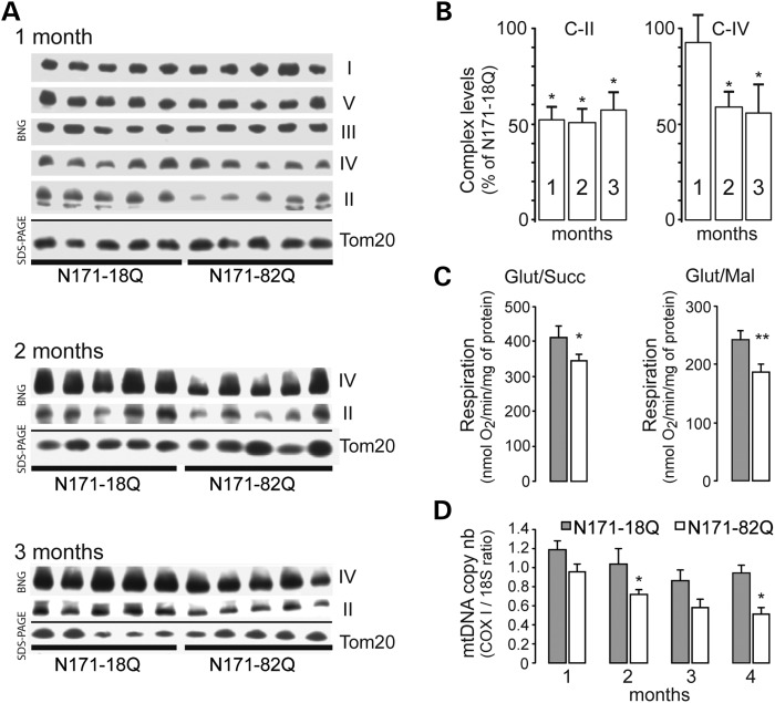
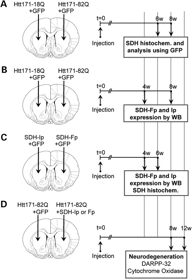
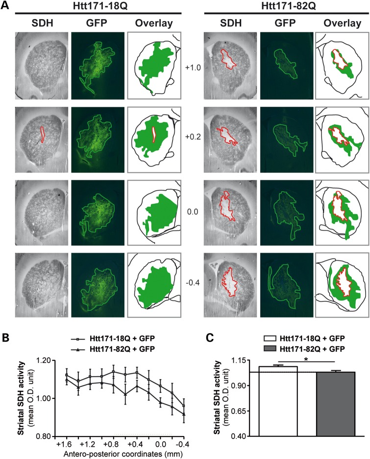
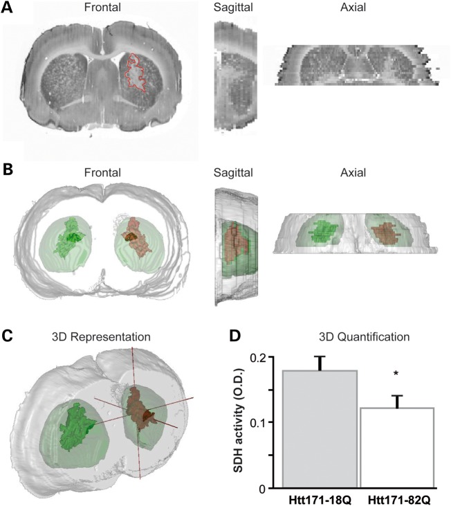
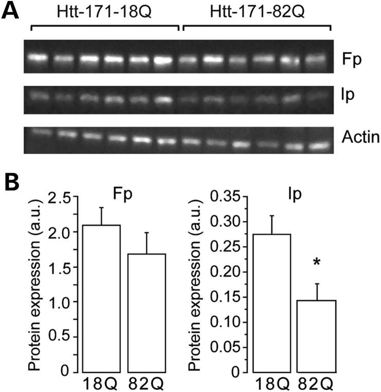
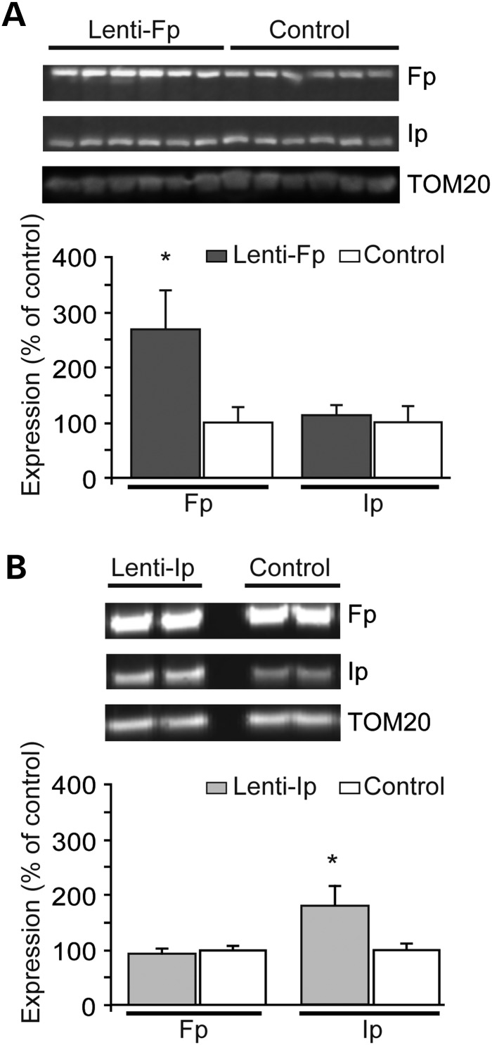
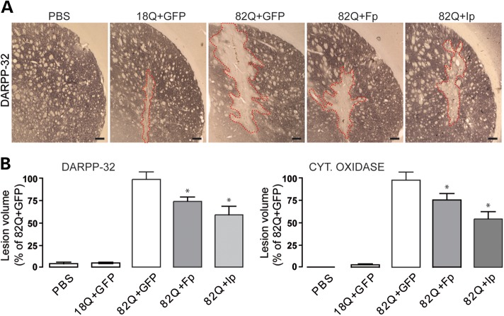
Huntington's disease (HD) is a neurodegenerative disorder caused by an abnormal expansion of a CAG repeat encoding a polyglutamine tract in the huntingtin (Htt) protein. The mutation leads to neuronal death through mechanisms which are still unknown. One hypothesis is that mitochondrial defects may play a key role. In support of this, the activity of mitochondrial complex II (C-II) is preferentially reduced in the striatum of HD patients. Here, we studied C-II expression in different genetic models of HD expressing N-terminal fragments of mutant Htt (mHtt). Western blot analysis showed that the expression of the 30 kDa Iron-Sulfur (Ip) subunit of C-II was significantly reduced in the striatum of the R6/1 transgenic mice, while the levels of the FAD containing catalytic 70 kDa subunit (Fp) were not significantly changed. Blue native gel analysis showed that the assembly of C-II in mitochondria was altered early in N171-82Q transgenic mice. Early loco-regional reduction in C-II activity and Ip protein expression was also demonstrated in a rat model of HD using intrastriatal injection of lentiviral vectors encoding mHtt. Infection of the rat striatum with a lentiviral vector coding the C-II Ip or Fp subunits induced a significant overexpression of these proteins that led to significant neuroprotection of striatal neurons against mHtt neurotoxicity. These results obtained in vivo support the hypothesis that structural and functional alterations of C-II induced by mHtt may play a critical role in the degeneration of striatal neurons in HD and that mitochondrial-targeted therapies may be useful in its treatment.


Figures

Figure 1.
Figure 1.
Change in C-II subunits in R6/1 mice. The striatum was dissected out from 16-week-old mice transgenic for human mHtt exon 1 and analyzed by western blot. Equal amount of proteins was loaded in each lane. Each lane represents a different mouse. (A) Typical western blot of samples showing the expression of Fp, Ip and actin in striatal samples. (B) Histograms corresponding to the quantification of the expression of the proteins by image analysis. Note that there is a significant loss of SDH subunit Ip, while only a trend is seen for the Fp subunit. Results are the mean ± SEM. *P < 0.02, unpaired Student's t-test.
Figure 2.
Figure 2.
Mitochondrial defects in mitochondria isolated from N171-82Q transgenic mice. Mitochondria were isolated from the forebrain of N171-82Q transgenic mice and controls (N171-18Q mice) and analyzed by blue native gel (BNG) electrophoresis to detect assembled mitochondrial complexes (A and B) and evaluate respiration (C) and mtDNA copy number (D). In (A) are shown representative BN-PAGE from samples prepared with mice at different ages. Levels of Tom20 as detected using western blot were used to control for the quantity of mitochondria in each lane. Note that at 1 month, C-II levels are lower in HD transgenic mice when compared with controls, while other complexes remained essentially unchanged. (B) Quantification of complexes levels is shown. At later time points, C-IV levels are also reduced. Results are the mean ± SEM. *P < 0.05. (C) Respiration of mitochondria preparation was determined using either glutamate/succinate 5:2 mm (complex II) or substrates glutamate/malate 5:1 mm (complex I) at 2 and 3 months of age, respectively. Results are the mean ± SEM. *P < 0.05; **P < 0.02. (D) Evaluation of the mtDNA copy number assessed by the ratio between COX1 mRNA and 18S mRNA levels measured by RT-PCR. Results are the mean ± SEM. *P < 0.05.
Figure 3.
Figure 3.
Experimental design to study mitochondrial complex II expression change induced by mHtt in rats. In all experiments, adult rats received a stereotaxic injection of lentiviral vectors (4 µl). Animals were infected with a lentiviral vector encoding the 171 N-terminal amino acids of mHtt with 82 polyQ repeats (Htt171-82Q), or the corresponding wild-type fragment with 18 polyQ repeats (Htt171-18Q) with the reporter fluorescent protein green fluorescent protein (GFP) or vectors coding the C-II Fp or Ip subunit. In the first experiment (A), rats were injected with lentiviral vectors (4 µl) encoding GFP (left and right striatum, 100 ng/µl of p24) plus either Htt-171-82Q (right striatum, 150 ng/µl of p24) or Htt-171-18Q (left striatum, 150 ng/µl of p24). Semi-quantitative evaluation of C-II activity using histochemistry of SDH activity was performed at 6 weeks to analyze the co-localization of SDH activity with GFP and the 3D measurement of SDH activity in the area showing SDH depletion. In the second experiment (B), rats were injected with lentiviral vectors (4 µl) encoding GFP (left and right striatum, 100 ng/µl of p24) plus either Htt-171-82Q (right striatum, 150 ng/µl of p24) or Htt-171-18Q (left striatum, 150 ng/µl of p24). Rats were killed at 6 weeks post-surgery. Striatal punches of the fluorescent area were made to prepared tissue homogenate to perform western blot analysis of Fp, Ip and actin expression. In the third experiment (C), rats were injected with lentiviral vectors (4 µl) encoding GFP (left and right striatum, 100 ng/µl of p24) plus either lenti-Fp (right striatum, 150 ng/µl of p24) or lenti-Ip (left striatum, 150 ng/µl of p24). Rats were killed at 4 weeks post-surgery. Striatal punches were made to prepared tissue homogenate to perform western blot analysis of C-II subunits Fp and Ip and Tom20. In addition, rats were also injected to perform SDH histochemistry. In the fourth experiment (D), rats were co-injected with lentiviral vectors (4 µl) encoding wt-Htt171-18Q fragment or mHtt171-82Q fragment (100 ng/µl of p24) plus either Fp (150 ng/µl of p24), Ip (150 ng/µl of p24) or GFP as a control of viral load. Rats were killed at 12 weeks post-surgery and processed for the evaluation of striatal degeneration using DARPP-32 immunohistochemistry and COX histochemistry.
Figure 4.
Figure 4.
Lentiviral vectors expressing mHtt fragment leads to a loco-regional reduction in SDH activity in vivo. Animals were infected with a mixture of lentiviral vector encoding the reporter fluorescent protein GFP mixed with vectors coding Htt171-18Q (left panel) or mHtt171-82Q (right panel). (A) Bright field (SDH histochemistry) or fluorescence images were taken from representative coronal brain sections at the levels of the striatum at 6 weeks post-infection. Red lines delineate areas of SDH activity loss. Overlay show the area of SDH loss (red) superimposed to area where the transgenes are expressed (green). Note minimal loss of SDH activity in the control group, in contrast to the striatum injected with lenti-Htt171-82Q. Note that the zone presenting the loss of SDH activity is within the area expressing mHtt. (B) Analysis of the total SDH activity in the striatum along the rostrocaudal. (C) Histograms corresponding to the total SDH activity measured in the striatum in both groups. Scale bar, 1 mm. Data are expressed as the mean ± SEM. *P < 0.002, n = 6 per group, two-way ANOVA and post hoc Bonferroni/Dunn test.
Figure 5.
Figure 5.
Estimation of the loss of SDH activity in the area expressing mHtt in the lentiviral rat model. (A) Bright field (SDH histochemistry) or fluorescence images were taken from representative coronal brain sections at the levels of the striatum at 6 weeks post-infection and stacked for 3D reconstruction. (B and C) 3D reconstruction of the brain anatomy (gray) with super-imposition of the volume presenting a loss of C-II activity (red) in the striatum infected with lenti-Htt171-82Q (mHtt) and its symmetrical projection in the striatum with wt-Htt (green). (D) Histograms corresponding to the regional SDH activity measured in the striatum expressing Htt171-82Q and symmetrically in the contra-lateral striatum. Scale bar, 1 mm. Data are expressed as the mean ± SEM. *P < 0.01, n = 6 per group, paired Student's t-test.
Figure 6.
Figure 6.
Loss of C-II subunits produced by mHtt in the rat lentiviral model. Rats were infected with a mixture of lentiviral vector encoding the reporter fluorescent protein GFP mixed with vectors coding either Htt171-18Q (left panel) or mHtt171-82Q (right panel). Small striatal samples expressing mHtt or wt-Htt were dissected out at 6 weeks and analyzed by western blotting. Equal amount of proteins was loaded in each lane. Each lane represents a different rat. (A) A typical western blot of samples obtained at 6 weeks post-infection showing expression of Fp, Ip and actin in striatal samples. (B) Histograms corresponding to the quantification of the expression of the proteins by image analysis. Note that there is a significant loss of SDH subunit Ip, while only a trend is seen for the Fp subunit. Results are the mean ± SEM. *P < 0.02, n = 6 per group, unpaired Student's t-test.
Figure 7.
Figure 7.
Study of the transduction efficacy of lentiviral vectors coding C-II Ip and Fp subunits in vivo. Rats received intrastriatal injection of lenti-Fp (A) and lenti-Ip (B) mixed with lenti-GFP and the infected area was dissected out 4 weeks later using fluorescence detection for western blot analysis. Representative western blots are shown in upper panel. Control rats have been injected with lentiviral vectors coding the reporter gene LacZ. Results are the mean ± SEM. *P < 0.01, n = 4–6 per group, unpaired Student's t-test.
Figure 8.
Figure 8.
Neuroprotective efficacy of Fp and Ip against mHtt neurotoxicity in vivo. Rats were co-injected with lentiviral vectors encoding wt-Htt171-18Q fragment or mHtt171-82Q plus either, Fp, Ip or GFP as a control of viral load. Rats were killed at 12 weeks post-surgery and processed for histological evaluation. (A) Photomicrographs of representative lesions seen in the different groups of rats o characterize striatal degeneration using DARPP-32 immunohistochemistry. Scale bar, 200 µm. Histograms in (B) represent the volume of lesions produced in the different conditions when assessed by the Cavalieri method using DARPP32 immunohistochemistry (left) and cytochrome c oxidase (Cyt. Oxidase) histochemistry (right). Note that the volume of striatal lesions produced by mHtt is significantly reduced by overexpressing Fp and Ip proteins when compared with control (GFP). Data are the mean ± SEM. *P < 0.01, n = 10–12 per group, one-way ANOVA followed by the Bonferroni post hoc test.

Comment in

  • Epigenetics of Huntington's disease.
    McLauchlan D, Robertson NP. McLauchlan D, et al. J Neurol. 2013 Nov;260(11):2938-41. doi: 10.1007/s00415-013-7158-x. J Neurol. 2013. PMID: 24141735 No abstract available.

Similar articles

Cited by

References

    1. Damiano M., Galvan L., Deglon N., Brouillet E. Mitochondria in Huntington's disease. Biochim. Biophys. Acta. 2010;1802:52–61. - PubMed
    1. Harper P.S. Huntington's Disease. WB Saunders Company Ltd, London. 1991
    1. Borrell-Pages M., Zala D., Humbert S., Saudou F. Huntington's disease: from huntingtin function and dysfunction to therapeutic strategies. Cell Mol. Life Sci. 2006;63:2642–2660. - PMC - PubMed
    1. Myers R.H., Vonsattel J.P., Stevens T.J., Cupples L.A., Richardson E.P., Martin J.B., Bird E.D. Clinical and neuropathologic assessment of severity in Huntington's disease. Neurology. 1988;38:341–347. - PubMed
    1. Sugars K.L., Rubinsztein D.C. Transcriptional abnormalities in Huntington disease. Trends Genet. 2003;19:233–238. - PubMed

Publication types

MeSH terms