

The role of the hippo pathway in melanocytes and melanoma
- PMID: 23720711
- PMCID: PMC3655322
- DOI: 10.3389/fonc.2013.00123
The role of the hippo pathway in melanocytes and melanoma
Abstract
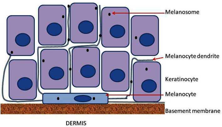
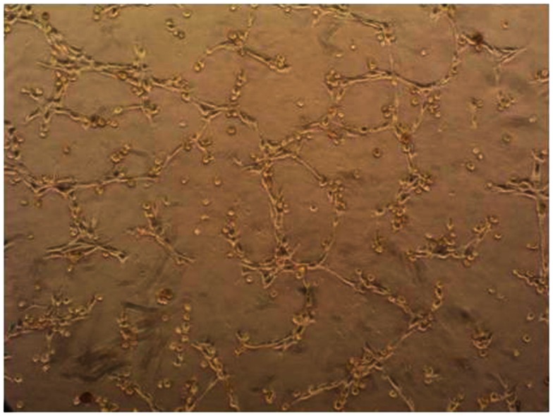
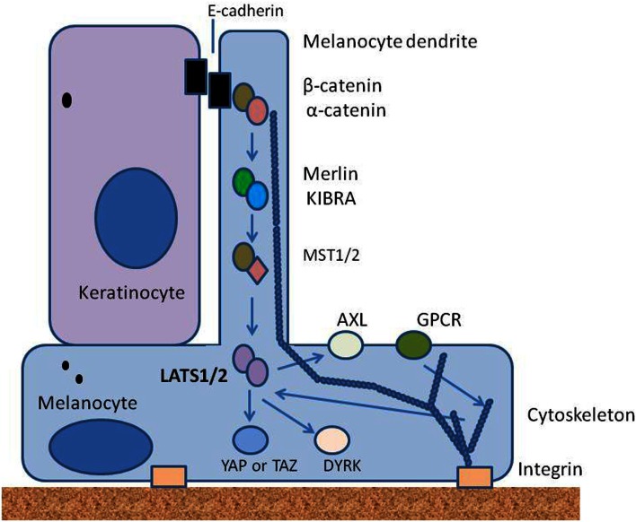
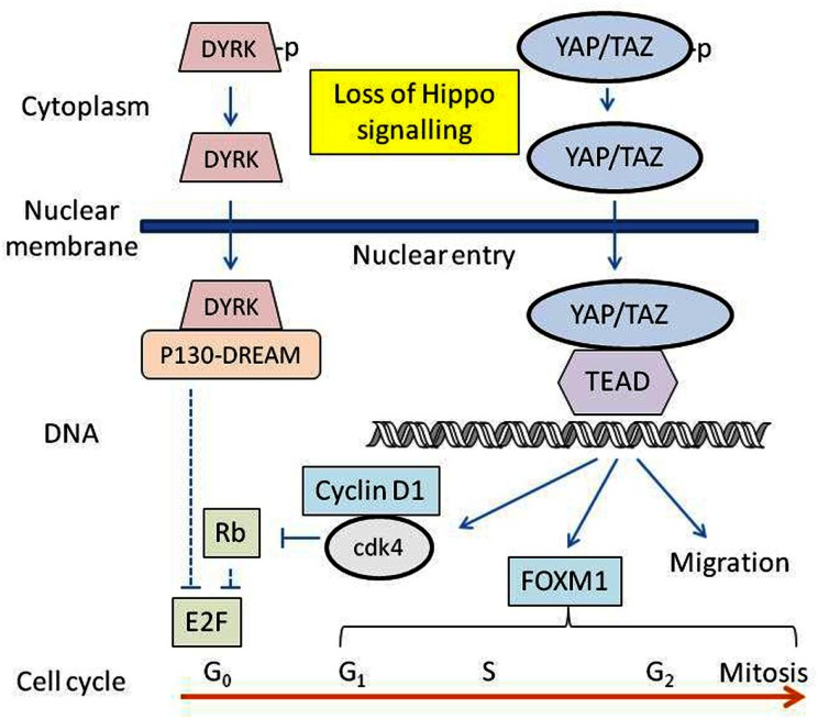
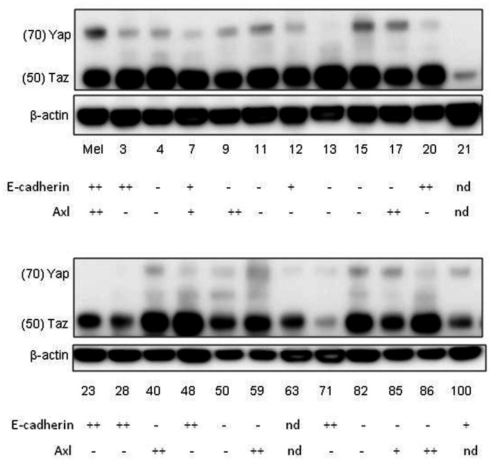
The Hippo signaling pathway comprises a series of cytoplasmic tumor suppressor proteins including Merlin and the Lats1/2 and MST1/2 kinases, and is thought to play a critical role in determining the sizes of organs and tissues. The Hippo pathway is regulated upstream by extracellular mechanosensory signaling arising from cell shape and polarity, as well as by a variety of extracellular signaling molecules. When active, the pathway maintains the transcriptional activators Yes-associated protein (YAP) and TAZ in phosphorylated forms in the cytoplasm, preventing cell proliferation. When the Hippo pathway is inactivated, YAP and TAZ are translocated to the nucleus and induce the expression of a variety of proteins concerned with entry into the cell division cycle, such as cyclin D1 and Fox M1, as well as the inhibition of apoptosis. The failure of the Hippo pathway has been implicated in the development of many different types of cancer but there is limited information available as to its involvement in melanoma. We hypothesize here firstly that the Hippo pathway is involved in maintaining density of cutaneous melanocytes on the basement membrane at the junction of the epidermis and the dermis, and secondly, that its function is disturbed in melanoma. We have analyzed a series of 23 low passage human melanoma lines as well as cultured normal melanoma, and find that melanocytes, as well as all melanoma cell lines examined express TAZ. Melanocytes and most melanoma lines also express YAP. E-cadherin, an upstream regulator of the Hippo pathway, and Axl, a receptor tyrosine kinase regulated by the Hippo pathway, are expressed in melanocytes and in several melanoma cell lines. These observations, together with published evidence for the presence of Merlin, Lats1/2, and MST1/2 in melanocytes and melanoma cells, support the hypothesis that the Hippo pathway is an important component of melanocyte and melanoma behavior.
Keywords: E-cadherin; cell proliferation; cytoskeleton; epidermal melanocytes; merlin.
Figures
Similar articles
-
Reciprocal regulation of YAP/TAZ by the Hippo pathway and the Small GTPase pathway.Small GTPases. 2020 Jul;11(4):280-288. doi: 10.1080/21541248.2018.1435986. Epub 2018 Apr 20. Small GTPases. 2020. PMID: 29457552 Free PMC article. Review.
-
Large tumor suppressor homologs 1 and 2 regulate mouse liver progenitor cell proliferation and maturation through antagonism of the coactivators YAP and TAZ.Hepatology. 2016 Nov;64(5):1757-1772. doi: 10.1002/hep.28768. Epub 2016 Sep 30. Hepatology. 2016. PMID: 27531557 Free PMC article.
-
MAP4K family kinases act in parallel to MST1/2 to activate LATS1/2 in the Hippo pathway.Nat Commun. 2015 Oct 5;6:8357. doi: 10.1038/ncomms9357. Nat Commun. 2015. PMID: 26437443 Free PMC article.
-
Hippo-Independent Regulation of Yki/Yap/Taz: A Non-canonical View.Front Cell Dev Biol. 2021 Apr 1;9:658481. doi: 10.3389/fcell.2021.658481. eCollection 2021. Front Cell Dev Biol. 2021. PMID: 33869224 Free PMC article. Review.
-
The Hippo Pathway Component TAZ Promotes Immune Evasion in Human Cancer through PD-L1.Cancer Res. 2018 Mar 15;78(6):1457-1470. doi: 10.1158/0008-5472.CAN-17-3139. Epub 2018 Jan 16. Cancer Res. 2018. PMID: 29339539
Cited by
-
LATS1 Is a Mediator of Melanogenesis in Response to Oxidative Stress and Regulator of Melanoma Growth.Int J Mol Sci. 2021 Mar 18;22(6):3108. doi: 10.3390/ijms22063108. Int J Mol Sci. 2021. PMID: 33803640 Free PMC article.
-
miR-375-3p suppresses tumorigenesis and partially reverses chemoresistance by targeting YAP1 and SP1 in colorectal cancer cells.Aging (Albany NY). 2019 Sep 22;11(18):7357-7385. doi: 10.18632/aging.102214. Epub 2019 Sep 22. Aging (Albany NY). 2019. PMID: 31543507 Free PMC article.
-
Long non-coding RNA expression identified by microarray analysis: Candidate biomarkers in human acral lentiginous melanoma.Oncol Lett. 2020 Feb;19(2):1465-1477. doi: 10.3892/ol.2019.11207. Epub 2019 Dec 11. Oncol Lett. 2020. PMID: 31966073 Free PMC article.
-
The role of transcriptional coactivator TAZ in gliomas.Oncotarget. 2016 Dec 13;7(50):82686-82699. doi: 10.18632/oncotarget.12625. Oncotarget. 2016. PMID: 27764783 Free PMC article.
-
Deregulation of the Hippo pathway in soft-tissue sarcoma promotes FOXM1 expression and tumorigenesis.Proc Natl Acad Sci U S A. 2015 Jun 30;112(26):E3402-11. doi: 10.1073/pnas.1420005112. Epub 2015 Jun 15. Proc Natl Acad Sci U S A. 2015. PMID: 26080399 Free PMC article.
References
-
- Arck P. C., Overall R., Spatz K., Liezman C., Handjiski B., Klapp B. F., et al. (2006). Towards a “free radical theory of graying”: melanocyte apoptosis in the aging human hair follicle is an indicator of oxidative stress induced tissue damage. FASEB J. 20, 1567–156910.1096/fj.05-4039fje - DOI - PubMed
LinkOut - more resources
Full Text Sources
Other Literature Sources
Research Materials
Miscellaneous