. 2013 Aug;87(15):8624-37.
doi: 10.1128/JVI.00899-13. Epub 2013 May 29.

Three infectious viral species lying in wait in the banana genome

Affiliations

Three infectious viral species lying in wait in the banana genome

Matthieu Chabannes et al. J Virol. 2013 Aug.

Abstract

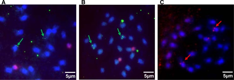
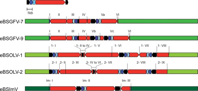
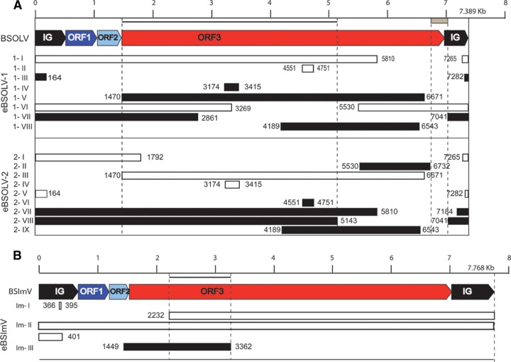
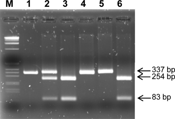
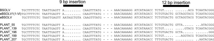
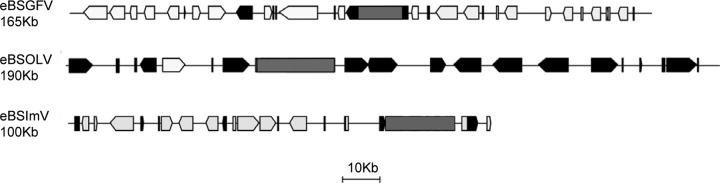
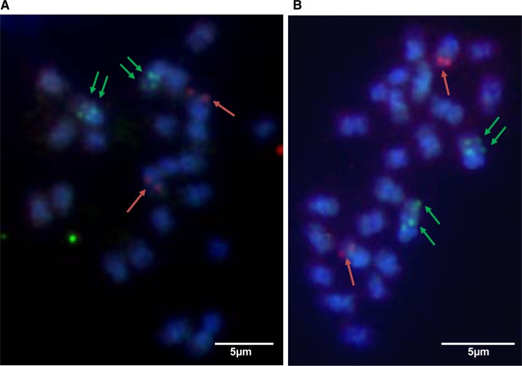
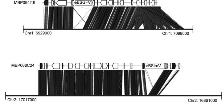
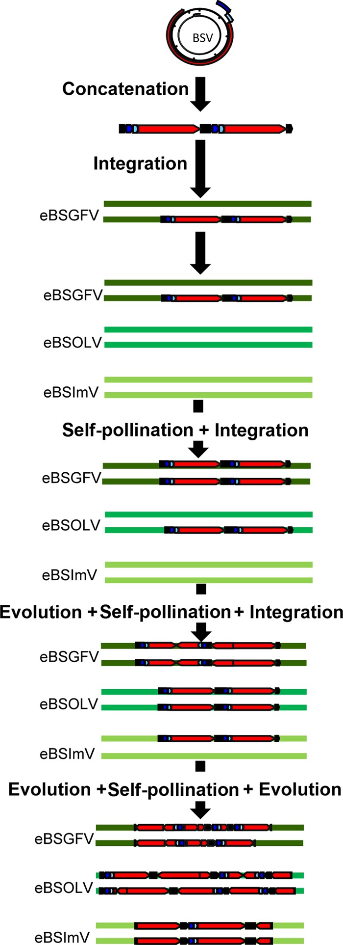
Plant pararetroviruses integrate serendipitously into their host genomes. The banana genome harbors integrated copies of banana streak virus (BSV) named endogenous BSV (eBSV) that are able to release infectious pararetrovirus. In this investigation, we characterized integrants of three BSV species-Goldfinger (eBSGFV), Imove (eBSImV), and Obino l'Ewai (eBSOLV)-in the seedy Musa balbisiana Pisang klutuk wulung (PKW) by studying their molecular structure, genomic organization, genomic landscape, and infectious capacity. All eBSVs exhibit extensive viral genome duplications and rearrangements. eBSV segregation analysis on an F1 population of PKW combined with fluorescent in situ hybridization analysis showed that eBSImV, eBSOLV, and eBSGFV are each present at a single locus. eBSOLV and eBSGFV contain two distinct alleles, whereas eBSImV has two structurally identical alleles. Genotyping of both eBSV and viral particles expressed in the progeny demonstrated that only one allele for each species is infectious. The infectious allele of eBSImV could not be identified since the two alleles are identical. Finally, we demonstrate that eBSGFV and eBSOLV are located on chromosome 1 and eBSImV is located on chromosome 2 of the reference Musa genome published recently. The structure and evolution of eBSVs suggest sequential integration into the plant genome, and haplotype divergence analysis confirms that the three loci display differential evolution. Based on our data, we propose a model for BSV integration and eBSV evolution in the Musa balbisiana genome. The mutual benefits of this unique host-pathogen association are also discussed.


Figures

Fig 1
Fig 1
Localization of the three BSV integrations by fluorescent in situ hybridization in root cells of PKW banana chromosomes. Hybridizations were performed with full-length genome probes for each BSV species and detected with FITC (green for BSGFV in panel A and BSImV in panel B) or rhodamine (red for BSOLV in panel C). Pink diffuse signals in panels A and B correspond to the 45S rDNA sequence labeled with Alexa 555. Chromosomes are counterstained with DAPI (blue).
Fig 2
Fig 2
Overview of eBSV structures in PKW. Banana genomic sequences are in green. The BSV genome is represented in linear view with dark blue, light blue, and red boxes indicating ORF 1, ORF2, and ORF3 of the virus, respectively. The intergenic region is in black. eBSV fragments are indicated.
Fig 3
Fig 3
Positions of eBSV fragments in the counterpart BSV genome. BSOLV (A) and BSImV (B) genomes are each represented in linear view, with a kilobase pair scale bar at the top. IG, ORF1, ORF2, and ORF3 are labeled. Boxes below the BSV genome represent fragments of eBSV in the same orientation (white boxes) or in reverse orientation (black boxes). Coordinates indicate boundaries of eBSV fragments in reference to the viral genome. The names of fragments are indicated on the left side. The gray box below the scale bar indicates the missing fragment of ORF3 in eBSOLV-2. Black lines below the scale bar represent the zone of ORF3 used for dating analysis of both BSOLV and BSImV.
Fig 4
Fig 4
Genotyping of eBSOLV-1 and eBSOLV-2 in the F1 triploid (AAB) population using a dCAP marker. The PCR product obtained from eBSOLV-1 is cut into two bands of 83 and 254 bp by the endonuclease HaeIII (New England BioLabs), whereas the one from eBSOLV-2 is not digested. Lane M, 1-kb DNA ladder (Invitrogen); lane 1, undigested PCR product of the diploid M. balbisiana PKW; lanes 2 to 6, digested PCR product from PKW carrying both eBSOLV alleles, BAC MBP_31007 carrying eBSOLV-1, BAC MBP_73B22 carrying eBSOLV-2, and two triploid (AAB) F1 progeny of a genetic cross between PKW and M. acuminata cv. IDN 110 4x (AAAA) carrying eBSOLV-2 and eBSOLV-1, respectively. Digested DNA was loaded onto a 2.5% agarose gel stained with ethidium bromide, and bands were visualized under UV light.
Fig 5
Fig 5
Comparison of IGs of BSOLV infecting triploid offspring and eBSOLV. Primers (BSV2, 5′-GTA TCA GAG CAA GGT TCG TTT TT-3′, and BSV525, 5′-ATC CCA AGT TTT CTC GAC CAT AA-3′) surrounding the 9- and 12-bp deletions in the IG of BSOLV were designed and used in IC-PCR on 5 independent infected interspecific AAB hybrids (plants 55, 139, 196, 199, and 207) using the following PCR program: denaturation stage at 94°C for 5 min followed by 30 cycles of 30 s at 94°C, 30 s at 60°C, and 1 min at 72°C and a final extension at 72°C for 10 min. PCR products were purified and sequenced. BSOLV, IG of episomal virus (accession number AJ002234); eBSOLV1 (1-VI), IG present in fragment VI of eBSOLV allele 1; eBSOLV, IG present in both alleles of the wild diploid M. balbisiana PKW (except in fragment VI of eBSOLV1); plants 55 to 207, IG of episomal virus produced from eBSOLV in the different hybrids.
Fig 6
Fig 6
Gene and TE contents surrounding eBSVs in the Musa genome present in BAC clones. The black boxes correspond to TE, the gray boxes to eBSV, and the white boxes to genes in BAC clones containing eBSGFV, eBSImV, and eBSOLV.
Fig 7
Fig 7
Localization of the three eBSV on the chromosomes of PKW. Two independent metaphases are shown (A and B). Hybridizations were performed with full-length genome probes for each BSV species and detected with FITC (green) for BSGFV and BSOLV and with Alexa 594 for BSImV. Chromosomes are counterstained with DAPI (blue).
Fig 8
Fig 8
eBSV locus synteny with the Musa acuminata reference genome. “MBP094I16” and “MBP068C24” refer to BACs containing eBSGFV and eBSImV, respectively. The black boxes correspond to TEs and the white boxes to genes. Thick black lines represent the part of chromosomes 1 and 2 of the reference M. acuminata genome (38) matching with BAC containing eBSGFV and eBSImV, respectively. Positions on chromosome are indicated with coordinates.
Fig 9
Fig 9
Proposed scenario for BSGFV, BSOLV, and BSImV integrations into the M. balbisiana nuclear genome. The life cycle of the virus produces concatenated viral genomes in the nucleus of the cell. This concatenated genome is integrated into the nuclear genome of PKW by illegitimate recombination. The three BSV species integrate sequentially into the PKW genome. After each BSV integration, the homozygous state is obtained by self-pollination and selected. Between each new integration and self-pollination, the previously integrated BSV evolves continuously by unequal recombination and point mutation insertions, leading to the present-day structure. The BSV genome is represented in linear view, with dark blue, light blue, and red boxes indicating the three ORFs of the virus. The IG is in black.

References

    1. Weiss RA. 2006. The discovery of endogenous retroviruses. Retrovirology 3:67. - PMC - PubMed
    1. Vogt PK. 1997. Historical introduction to the general properties of retroviruses, p 1–26 In Coffin JM, Hughes SH, Varmus HE. (ed), Retroviruses. Cold Spring Harbor Laboratory Press, Cold Spring Harbor, NY - PubMed
    1. Belyi VA, Levine AJ, Skalka AM. 2010. Unexpected inheritance: multiple integrations of ancient bornavirus and ebolavirus/marburgvirus sequences in vertebrate genomes. PLoS Pathog. 6:e1001030. - PMC - PubMed
    1. Taylor DJ, Leach RW, Bruenn J. 2010. Filoviruses are ancient and integrated into mammalian genomes. BMC Evol. Biol. 10:193. - PMC - PubMed
    1. Belyi VA, Levine AJ, Skalka AM. 2010. Sequences from ancestral single-stranded DNA viruses in vertebrate genomes: the Parvoviridae and Circoviridae are more than 40 to 50 million years old. J. Virol. 84:12458–12462 - PMC - PubMed

Publication types

LinkOut - more resources