. 2013 Aug;87(15):8591-605.
doi: 10.1128/JVI.01081-13. Epub 2013 May 29.

Protection against lethal influenza with a viral mimic

Affiliations

Protection against lethal influenza with a viral mimic

Steven F Baker et al. J Virol. 2013 Aug.

Abstract

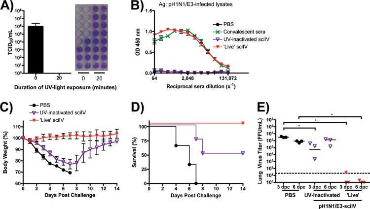
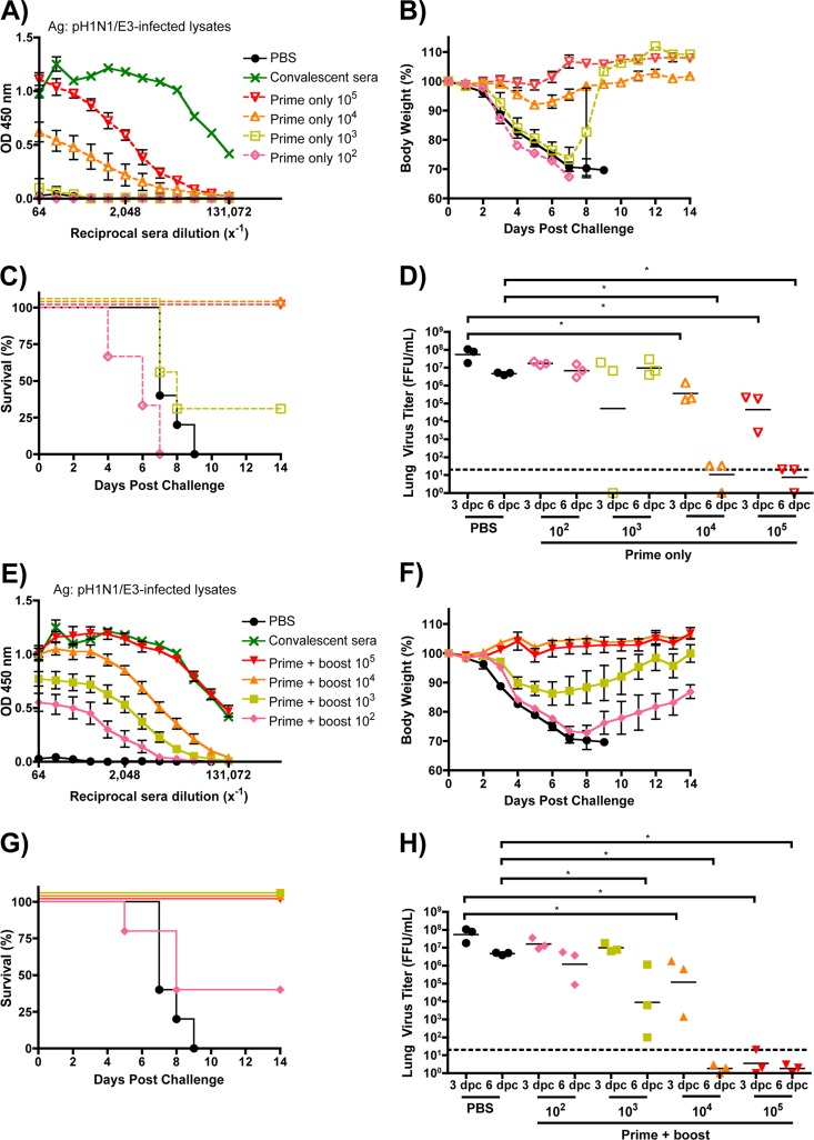
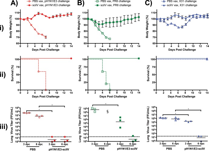
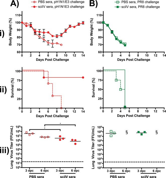
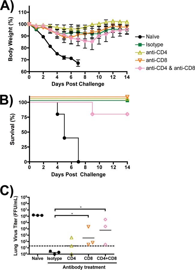
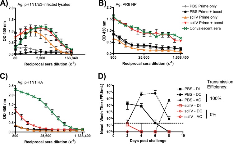
Despite countermeasures against influenza virus that prevent (vaccines) and treat (antivirals) infection, this upper respiratory tract human pathogen remains a global health burden, causing both seasonal epidemics and occasional pandemics. More potent and safe new vaccine technologies would contribute significantly to the battle against influenza and other respiratory infections. Using plasmid-based reverse genetics techniques, we have developed a single-cycle infectious influenza virus (sciIV) with immunoprotective potential. In our sciIV approach, the fourth viral segment, which codes for the receptor-binding and fusion protein hemagglutinin (HA), has been removed. Thus, upon infection of normal cells, although no infectious progeny are produced, the expression of other viral proteins occurs and is immunogenic. Consequently, sciIV is protective against influenza homologous and heterologous viral challenges in a mouse model. Vaccination with sciIV protects in a dose- and replication-dependent manner, which is attributed to both humoral responses and T cells. Safety, immunogenicity, and protection conferred by sciIV vaccination were also demonstrated in ferrets, where this immunization additionally blocked direct and aerosol transmission events. All together, our studies suggest that sciIV may have potential as a broadly protective vaccine against influenza virus.


Figures

Fig 1
Fig 1
Three mutations in the receptor-binding domain of pH1N1 HA allow for complementation of sciIV. (A) Transient HA complementation in MDCK cells. MDCK cells were transfected with pCAGGS protein expression plasmids for HA from pH1N1 (pH1N1-HA) or pH1N1/E3 (pH1N1/E3-HA) or empty plasmid and, 24 h posttransfection, infected with WSN-sciIV (12) (MOI, 0.01) to evaluate trans-complementation by GFP focus formation. GFP-expressing cells were visualized by fluorescence microscopy 24 and 48 h postinfection. Representative images at ×10 magnification are illustrated. Scale bars, 40 μM. (B) HA protein expression by MDCK stable cell lines. Monolayers of MDCK cells stably transfected with WSN-HA or pH1N1/E3-HA or parental cells were fixed and incubated with MAbs specific for WSN (2G9) or pH1N1 (31C2) HA and with DAPI for nuclear staining. Representative images are shown at ×40 magnification. Scale bars, 10 μM. (C and D) Multicycle growth of sciIV. Parental, WSN-HA, or pH1N1/E3-HA MDCK cells were infected with WSN-sciIV at an MOI of 0.001. At various times postinfection, GFP expression was visualized by fluorescence microscopy (×10 representative images; scale bars, 40 μM) (C), and TCSs were collected for titration in MDCK WSN-HA cells (D). Data represent means ± SD of the results determined for triplicates. (E) Plaque morphology of sciIV in HA-expressing cells. WSN-HA, pH1N1/E3-HA, or parental MDCK cells were infected with WSN-sciIV. Three days postinfection, monolayers were stained with crystal violet.
Fig 2
Fig 2
Characterization of pH1N1/E3-sciIV. Plasmid-based reverse genetics techniques were used to rescue a sciIV with genes derived from pH1N1, except the fourth segment which codes for HA(45)GFP(80), as previously described (12). (A) GFP is expressed by cells infected with pH1N1/E3-sciIV. MDCK cells were mock infected or infected with pH1N1 or pH1N1/E3-sciIV at an MOI of 5. Ten hours postinfection, monolayers were fixed, permeabilized, and incubated with a MAb against NP (HT103) and subjected to nuclear counterstaining with DAPI. Representative ×20 images (NP, GFP, DAPI, and merged [bottom row]) are shown. Scale bars, 20 μM. (B and C) Multicycle growth of pH1N1/E3-sciIV. WSN-HA, pH1N1/E3-HA, or parental MDCK cells were infected with pH1N1/E3-sciIV at an MOI of 0.001. At various times postinfection, GFP expression was visualized by fluorescence microscopy (B) (×10 representative images shown; scale bars, 40 μM), and TCSs were collected for titration in MDCK WSN-HA cells (C). Data represent means ± SD of the results determined for triplicates. (D) Multicycle growth of pH1N1/E3-sciIV and pH1N1/E3. Parental or pH1N1/E3-HA-expressing MDCK cells were infected with pH1N1/E3 or pH1N1/E3-sciIV at an MOI of 0.001. At various times postinfection, TCSs were collected for titration on MDCK cells by IFA using the NP MAb HT103. Data represent means ± SD of the results determined for triplicates. (E) Plaque morphology of pH1N1/E3-sciIV. WSN-HA, pH1N1/E3-HA, or parental MDCK cells were infected with pH1N1 or pH1N1/E3-sciIV for plaque assay. Three days postinfection, monolayers were stained with crystal violet. (F) pH1N1/E3-sciIV can infect cells that do not express HA. Parental, WSN-HA, or pH1N1/E3-HA MDCK cells were infected at an MOI of 5 with pH1N1/E3-sciIV. Ten hours postinfection, monolayers were analyzed by fluorescence microscopy for GFP expression. Representative images (×10 magnification) are shown. Scale bars, 40 μM.
Fig 3
Fig 3
Immunogenicity of pH1N1/E3-sciIV in mice. (A to C) Humoral immune response to vaccination. Female 6-to-8-week-old C56BL/6 mice (n = 5) were inoculated at 2-week intervals i.n. with 105 FFU of pH1N1/E3-sciIV once (orange triangles, prime only) or twice (red triangles, prime plus boost) or with PBS (black circles) as a negative control. Two weeks postvaccination, sera were collected and evaluated for IgG Abs against total influenza virus protein (A), recombinant NP from PR8 (B), or recombinant HA from pH1N1 (C) by ELISA. Convalescent-phase sera (green) collected 2 weeks after naive mice were infected with 0.02 MLD50 pH1N1/E3 were used as a positive control (n = 3). Data represent means ± standard errors of the means (SEM) of the results from 4 independent experiments. Ag, antigen; OD, optical density. (D) CD8+ T cells present in the lungs after immunization with pH1N1/E3-sciIV. Female 6-to-8-week-old C56BL/6 mice were inoculated twice at 2-week intervals i.n. with 105 FFU of pH1N1/E3-sciIV or PBS (Naive) or were infected with 0.02 MLD50 pH1N1/E3 (n = 3). Ten days after the final vaccination or infection, lungs were extracted and resident cells were pooled and prepared for flow cytometry. Live, CD3+ CD4 CD8+ cells specific for pH1N1 NP366-374 tetramer were counted. Data represent means ± SEM of the results from 4 independent experiments. Statistical analysis was performed using Student's one-tailed paired t test. * = P ≤ 0.05; ** = P ≤ 0.01; ns = not significant.
Fig 4
Fig 4
pH1N1/E3-sciIV vaccination protects mice from lethal homologous challenge. (A) Representation of the immunization and challenge schedule. (B) Tolerability of pH1N1/E3-sciIV. Female 6-to-8-week-old C56BL/6 mice were inoculated at 2-week intervals i.n. with 105 FFU of pH1N1/E3-sciIV once or twice or with PBS as a negative control (n = 5). Weight loss was monitored daily for 2 weeks after the PBS or first (or ange triangles, prime only) or second (red triangles, prime plus boost) sciIV inoculation. Plotted data represent means ± SEM. (C and D) Protection from lethal challenge conferred by pH1N1/E3-sciIV vaccination. Immunized mice from the experiment described for panel B were challenged with 10 MLD50 pH1N1/E3, and weight loss (plotted data represent means ± SEM) (C) and survival (D) were monitored daily for 2 weeks postchallenge. (E) Virus lung titers. Mice immunized and challenged as described for panels B and D were sacrificed at 3 and 6 days postchallenge (dpc) to evaluate levels of replicating challenge virus in the lungs by IFA as described for Fig. 2D (n = 3). Symbols represent data from individual mice; bars represent geometric mean lung virus titers, and the dotted line denotes the limit of detection (20 FFU/ml). Statistical analysis was performed using the Mann-Whitney test; * = P ≤ 0.05.
Fig 5
Fig 5
UV-inactivated pH1N1-sciIV does not fully protect mice from lethal homologous challenge. (A) UV light treatment renders sciIV noninfectious. SciIV was exposed to UV light for 20 min on ice, and infectivity was determined by TCID50 assay on MDCK WSN-HA cells. (B) Immunogenicity of UV-inactivated sciIV. Female 6-to-8-week-old C56BL/6 mice were inoculated twice at 2-week intervals i.n. with 105 FFU of pH1N1/E3-sciIV that was (purple triangles; UV-inactivated sciIV) or was not (red triangles; ‘Live’ sciIV) exposed to UV light on ice for 20 min or with PBS (black circles) as a negative control (n = 5). Two weeks postvaccination, sera were collected and evaluated for IgG Abs against total influenza virus protein by ELISA. Convalescent-phase sera (green) were used as a positive control (n = 3); plotted data represent means ± SEM. (C and D) Protection from lethal challenge by pH1N1/E3-sciIV vaccination. Immunized mice from the experiment described for panel B were challenged with 10 MLD50 pH1N1/E3, and weight loss (C) (plotted data represent meana ± SEM) and survival (D) were monitored daily for 2 weeks postchallenge. (E) Virus lung titers. Mice immunized and challenged as described for panels C and D were sacrificed at 3 and 6 days postchallenge to evaluate levels of replicating challenge virus in the lungs by IFA (n = 3). Symbols represent individual mice; bars represent geometric mean lung virus titers, and the dotted line denotes the limit of detection (20 FFU/ml). Statistical analysis was performed using the Mann-Whitney test; * = P ≤ 0.05.
Fig 6
Fig 6
Dose-dependent immunogenicity and protective efficacy conferred by sciIV vaccination. Female 6-to-8-week-old C56BL/6 mice were inoculated once (A to D, dotted lines) or twice (E to H, solid lines) at 2-week intervals i.n. with 10-fold dilutions of pH1N1/E3-sciIV (red, 105; orange, 104; yellow, 103; pink, 102) or with PBS (black) as a negative control (n = 5). Two weeks postvaccination, sera were collected and evaluated for IgG Abs against total influenza virus protein by ELISA (A and E; plotted data represent means ± SEM). Convalescent-phase sera (green) were used as a positive control (n = 3). Immunized mice from the experiment described for panels A and E were challenged with 10 MLD50 pH1N1/E3, and weight loss (B and F; plotted data represent means ± SEM) and survival (C and G) were monitored daily for 2 weeks postchallenge. (D and H) Virus lung titers. Mice immunized and challenged as described above were sacrificed at 3 and 6 days postchallenge to evaluate levels of replicating challenge virus in the lungs by IFA (n = 3). The experiments represented in panels D and H were performed simultaneously; thus, the same PBS-vaccinated controls were plotted in both graphs. Symbols represent individual mice; bars represent geometric mean lung virus titers, and the dotted line denotes the limit of detection (20 FFU/ml). Statistical analysis was performed using the Mann-Whitney test; * = P ≤ 0.05.
Fig 7
Fig 7
Homosubtypic and heterosubtypic immunity conferred by sciIV vaccination. (i and ii) Female 6-to-8-week-old C56BL/6 mice were inoculated (vax) twice at 2-week intervals i.n. with 105 FFU of pH1N1/E3-sciIV (filled symbols) or with PBS (open symbols) as a negative control (n = 5). Two weeks postvaccination, mice were challenged with 10 MLD50 pH1N1/E3 (A, red circles) or PR8 (B, green squares) or with 104 PFU X31 (C, blue triangles) and weight loss (panels i; plotted data represent means ± SEM) and survival (panels ii) were monitored daily for 2 weeks postchallenge. (iii) Virus lung titers. Mice immunized and challenged as described above were sacrificed at 3 and 6 days postchallenge to evaluate levels of replicating challenge virus in the lungs by IFA (n = 3). Symbols represent individual mice; bars represent geometric mean lung virus titers, and the dotted line denotes the limit of detection (20 FFU/ml). Statistical analysis was performed using the Mann-Whitney test; * = P ≤ 0.05. §, no mice surviving at time of lung extraction.
Fig 8
Fig 8
Passive immune transfer does not protect from lethal heterosubtypic challenge. (i and ii) Female 6-to-8-week-old C56BL/6 mice were inoculated twice at 2-week intervals i.n. with 105 FFU of pH1N1/E3-sciIV or with PBS as a negative control (n = 5). Two weeks after the final vaccination, sera were collected over the course of 1 week, checked for seroconversion by ELISA against total influenza virus protein (data not shown), and pooled. Naive mice received 100 μl of immune (sciIV sera, filled symbols) or nonimmune (PBS sera, open symbols) sera (n = 11) i.p. 1 day prior to challenge with 10 MLD50 pH1N1/E3 (A, red circles) or PR8 (B, green squares). Weight loss (panels i; plotted data represent means ± SEM) and survival (panels ii) were monitored daily for 2 weeks postchallenge. (iii) Virus lung titers. Mice passively immunized and challenged as described above were sacrificed at 3 and 6 days postchallenge to evaluate levels of replicating challenge virus in the lungs by IFA (n = 3). Symbols represent individual mice; bars represent geometric mean lung virus titers, and the dotted line denotes the limit of detection (20 FFU/ml). Statistical analysis was performed using the Mann-Whitney test; * = P ≤ 0.05. §, no mice surviving at time of lung extraction.
Fig 9
Fig 9
T cells in sciIV-vaccinated mice enhance clearance of heterologous challenge. (A and B) Female 6-to-8-week-old C56BL/6 mice were inoculated twice at 2-week intervals i.n. with 105 FFU of pH1N1/E3-sciIV or with PBS (naïve; black circles) as a negative control (n = 11). Two weeks postvaccination, immunized mice were depleted of CD4 (yellow open triangles) or CD8 (orange open triangles) or both CD4 and CD8 (pink open diamonds) T cells by i.p. injection with 3 doses of blocking Abs (−2, 0, and 2 days postchallenge) or were given an IgG2b isotype control (green filled squares). All mice were challenged with 10 MLD50 PR8, and weight loss (A; plotted data represent means ± SEM) and survival (B) were monitored daily for 2 weeks postchallenge. (C) Virus lung titers. Mice immunized, depleted of T cells, and challenged as described above were sacrificed at 3 and 6 days postchallenge to evaluate levels of replicating challenge virus in the lungs by IFA (n = 3). Symbols represent individual mice; bars represent geometric mean lung virus titers, and the dotted line denotes the limit of detection (20 FFU/ml). Statistical analysis was performed using the Mann-Whitney test; * = P ≤ 0.05. Statistical significance (P ≤ 0.05) was also achieved in comparisons of each treatment group with the naive group (not shown).
Fig 10
Fig 10
SciIV are immunogenic, induce protection from homologous infection, and prevent transmission in ferrets. (A to C) Five-month-old male ferrets were inoculated twice at 4-week intervals i.n. with 106 FFU of pH1N1/E3-sciIV or with PBS as a negative control (n = 3). Sera were collected 4 weeks postprime and 4 weeks postboost and evaluated for IgG Abs against total influenza virus protein (A; plotted data represent means ± SEM), recombinant NP from PR8 (B; plotted data represent means ± SEM), or recombinant HA from pH1N1 (C; plotted data represent means ± SEM) by ELISA. Sera from ferrets convalescing from A/California/07/2009 influenza virus infection were used as a positive control (n = 3). (D) Protection from challenge and transmission conferred by pH1N1/E3-sciIV vaccination. Immunized ferrets from the experiments represented in panels A to C were then divided among 6 total housing units and challenged with 106 pH1N1 FFU (direct infection [DI]; squares) 1 day prior to being cohoused with a direct contact (DC; circles) and adjacent to an aerosol contact (AC; diamonds; n = 3/group). Starting 2 days postchallenge and every other day following, nasal washes were collected and used to evaluate levels of replicating challenge virus by IFA. Symbols represent mean virus titers ± SD; the dotted line denotes the limit of detection (20 FFU/ml).

References

    1. Lemey P, Suchard M, Rambaut A. 2009. Reconstructing the initial global spread of a human influenza pandemic: a Bayesian spatial-temporal model for the global spread of H1N1pdm. PLoS Curr. 1:RRN1031.10.1371/currents.RRN1031 - DOI - PMC - PubMed
    1. Wright PF, Neumann G, Kawaoka Y. 2007. Orthomyxoviruses, p 1691–1740 In Knipe DM, Howley PM, Griffin DE, Lamb RA, Martin MA, Roizman B, Straus SE. (ed), Fields virology, 5th ed Lippincott Williams & Wilkins, Philadelphia, PA
    1. Commission on Influenza, Board for the Investigation and Control of Influenza and Other Epidemic Diseases in the Army, Preventive Medicine Service, Office of the Surgeon General, United States Army 1944. A clinical evaluation of vaccination against influenza. Joint Report with Members of the Commission on Influenza, Board for the Investigation and Control of Influenza and Other Epidemic Diseases in the Army, Preventive Medicine Service, Office of the Surgeon General, United States Army. JAMA 124:982–985
    1. Osterholm MT, Kelley NS, Sommer A, Belongia EA. 2012. Efficacy and effectiveness of influenza vaccines: a systematic review and meta-analysis. Lancet Infect. Dis. 12:36–44 - PubMed
    1. Palese PS, Shaw ML. 2007. Orthomyxoviridae: the viruses and their replication, p 1647–1689 In Knipe DM, Howley PM, Griffin DE, Lamb RA, Martin MA, Roizman B, Straus SE. (ed), Fields virology, 5th ed Lippincott Williams & Wilkins, Philadelphia, PA

Publication types

MeSH terms