. 2013 Jul 12;288(28):20248-60.
doi: 10.1074/jbc.M113.455956. Epub 2013 May 28.

Calcium and calmodulin-dependent serine/threonine protein kinase type II (CaMKII)-mediated intramolecular opening of integrin cytoplasmic domain-associated protein-1 (ICAP-1α) negatively regulates β1 integrins

Affiliations

Calcium and calmodulin-dependent serine/threonine protein kinase type II (CaMKII)-mediated intramolecular opening of integrin cytoplasmic domain-associated protein-1 (ICAP-1α) negatively regulates β1 integrins

Angélique Millon-Frémillon et al. J Biol Chem. .

Abstract

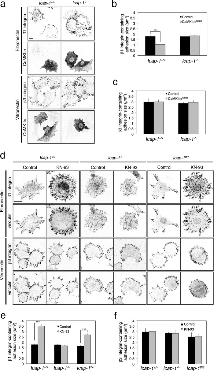
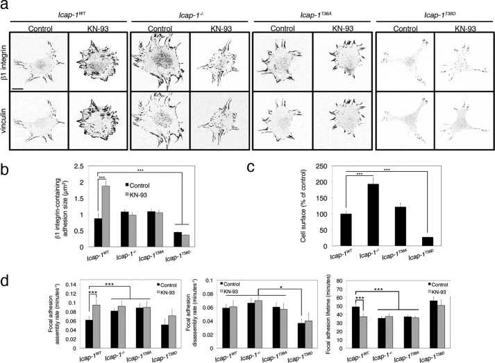
Focal adhesion turnover during cell migration is an integrated cyclic process requiring tight regulation of integrin function. Interaction of integrin with its ligand depends on its activation state, which is regulated by the direct recruitment of proteins onto the β integrin chain cytoplasmic domain. We previously reported that ICAP-1α, a specific cytoplasmic partner of β1A integrins, limits both talin and kindlin interaction with β1 integrin, thereby restraining focal adhesion assembly. Here we provide evidence that the calcium and calmodulin-dependent serine/threonine protein kinase type II (CaMKII) is an important regulator of ICAP-1α for controlling focal adhesion dynamics. CaMKII directly phosphorylates ICAP-1α and disrupts an intramolecular interaction between the N- and the C-terminal domains of ICAP-1α, unmasking the PTB domain, thereby permitting ICAP-1α binding onto the β1 integrin tail. ICAP-1α direct interaction with the β1 integrin tail and the modulation of β1 integrin affinity state are required for down-regulating focal adhesion assembly. Our results point to a molecular mechanism for the phosphorylation-dependent control of ICAP-1α function by CaMKII, allowing the dynamic control of β1 integrin activation and cell adhesion.

Keywords: Adhesion; CaMKII; Focal Adhesion; ICAP-1; Integrins; Membrane Proteins; Signal Transduction.


Figures

FIGURE 1.
FIGURE 1.
CaMKIIα negatively regulates the formation of β1 integrin-dependent adhesion complexes in an ICAP-1α-dependent manner. a, Icap-1+/+ and Icap-1−/− osteoblasts expressing or not the constitutively activated CaMKIIαT286D and spread on FN (10 μg/ml) or on VN (5 μg/ml) for 2 h were immunostained to visualize β1 integrin (using 9EG7 antibody) and CaMKIIα. Scale bar, 10 μm. b and c, quantification of β1 (b) and β3 (c) integrin-containing FA size in Icap-1+/+ and Icap-1−/− osteoblasts expressing or not the constitutively activated CaMKIIαT286D. ***, p < 0.0001. d, Icap-1+/+, Icap-1−/−, and Icap-1WT osteoblasts spread on FN (10 μg/ml) or on VN (5 μg/ml) and treated or not with KN-93 (10 μm) for 2 h were immunostained to visualize vinculin and either activated β1 integrin (using 9EG7 antibody, for FN coating) or β3 integrin (for VN coating). Scale bar, 10 μm. e and f, quantification of β1 (e) and β3 (f) integrin-containing FA size in Icap-1+/+, Icap-1−/−, and Icap-1WT osteoblasts treated or not with KN-93 (10 μm) for 2 h. ***, p value < 0.0001. Error bars, S.D.
FIGURE 2.
FIGURE 2.
CaMKIIα regulates FA dynamics upstream of ICAP-1α. a, time lapse sequences of eGFP-VASP-expressing osteoblasts treated or not with KN-93 (10 μm). The arrows indicate the turnover of a single adhesion complex. Scale bar, 5 μm. b, Icap-1+/+ and Icap-1−/− osteoblasts expressing eGFP-VASP were treated or not with KN-93 (10 μm) for 2 h, and focal adhesion dynamics were recorded. Four parameters were measured: assembly and disassembly rates, adhesion steady state duration, and total lifetime. At least 20 cells and 10 FAs in each cell type were recorded. ***, p < 0.0001. c, Icap-1+/+ and Icap-1−/− osteoblasts spread on FN (10 μg/ml) and treated or not with KN-93 (10 μm) for 16 h were immunostained to visualize vinculin and activated β1 integrin (using 9EG7 antibody). Scale bar, 10 μm. d, quantification of FA numbers in Icap-1+/+ and Icap-1−/− osteoblasts spread on FN (10 μg/ml) and treated or not with KN-93 (10 μm) for 16 h. ***, p value < 0.0001. e, quantification of FA dynamics in Icap-1+/+, Icap-1−/−, and Icap-1WT osteoblasts expressing either eGFP- or mRFP-VASP and treated or not with KN-93 (10 μm) for 16 h. Three parameters were measured: assembly and disassembly rates and the adhesion lifetime. At least 20 cells and 10 FAs in each cell type were recorded. ***, p < 0.0001. Error bars, S.D.
FIGURE 3.
FIGURE 3.
CaMKIIα regulates FA formation and dynamics through ICAP-1α binding to β1 cytodomain. a, β1wild-type, β1D759A, and β1V787T osteoblast cells expressing CaMKIIαT286D mutant were labeled with anti-activated β1 integrin and anti-CaMKIIα antibodies. Scale bar, 10 μm. b, quantification of β1 integrin-containing FA size in β1wild-type, β1D759A, and β1V787T osteoblast cells expressing or not the constitutively activated CaMKIIαT286D. ***, p < 0.0001. c, β1wild-type, β1D759A, and β1V787T osteoblast cells were treated or not with 10 μm KN-93 for 2 h and immunostained to visualize vinculin and activated β1 integrin. Scale bar, 10 μm. d, quantification of β1 integrin-containing FA size in β1wild-type, β1D759A, and β1V787T osteoblast cells treated or not with KN-93 (10 μm) for 2 h. ***, p < 0.0001. e, quantification of FA assembly and disassembly rates and total lifetime in β1wild-type, β1D759A, and β1V787T osteoblast cells expressing eGFP-VASP and treated or not with 10 μm KN-93 for 16 h. ***, p value < 0.0001. Error bars, S.D.
FIGURE 4.
FIGURE 4.
CaMKIIα-mediated down-regulation of FA formation and turnover requires ICAP-1α threonine 38 phosphorylation. a, Icap-1−/−, Icap-1rescue, Icap-1T38A, and Icap-1T38D osteoblasts were treated or not with 10 μm KN-93 for 2 h and immunostained to visualize vinculin and activated β1 integrin. Scale bar, 10 μm. b, quantification of β1 integrin-containing FA size in Icap-1−/−, Icap-1rescue, Icap-1T38A, and Icap-1T38D osteoblasts treated or not with KN-93 (10 μm) for 2 h. ***, p < 0.0001. c, quantification of the surface of Icap-1−/−, Icap-1rescue, Icap-1T38A, and Icap-1T38D osteoblasts. ***, p < 0.0001. d, quantification of FA assembly and disassembly rates and total lifetime in Icap-1−/−, Icap-1rescue, Icap-1T38A, and Icap-1T38D osteoblasts expressing eGFP- or mRFP-VASP and treated or not with 10 μm KN-93 for 16 h. ***, p < 0.0001. Error bars, S.D.
FIGURE 5.
FIGURE 5.
ICAP-1α exists in a close, inactive conformation opened upon threonine 38 phosphorylation. a, pull-down assays between ICAP-1α and β1A integrin cytodomain. Left, CHO cell lysates expressing ICAP-1αwild-type and treated with phosphatase inhibitors or alkaline phosphatase were incubated with either GST alone or fused to the cytoplasmic tail of β1A or β3 integrin. Alternatively (bottom), CHO cell lysates expressing either ICAP-1αwild-type, ICAP-1αT38A, or ICAP-1αT38D mutants were incubated with GST alone or GST-integrin tail fusion proteins. Bound fraction of ICAP-1α was revealed by immunoblotting. b, pull-down assays between kindlin-2 and β1A integrin cytodomain. HEK 293 cells were transiently transfected with pcDNA3.1-ICAPwt or pcDNA3.1-ICAPT38D, respectively, and kindlin-2 associated with β1A and β3 integrin tails fused to GST or GST alone were visualized by Western blotting. c, solid phase assay of the intramolecular interaction between N- and C-terminal ICAP-1α. Increased amounts of either N-terminal ICAP-1wild-type, N-terminal ICAP-1T38A, or N-terminal ICAP-1T38D (1–400 ng) were added to C-terminal ICAP-1 (10 μg) coated onto a 96-well microplate. After washes, the interaction between ICAP-1 fragments was revealed with an anti-ICAP-1 antibody (9B10H) that recognizes the N-terminal region. As a negative control, BSA-coated wells were used to estimate the nonspecific binding of N-terminal ICAP-1. Error bars, S.D. of three independent experiments. d, cells were co-microinjected with the recombinant C-terminal ICAP-1 (residues 100–200) and dextran rhodamine alone or with N-terminal ICAP-1 bearing the Thr-38 phosphomimetic mutations (amino acids 1–99). Cells were fixed and stained for vinculin to visualize adhesion sites. Numbers of cells devoid or not of FAs were counted, and FA disruption efficiency was expressed as a percentage of total cells.
FIGURE 6.
FIGURE 6.
In vitro phosphorylation of ICAP-1α by CaMKIIα. a, ICAP-1α association with αCaMKII was analyzed by immunoprecipitation of FLAG-ICAP-1α in CHO cells expressing or not expressing αCaMKIIT286D. Interaction between CaMKII and ICAP was analyzed after immunoprecipitation of FLAG-ICAP-1, and the presence of CaMKII was detected by Western blotting analysis using polyclonal CaMKII antibody. FLAG-ICAP co-immunoprecipitated ectopically expressed CaMKII (left) or endogenous CaMKII (middle), whereas control immunoprecipitation (when FLAG-ICAP was not expressed) did not immunoprecipitate CaMKII as expected (right). b, CaMKIIα immunoprecipitated from rat brain and recombinant N-terminal ICAP-1 (residues 1–99) were incubated with calmodulin, Ca2+, and [γ-32P]ATP. ICAP-1 phosphorylation was revealed by autoradiography. EGTA was added as a negative control of CaMKII-dependent ICAP-1 phosphorylation. c, schematic representation of ICAP-1α N- and C-terminal structures illustrating ICAP-1 head domain with the kinase phosphorylation sites and the PTB domain containing the β1A integrin binding site. c, His-ICAP-1wild-type or mutated form T38A, immobilized on nickel-nitrilotriacetic acid beads, was incubated with purified CaMKII in the presence of [γ-32P]ATP. GST protein immobilized on glutathione-Sepharose beads was used as control. CaMKII-induced phosphorylation was analyzed by autoradiography (top). The amount of recombinant proteins was determined with Coomassie staining (bottom).
FIGURE 7.
FIGURE 7.
Model of the signaling pathway regulating ICAP-1α function in the dynamics of β1 integrin-mediated FAs. a, schematic representation of ICAP-1. b, ICAP-1 phosphorylation controls β1 integrin activation. In the cytoplasm, ICAP-1α adopts a bent conformation in which the β1 integrin binding site (IBS) is masked by an intramolecular interaction between the N- and the C-terminal moieties. Phosphorylation of threonine 38 by CaMKIIα disrupts this association, releases the PTB domain, and promotes ICAP-1α association with the β1A cytoplasmic tail. ICAP-1α maintains low affinity of β1 integrin to slow down FA assembly and turnover. Illustrated proteins are not to scale.

References

    1. Hynes R. O. (2002) Integrins. Bidirectional, allosteric signaling machines. Cell 110, 673–687 - PubMed
    1. Lauffenburger D. A., Horwitz A. F. (1996) Cell migration. A physically integrated molecular process. Cell 84, 359–369 - PubMed
    1. Xiong J. P., Stehle T., Diefenbach B., Zhang R., Dunker R., Scott D. L., Joachimiak A., Goodman S. L., Arnaout M. A. (2001) Crystal structure of the extracellular segment of integrin α Vβ3. Science 294, 339–345 - PMC - PubMed
    1. Shimaoka M., Takagi J., Springer T. A. (2002) Conformational regulation of integrin structure and function. Annu. Rev. Biophys. Biomol. Struct. 31, 485–516 - PubMed
    1. Takagi J., Petre B. M., Walz T., Springer T. A. (2002) Global conformational rearrangements in integrin extracellular domains in outside-in and inside-out signaling. Cell 110, 599–611 - PubMed

Publication types

MeSH terms

LinkOut - more resources