. 2013 Aug 2;288(31):22681-92.
doi: 10.1074/jbc.M113.472365. Epub 2013 May 28.

Molecular mechanisms, thermodynamics, and dissociation kinetics of knob-hole interactions in fibrin

Affiliations

Molecular mechanisms, thermodynamics, and dissociation kinetics of knob-hole interactions in fibrin

Olga Kononova et al. J Biol Chem. .

Abstract

Polymerization of fibrin, the primary structural protein of blood clots and thrombi, occurs through binding of knobs 'A' and 'B' in the central nodule of fibrin monomer to complementary holes 'a' and 'b' in the γ- and β-nodules, respectively, of another monomer. We characterized the A:a and B:b knob-hole interactions under varying solution conditions using molecular dynamics simulations of the structural models of fibrin(ogen) fragment D complexed with synthetic peptides GPRP (knob 'A' mimetic) and GHRP (knob 'B' mimetic). The strength of A:a and B:b knob-hole complexes was roughly equal, decreasing with pulling force; however, the dissociation kinetics were sensitive to variations in acidity (pH 5-7) and temperature (T = 25-37 °C). There were similar structural changes in holes 'a' and 'b' during forced dissociation of the knob-hole complexes: elongation of loop I, stretching of the interior region, and translocation of the moveable flap. The disruption of the knob-hole interactions was not an "all-or-none" transition as it occurred through distinct two-step or single step pathways with or without intermediate states. The knob-hole bonds were stronger, tighter, and more brittle at pH 7 than at pH 5. The B:b knob-hole bonds were weaker, looser, and more compliant than the A:a knob-hole bonds at pH 7 but stronger, tighter, and less compliant at pH 5. Surprisingly, the knob-hole bonds were stronger, not weaker, at elevated temperature (T = 37 °C) compared with T = 25 °C due to the helix-to-coil transition in loop I that helps stabilize the bonds. These results provide detailed qualitative and quantitative characteristics underlying the most significant non-covalent interactions involved in fibrin polymerization.

Keywords: A:a and B:b Knob-Hole Bonds; Biophysics; Dissociation Kinetics; Fibrin; Fibrinogen; Free Energy Landscape; Molecular Dynamics; Protein-Protein Interactions.


Figures

FIGURE 1.
FIGURE 1.
Ribbon structures of fibrin(ogen) (A), the A:a knob-hole bond (B and C), and the B:b knob-hole bond (D and E). The structures correspond to the A:a knob-hole complex (model system Aa1) and B:b knob-hole complex (system Bb1), respectively, at pH 7 and T = 25 °C. B and D, the interface of the A:a knob-hole complex (B) and B:b knob-hole complex (D) in which the binding determinants, loop I (region I shown in blue), interior region (region II shown in green), and moveable flap (region III shown in red), interact with peptides GPRP and GHRP (shown in orange), respectively. C and E, simulation setup. Holes ‘a’ and ‘b’ are constrained through fixing the C termini of the γ chain (residue γGly160) and β chain (residue βVal205), respectively (see “Experimental Procedures”). A constant pulling force f (represented by the black arrow) is applied to the Pro4 residue of GPRP peptide and Pro4 residue of GHRP peptide in the direction perpendicular to the binding interface to dissociate the knob-hole bond. Also shown are structural details of A:a and B:b knob-hole bonds in which residues in binding regions I–III in holes ‘a’ and ‘b’ establish binding contacts with peptides GPRP and GHRP.
FIGURE 2.
FIGURE 2.
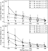
Kinetics of the forced dissociation of the A:a and B:b knob-hole complexes. The average bond lifetimes (τ) with standard deviations (error bars) for the A:a knob-hole complex (model systems Aa1–Aa4; A) and for the B:b knob-hole complex (model systems Bb1–Bb4; B) as a function of pulling force (f) are compared for different ambient conditions (at pH 5 and 7 and T = 25 and 37 °C; see supplemental Tables S1 and S2).
FIGURE 3.
FIGURE 3.
Dependence of kinetic pathways for forced dissociation of the A:a and B:b knob-hole bonds on pH and temperature. Shown are the time-dependent profiles of the total number of binary contacts (Q) stabilizing the A:a knob-hole complex for model systems Aa1 and Aa2 (A) and Aa3 and Aa4 (B) and the B:b knob-hole complex for model systems Bb1 and Bb2 (C) and Bb3 and Bb4 (D). The profiles of Q indicate two distinct dissociation pathways: the one-step pathway of unbinding (BU) from the bound state (B) to the unbound state (U) and the two-step pathway of unbinding (BIU) in which formation of the intermediate state (I) occurs. The time-dependent maps of binary contacts for A:a and B:b knob-hole bond complexes for different pH values and temperature are presented in supplemental Figs. S2 and S3, respectively.
FIGURE 4.
FIGURE 4.
Free energy landscape underlying the thermodynamics of A:a and B:b knob-hole interactions in fibrin. The Gibbs free energy for unbinding, ΔG, for model systems Aa1 and Aa2 (A) and Aa3 and Aa4 (B) and for model systems Bb1 and Bb2 (C) and Bb3 and Bb4 (D) as a function of knob-hole interaction range X are compared for different ambient conditions (at pH 7 and 5 and T = 25 and 37 °C; see supplemental Tables S1 and S2). The standard deviations (error bars) of ΔG are shown. The values of the equilibrium binding energy Gb, the width of the bound state Δx, and the distance between the bound state and transition state Δx shown in A are given in Table 2.
FIGURE 5.
FIGURE 5.
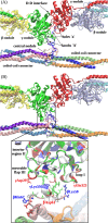
Computational reconstruction of the non-covalent coupling of the central nodule (bearing sites ‘A’) and the γ-nodules (bearing sites ‘a’). A, ribbon representation of the initial structure (before equilibration) of the double-D fragment of abutted fibrin molecules containing two γ- and two β-nodules. The residues in site ‘a’ form binding contacts with the residues of site ‘A’ emanating from the central nodule of the third fibrin monomer between the coiled coil connectors. B shows the translocation of the central nodule following formation of the A:a knob-hole bonds observed at the end of the simulation run. Also shown is the magnified view of electrostatic contacts between residues γGlu323 in loop I, γLys356 in the interior region, and γAsp297 in the moveable flap (all residues belong to site ‘a’ in the γ-nodule) and residues βLys58, βAsp61, and βHis67 in the N-terminal portion of the β chain in the central nodule (the GPR motif has been suppressed for clarity). In the central nodule, the residues colored in red represent negatively charged amino acids, whereas the residues colored in blue represent positively charged amino acids.
FIGURE 6.
FIGURE 6.
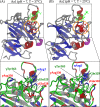
Comparison of the A:a knob-hole interactions in neutral solution (pH 7) at T = 37 °C (model system Aa1; A) and T = 25 °C (model system Aa2; B). Shown are the ribbon structures of binding site ‘a’ interacting with knob ‘A’. Color denotation is as follows: in hole ‘a’, α-helices are shown in red color, β-strands are in blue color, and coils and turns are shown in gray color; knob ‘A’ is displayed in green color. Residues γ327–330 in loop I form an α-helix at 25 °C but transition to a random coil structure at 37 °C. Interacting residues in loop I and GPRP are magnified below. The electrostatic coupling among residue γTyr363; residues γGlu328, γGln329, and γAsp330 in loop I; and residue Arg3 in the GPRP peptide is indicated.

Similar articles

Cited by

References

    1. Weisel J. W. (2004) The mechanical properties of fibrin for basic scientists and clinicians. Biophys. Chem. 112, 267–276 - PubMed
    1. Ferry J. D. (1988) in Biological and synthetic polymer networks (Kramer O., ed) pp. 41–55, Elsevier, Amsterdam
    1. Liu W., Jawerth L. M., Sparks E. A., Falvo M. R., Hantgan R. R., Superfine R., Lord S. T., Guthold M. (2006) Fibrin fibers have extraordinary extensibility and elasticity. Science 313, 634. - PMC - PubMed
    1. Weisel J. W. (2008) Enigmas of blood clot elasticity. Science 320, 456–457 - PubMed
    1. Weisel J. W. (2005) Fibrinogen and fibrin. Adv. Protein Chem. 70, 247–299 - PubMed

Publication types

LinkOut - more resources