Comparative Study
. 2013 Sep;12(9):2640-56.
doi: 10.1074/mcp.M112.024109. Epub 2013 May 29.

Comparing and combining capillary electrophoresis electrospray ionization mass spectrometry and nano-liquid chromatography electrospray ionization mass spectrometry for the characterization of post-translationally modified histones

Affiliations
Comparative Study

Comparing and combining capillary electrophoresis electrospray ionization mass spectrometry and nano-liquid chromatography electrospray ionization mass spectrometry for the characterization of post-translationally modified histones

Bettina Sarg et al. Mol Cell Proteomics. 2013 Sep.

Abstract

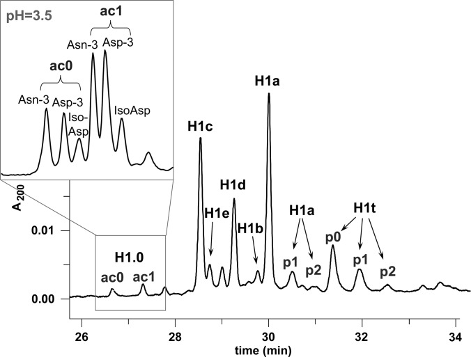
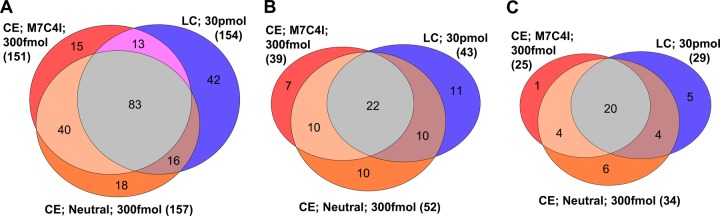
We present the first comprehensive capillary electrophoresis electrospray ionization mass spectrometry (CESI-MS) analysis of post-translational modifications derived from H1 and core histones. Using a capillary electrophoresis system equipped with a sheathless high-sensitivity porous sprayer and nano-liquid chromatography electrospray ionization mass spectrometry (nano-LC-ESI-MS) as two complementary techniques, we characterized H1 histones isolated from rat testis. Without any pre-separation of the perchloric acid extraction, a total of 70 different modified peptides, including 50 phosphopeptides, were identified in the rat linker histones H1.0, H1a-H1e, and H1t. Out of the 70 modified H1 histone peptides, 27 peptides could be identified with CESI-MS only, and 11 solely with LC-ESI-MS. Immobilized metal-affinity chromatography enrichment prior to MS analysis yielded a total of 55 phosphopeptides; 22 of these peptides could be identified only by CESI-MS, and 19 only by LC-ESI-MS, showing the complementarity of the two techniques. We mapped 42 H1 modification sites, including 31 phosphorylation sites, of which 8 were novel sites. For the analysis of core histones, we chose a different strategy. In a first step, the sulfuric-acid-extracted core histones were pre-separated using reverse-phase high-performance liquid chromatography. Individual rat testis core histone fractions obtained in this way were digested and analyzed via bottom-up CESI-MS. This approach yielded the identification of 42 different modification sites including acetylation (lysine and N(α)-terminal); mono-, di-, and trimethylation; and phosphorylation. When we applied CESI-MS for the analysis of intact core histone subtypes from butyrate-treated mouse tumor cells, we were able to rapidly detect their degree of modification, and we found this method very useful for the separation of isobaric trimethyl and acetyl modifications. Taken together, our results highlight the need for additional techniques for the comprehensive analysis of post-translational modifications. CESI-MS is a promising new proteomics tool as demonstrated by this, the first comprehensive analysis of histone modifications, using rat testis as an example.


Figures

Fig. 1.
Fig. 1.
CZE separation of H1 histones from rat testis carried out in 0.5 m sodium phosphate buffer at pH 2.0 containing 0.02% HPMC and (inset) the separation of histone H1.0 carried out in 0.1 m sodium phosphate buffer at pH 3.5 containing 0.02% HPMC. Running conditions were as follows: injection time, 2 s; UV detection at 200 nm; voltage, 12 kV; untreated capillary (50 cm in length; 75 μm inner diameter). ac0, ac1: non-acetylated and monoacetylated forms; p0, p1, p2: non-, mono-, di-, and triphosphorylated forms.
Fig. 2.
Fig. 2.
Base peak electropherograms of rat testis H1 histones digested with endoproteinase Arg-C using positively charged capillaries. A, PEI-coated capillary, separation voltage of −25 kV. B, M7C4I-coated capillary, separation voltage of −25 kV. C, M7C4I-coated capillary, separation voltage of −12.5 kV. BGE: 0.1% (v/v) formic acid. Sample amount: 6.15 ng (300 fmol). Capillary length: 100 cm with porous tip. Inner diameter 30 μm, outer diameter 150 μm.
Fig. 3.
Fig. 3.
Base peak chromatograms of rat testis H1 histones digested with endoproteinase Arg-C using LC-ESI-MS. A, sample amount: 6.15 ng (300 fmol). B, sample amount: 61.5 ng (3 pmol). C, sample amount: 615 ng (30.0 pmol). LC-ESI-MS was performed using a homemade fritless column packed 10 cm with 3 μm reversed-phase C18 (Reprosil). The gradient (solvent A: 0.1% formic acid; solvent B: 0.1% formic acid in 85% acetonitrile) started at 4% B. The concentration of solvent B was increased linearly from 4% to 50% over 50 min and from 50% to 100% over 5 min. A flow rate of 250 nl/min was applied.
Fig. 4.
Fig. 4.
Base peak electropherograms of rat testis H1 histones digested with endoproteinase Arg-C using (A) an M7C4I-coated capillary, a separation voltage of −12.5 kV, and a BGE of 0.3% (v/v) formic acid; (B) an M7C4I-coated capillary, a separation voltage of −12.5 kV, and a BGE of 0.6% (v/v) formic acid; and (C) a neutrally coated capillary, a separation voltage of +30 kV, and a BGE of 10% (v/v) acetic acid. Sample amount: 6.15 ng (300 fmol). Capillary length: 100 cm with porous tip. Inner diameter: 30 μm; outer diameter: 150 μm.
Fig. 5.
Fig. 5.
Number of unmodified and modified histone H1 peptides identified via CESI-MS and LC-ESI-MS/MS analysis. A, number of identified histone H1 peptides (modified and non-modified) merged from triplicate analyses. B, the total number of modified peptides and the distribution of specific types of modifications within each column is shown. The numbers presented in the diagram are the sum of unique modified peptides found in triplicate runs. The overlap of peptides identified with CESI-MS ranged from 75.0% to 84.1%; overlap with LC-ESI-MS ranged from 65.8% to 71.4%.
Fig. 6.
Fig. 6.
Venn diagram showing the overlap of (A) total histone H1 peptides, (B) modified H1 peptides, and (C) modification sites identified by means of CESI-MS and LC-ESI-MS from triplicate runs.
Fig. 7.
Fig. 7.
Mass distribution of histone H1 phosphopeptides obtained with IMAC enrichment using CESI-MS and LC-ESI-MS. Data originate from Table I. Each analysis was performed three times.
Fig. 8.
Fig. 8.
CESI-MS analysis of intact hyperacetylated core histones using an M7C4I-coated capillary (conditions as described in Fig. 2B). A, base peak electropherogram of intact H4. B, mass deconvoluted spectra of individual H4 peaks (1–5). Average mass of histone H4 calculated for ac0me2: 11,330.27 Da (UniProt accession P62806). C, base peak electropherogram of intact H2A.2. D, base peak electropherogram of intact H2A.1. E, base peak electropherogram of intact H2B. F, base peak electropherogram of intact H3.2 + H3.3. ac0, ac1, ac2, ac3, ac4: non-, mono-, di-, tri-, and tetraacetylated forms; me0, me1, me2, me3: non-, mono-, di-, and trimethylated forms.
Fig. 9.
Fig. 9.
Acetylation (A) and methylation (B) levels of hyperacetylated histone H4 in mouse erythroleukemia cells. Acetylation levels were determined by integrating the peak areas of MS1 precursors shown in Fig. 8A and were calculated as the percent area relative to the entire area of H4. The methylation status of each acetylated proteoform was determined individually by computing the monoisotopic mass intensities for the non-, mono-, di-, and trimethylated H4 present in each peak. Analyses are based on triplicate runs.

Similar articles

Cited by

References

    1. Izzo A., Kamieniarz K., Schneider R. (2008) The histone H1 family: specific members, specific functions? Biol. Chem. 389, 333–343 - PubMed
    1. Happel N., Doenecke D. (2009) Histone H1 and its isoforms: contribution to chromatin structure and function. Gene 431, 1–12 - PubMed
    1. Lindner H., Helliger W., Puschendorf B. (1990) Separation of rat tissue histone H1 subtypes by reverse-phase h.p.l.c. identification and assignment to a standard H1 nomenclature. Biochem. J. 269, 359–363 - PMC - PubMed
    1. Wood C., Snijders A., Williamson J., Reynolds C., Baldwin J., Dickman M. (2009) Post-translational modifications of the linker histone variants and their association with cell mechanisms. FEBS J. 276, 3685–3697 - PubMed
    1. Turner B. M. (2000) Histone acetylation and an epigenetic code. Bioessays 22, 836–845 - PubMed

Publication types

MeSH terms

LinkOut - more resources